Safety Evaluation Of Cistanche Tubulosa For Food Use (Oral Toxicology & Genotoxicity)
Jul 22, 2026
If you are developing a Cistanche supplement, functional food, beverage, or cosmetic ingredient for the European or North American market, the first question you will face from regulatory teams, brand partners, and sophisticated distributors is not "Does it work?" but:
Does cistanche safe-especially for repeated oral use and at meaningful doses?
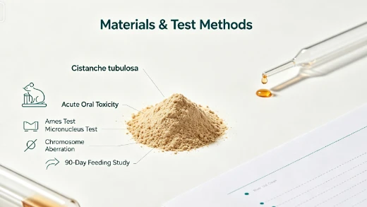
For botanical ingredients, the biggest commercial bottleneck is often toxicology data quality: acute toxicity, genetic toxicology (mutagenicity/clastogenicity), and repeated-dose studies (e.g., 90-day feeding). The Chinese study translated below addresses exactly these points for Cistanche tubulosa (Schrenk) R. Wight using recognized toxicological approaches, including:
Acute oral toxicity (mice and rats)
Three genotoxicity assays
A 90-day oral feeding toxicity study in rats plus a 28-day recovery observation
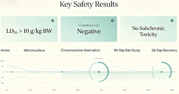
For companies that still worry whether cistanche is safe to take, these results provide a structured safety signal: no acute toxicity, no genotoxicity, and no observed subchronic toxicity under the test conditions.
About our supply chain
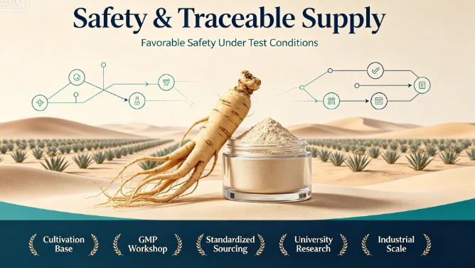
For brands seeking consistent raw material and scalable manufacturing, our upstream and processing capabilities are aligned with industrial compliance and quality needs. According to our company profile, Chengdu Wecistanche Bio‑Tech Co., Ltd. (established 2003) states it operates a complete Cistanche industrial chain, including large-scale cultivation bases, collection and storage capacity, and a GMP production workshop with advanced purification facilities, and multiple certifications/patents; it also describes long-term research collaborations with major universities. This matters because high-quality safety data is best supported by traceable, controlled, and standardized botanical sourcing and processing.
Click The Picture To Get More Details About Wecistanche Factory


Abstract
Cistanche tubulosa (Schrenk) R. Wight, a perennial herbaceous plant of the genus Cistanche (family Orobanchaceae), has a long history of use in China. However, because it has not yet been included in the official catalog of approved food raw materials, industrial development and product innovation have been severely restricted. To verify its safety for consumption, this study systematically conducted a food-safety evaluation of C. tubulosa in reference to relevant Chinese national standards and regulations. The evaluation included acute oral toxicity tests (mice and rats), three genotoxicity tests, and a 90‑day oral feeding toxicity test in rats.
Results showed that the median lethal dose for both male and female mice and rats was LD₅₀ > 10 g/(kg·BW), indicating a non‑toxic classification. The bacterial reverse mutation (Ames) test was negative at all dose levels. In the mammalian erythrocyte micronucleus test, no significant differences were observed in micronucleated polychromatic erythrocyte frequencies between treated groups [7.14,3.57,and1.79g/(kg⋅BW)][7.14, 3.57, and 1.79 g/(kg·BW)][7.14,3.57,and1.79g/(kg⋅BW)] and the solvent control (P > 0.05). The in vitro mammalian cell chromosome aberration test was negative. In the 90‑day oral toxicity study in rats (dietary concentrations of 100 g/kg, 50 g/kg, and 25 g/kg feed), and during the additional 28‑day recovery observation, no delayed toxicity was observed.
In summary, under the experimental conditions of this study, C. tubulosa showed no acute toxicity, no genotoxicity, and no subchronic toxicity, suggesting favorable safety. This research provides experimental support for adding C. tubulosa to China's national "food–medicine substances" catalog and for broader application in functional foods and health products.
Keywords: Cistanche tubulosa; acute oral toxicity; genotoxicity; 90‑day oral toxicity
0 Introduction
Cistanche tubulosa (Schrenk) R. Wight is a perennial herbaceous plant in the genus Cistanche (Orobanchaceae). It is also known by various traditional names such as "Rou Cong Rong," "Zong Rong," "Di Jing," and others [1–2]. Cistanche has been used in China for a long time. The Shennong's Herbal Classic lists it as a "superior grade" herb, describing functions such as supporting the middle, nourishing the five organs, strengthening yin, and benefiting essence [3].
C. tubulosa is one of the botanical sources for Cistanche in the Pharmacopoeia of the People's Republic of China. The Pharmacopoeia records its traditional functions as tonifying kidney yang, replenishing essence and blood, and moistening the intestine to relieve constipation [4]. Modern pharmacological studies indicate broad bioactivities, including kidney support [5], antioxidant and anti‑aging effects [6–9], neuroprotection (e.g., against Alzheimer's and Parkinson's diseases) [10–11], anti‑inflammatory and immunomodulatory effects [12], anti‑viral pneumonia potential [13], inhibition of tumor cells [14], improvement in ischemic stroke outcomes [15], cognitive support [16], liver protection [17], and relief of constipation [18–19].
C. tubulosa is a specialty variety in Xinjiang, China, mainly distributed along the edge of the Taklamakan Desert in southern Xinjiang. Locally it is commonly consumed in diverse ways: brewed as tea, infused in liquor, stewed with meat, served cold as a salad, stir‑fried, or used as filling for meat pies. Public data from China's State Administration for Market Regulation indicates that health foods developed with C. tubulosa as a raw material have reached 14 products, including well‑known brands such as Jinjiu and Amway's ginkgo–cistanche tablets. Xinjiang Hotan is the largest artificial planting area for Cistanche in China, with C. tubulosa exceeding 400,000 mu (over 40,000 hectares), supporting more than 100,000 households and about 200,000 jobs, helping over 200,000 people in desert regions alleviate poverty. A survey estimated that in 2022, C. tubulosa production was about 3,800 tons [20]. However, as output increased, prices continued to decline, largely because it has not been included in the food raw material catalog, limiting deep product development.
On November 17, 2023, China's National Health Commission and the State Administration for Market Regulation issued the "2023 No. 9 Announcement," adding nine substances (including Codonopsis and Cistanche deserticola) to the list of "substances traditionally both food and Chinese medicinal materials" ("food–medicine substances"). However, C. tubulosa was not included. Therefore, without deeper safety studies and progress toward inclusion in the national food–medicine catalog, industrial development of C. tubulosa may be constrained.
Accordingly, this study followed relevant national food safety standards and regulations to systematically conduct acute oral toxicity testing, three genotoxicity studies, and a 90‑day oral toxicity study, aiming to provide comprehensive scientific evidence for inclusion into the food–medicine substances list and broader application in foods and functional foods.
1 Materials and Methods
1.1 Test materials
Cistanche tubulosa was provided by the Xinjiang Uygur Autonomous Region Institute of Materia Medica. Batch No.: XJGH20240203. It was collected in Yutian County, Hotan Prefecture, Xinjiang, and identified as Cistanche tubulosa by Researcher Wang Guoping. The material was pulverized into powder; total weight 18.3 kg.
SPF Kunming mice were purchased from Shanghai JSJ Laboratory Animal Co., Ltd. Production license No.: SCXK (Hu) 2023‑0004.
SPF SD rats (equal numbers of males and females) were purchased from Zhejiang Viton‑Lihua Laboratory Animal Technology Co., Ltd. Production license No.: SCXK (Zhe) 2024‑0001.
Animal room conditions: temperature 20–25 °C, relative humidity 41%–68%. Animal use license No.: SYXK (Hu) 2023‑0046.
Animal diet provided by Shanghai Zhouyu Biotechnology Co., Ltd. Registration No.: Hu Feed Certificate (2021) 04027.
Reagents:
Cyclophosphamide (Sigma‑Aldrich; batch No.: WXBD0289V)
S9 (Molecular Toxicology Inc., USA; batch No.: 4807)
Calf serum (Thermo Fisher Scientific)
Giemsa stain (Beijing Solarbio)
Colchicine (Shanghai Dibai Biotechnology)
1.2 Instruments and equipment
Electronic balances BP310S and BP3100S (Sartorius, Germany); AU680 automatic biochemical analyzer (Beckman Coulter, USA); XN‑1000V automatic hematology analyzer (Sysmex, Japan); Compact X coagulation analyzer (Bayer, Germany); Cobas 6500 urine analyzer (Roche, Shanghai); YZ25B binocular indirect ophthalmoscope (Suzhou 66 Vision); ACS‑3 electronic scale (Shanghai Yamato); STP120 tissue processor, HistoStar embedding station, Varistain Gemini stainer (Thermo Fisher); RM2245 microtome and DM1000 microscope (Leica, Germany).
1.3 Test methods
1.3.1 Acute oral toxicity test
A limit test method was used with SPF Kunming mice and SPF SD rats after 3 days of acclimation. Animals were fasted for 16 hours (water allowed). Ten males and ten females were selected for each species, numbered 1–10 by sex and housed in cages.
A test suspension was prepared by mixing 10 g of C. tubulosa powder with distilled water up to 40 mL. Within 24 hours, animals received two oral gavage administrations separated by 6 hours, at a gavage volume of 20 mL/kg body weight. Body weight was measured weekly. Necropsy was performed on deceased animals and animals sacrificed at the end of observation, with gross pathological examination; histopathology was performed if gross lesions were observed.
1.3.2 Bacterial reverse mutation (Ames) test
A plate incorporation method was used with identified strains TA97a, TA98, TA100, TA102, TA1535, with and without S9 metabolic activation.
First test: 5 doses: 5,000; 1,580; 500; 158; 50 μg/plate
If negative, a second confirmatory test: 5,000; 1,000; 200; 40; 8 μg/plate
Positive, negative, and solvent (DMSO) controls were included, with three replicates per group. Positive controls used (with/without S9) were specified for each strain in the original text.
1.3.3 Mammalian erythrocyte micronucleus test
Based on the mouse acute oral toxicity test, the maximum gavage concentration was 0.357 g/mL. With a maximum gavage volume of 20 mL/kg BW, high/mid/low doses were set at 7.14, 3.57, and 1.79 g/kg BW.
Solutions were prepared at 357 mg/mL (high), then diluted to 179 mg/mL (mid) and 89 mg/mL (low). Cyclophosphamide served as the positive control.
Animals were randomized into 5 groups (10 mice per group, equal sexes): high/mid/low dose, solvent control, and positive control. A 30‑hour two‑dose gavage method was used at 20 mL/kg BW. Six hours after the second gavage, animals were sacrificed; femoral bone marrow was collected, mixed with calf serum, smeared, fixed, and Giemsa‑stained.
For each mouse, 2,000 polychromatic erythrocytes (PCEs) were counted to record micronucleated PCEs and calculate the micronucleus rate. Additionally, 200 erythrocytes were counted to calculate the proportion of PCEs among total erythrocytes.
1.3.4 In vitro mammalian cell chromosome aberration test
This test followed the Chinese national standard [21]. A pretest showed that at 5,000 μg/mL, the cell division index was inhibited by about 50%. The formal test used three doses: 5,000; 2,500; 1,250 μg/mL.
Negative control: serum‑free MEM. Positive controls: mitomycin C (0.25 μg/mL) without S9; cyclophosphamide (20 μg/mL) with S9. After 4 hours exposure, colchicine (1 μg/mL) was added to arrest metaphase. Slides were prepared and Giemsa‑stained. For each group, 100 metaphase cells were analyzed; aberration rate was calculated and compared using χ² tests.
1.3.5 90‑day oral toxicity test in rats
Eighty SPF mature SD rats (equal sexes) were randomized into four groups. The test material was mixed into feed at:
High: 100 g/kg feed
Mid: 50 g/kg feed
Low: 25 g/kg feed
Control: maintenance feed
Each group had 20 rats (equal sexes). Body weight and food intake were recorded weekly; general clinical observation was recorded daily; animals fasted overnight before each blood collection. Ophthalmic examination was conducted in the control and high‑dose groups before and after the dosing period. After stopping administration, a recovery group was observed for 28 days; at the end, urinalysis, hematology and biochemistry were performed, along with necropsy and organ weight measurement.
1.4 Data processing
Excel and SPSS 21.0 were used. Data were expressed as mean ± SD. If normality and homogeneity of variance were met, one-way ANOVA was used, with LSD‑t post‑hoc tests; otherwise, Kruskal–Wallis H tests were used. The micronucleus test used Poisson distribution processing in SPSS 19.0. The chromosome aberration test used χ² tests. Statistical significance: P < 0.05; highly significant: P < 0.01.
2 Results and Analysis
2.1 Acute oral toxicity
After gavage at 20 mL/kg BW, neither mice nor rats exhibited adverse reactions. During the 14‑day observation period, animals showed normal activity and glossy fur, with no signs of poisoning or death. Gross necropsy revealed no abnormalities. The oral LD₅₀ > 10 g/(kg·BW). According to acute toxicity grading, C. tubulosa was classified as non‑toxic.
2.2 Bacterial reverse mutation (Ames) test
The test substance formed a uniformly dispersed suspension in water. At the highest dose (5,000 μg/plate), only minor sediment was observed and did not affect colony counting. Therefore, sterile distilled water was selected as the solvent. A toxicity pretest in TA98 with plate incorporation indicated no bacterial toxicity at 625–5,000 μg/plate.
In both formal tests, for all five strains with and without S9 activation, revertant colony counts in all dose groups were less than twice the solvent control and showed no dose–response relationship. Therefore, the Ames test result was negative.
Table 1. Results of acute oral toxicity test of Cistanche tubulosa
| Species | Sex | Dose (mg/kg) | No. of animals | Body weight (g), Day 0 (mean ± SD) | Day 7 (mean ± SD) | Day 14 (mean ± SD) | Deaths (n) | Mortality (%) |
|---|---|---|---|---|---|---|---|---|
| Mouse | Female | 10,000 | 10 | 20.5 ± 1.1 | 28.1 ± 1.2 | 31.8 ± 1.5 | 0 | 0 |
| Mouse | Male | 10,000 | 10 | 20.6 ± 1.0 | 32.7 ± 1.4 | 38.6 ± 1.8 | 0 | 0 |
| Rat | Female | 10,000 | 10 | 195.2 ± 3.9 | 227.7 ± 9.2 | 245.8 ± 10.1 | 0 | 0 |
| Rat | Male | 10,000 | 10 | 188.0 ± 6.3 | 268.6 ± 12.2 | 327.0 ± 22.5 | 0 |
2.3 Mammalian erythrocyte micronucleus test
Compared with the solvent control, micronucleus rates showed no significant difference in any dose group (P > 0.05). The proportion of PCEs among total erythrocytes was not lower than 20% of the control group. Statistical analysis indicated a negative result under the test conditions.
Table 2. Bacterial reverse mutation assay results (revertants/plate, mean ± SD)
| Dose (μg/plate) | TA97a −S9 | TA97a +S9 | TA98 −S9 | TA98 +S9 | TA100 −S9 | TA100 +S9 | TA102 −S9 | TA102 +S9 | TA1535 −S9 | TA1535 +S9 |
|---|---|---|---|---|---|---|---|---|---|---|
| 50 | 119 ± 12 | 111 ± 7 | 32 ± 3 | 30 ± 1 | 122 ± 12 | 120 ± 10 | 292 ± 16 | 287 ± 15 | 11 ± 2 | 8 ± 3 |
| 158 | 112 ± 8 | 118 ± 13 | 33 ± 4 | 37 ± 6 | 116 ± 8 | 123 ± 7 | 299 ± 15 | 303 ± 17 | 8 ± 1 | 10 ± 3 |
| 500 | 114 ± 10 | 121 ± 10 | 36 ± 3 | 31 ± 6 | 127 ± 11 | 115 ± 12 | 297 ± 16 | 301 ± 16 | 15 ± 2 | 11 ± 5 |
| 1,580 | 119 ± 7 | 133 ± 9 | 31 ± 3 | 36 ± 4 | 120 ± 11 | 135 ± 8 | 296 ± 18 | 300 ± 16 | 10 ± 4 | 12 ± 5 |
| 5,000 | 118 ± 14 | 123 ± 12 | 32 ± 8 | 32 ± 8 | 114 ± 10 | 124 ± 11 | 301 ± 13 | 293 ± 13 | 11 ± 2 | 10 ± 1 |
| Untreated control | 120 ± 12 | 121 ± 12 | 31 ± 3 | 37 ± 3 | 123 ± 8 | 133 ± 7 | 294 ± 11 | 293 ± 16 | 10 ± 2 | 11 ± 2 |
| Solvent control | 112 ± 5 | 123 ± 19 | 35 ± 6 | 39 ± 3 | 118 ± 11 | 121 ± 12 | 291 ± 9 | 302 ± 15 | 7 ± 4 | 12 ± 3 |
| Positive control | 1,908 ± 110 | 2,383 ± 162 | 272 ± 15 | 3,297 ± 199 | 2,325 ± 176 | 2,424 ± 185 | 3,424 ± 284 | 1,194 ± 96 | 1,052 ± 123 | 277 ± 14 |
Table 3. Bacterial reverse mutation assay results (verification; revertants/plate, mean ± SD)
| Dose (μg/plate) | TA97a −S9 | TA97a +S9 | TA98 −S9 | TA98 +S9 | TA100 −S9 | TA100 +S9 | TA102 −S9 | TA102 +S9 | TA1535 −S9 | TA1535 +S9 |
|---|---|---|---|---|---|---|---|---|---|---|
| 50 | 118 ± 9 | 115 ± 8 | 34 ± 5 | 36 ± 6 | 128 ± 7 | 121 ± 6 | 285 ± 21 | 310 ± 10 | 12 ± 5 | 14 ± 1 |
| 158 | 114 ± 8 | 121 ± 7 | 31 ± 1 | 31 ± 2 | 120 ± 5 | 127 ± 7 | 290 ± 12 | 300 ± 14 | 8 ± 7 | 8 ± 2 |
| 500 | 120 ± 8 | 117 ± 10 | 35 ± 5 | 33 ± 3 | 124 ± 11 | 124 ± 9 | 296 ± 17 | 297 ± 13 | 9 ± 2 | 12 ± 3 |
| 1,580 | 115 ± 7 | 114 ± 9 | 32 ± 4 | 36 ± 4 | 115 ± 9 | 123 ± 8 | 294 ± 21 | 295 ± 17 | 11 ± 1 | 9 ± 4 |
| 5,000 | 114 ± 12 | 119 ± 5 | 33 ± 3 | 29 ± 5 | 123 ± 1 | 119 ± 10 | 283 ± 19 | 296 ± 16 | 11 ± 5 | 10 ± 2 |
| Untreated control | 112 ± 5 | 129 ± 10 | 31 ± 2 | 33 ± 2 | 118 ± 3 | 128 ± 13 | 299 ± 14 | 290 ± 19 | 9 ± 5 | 12 ± 4 |
| Solvent control | 113 ± 6 | 118 ± 9 | 30 ± 3 | 35 ± 3 | 121 ± 9 | 119 ± 16 | 298 ± 15 | 293 ± 15 | 7 ± 3 | 10 ± 3 |
| Positive control | 1,956 ± 254 | 2,205 ± 162 | 277 ± 19 | 3,297 ± 271 | 2,289 ± 178 | 2,328 ± 172 |
2.4 In vitro chromosome aberration test
In the pretest, after 4 hours exposure with or without S9, the dose that produced approximately 50% inhibition of cell division index was 5,000 μg/mL. In the formal test, with or without S9 activation, the chromosome aberration rates at high/mid/low doses did not differ significantly from the negative control (P > 0.05). The result was negative.
2.5 90‑day oral toxicity study in rats
2.5.1 Effects on growth and body weight
During dosing, rats were in good condition with no obvious poisoning signs; no delayed toxicity was observed during the 28‑day recovery period. Over 90 days, body weight increased with age in both sexes. Overall body weight, weight gain, and feed utilization showed no significant differences compared with controls (P > 0.05).
Table 4. Mammalian erythrocyte micronucleus test results
| Group | Dose | Sex | No. of animals | PCE counted (cells/animal) | Micronucleated PCE (mean ± SD) | PCE / total erythrocytes (%) (mean ± SD) | Micronucleus rate (‰) (mean ± SD) |
|---|---|---|---|---|---|---|---|
| Test substance | 7.14 g/kg | Female | 5 | 10,000 | 3.20 ± 1.10 | 54.20 ± 1.60 | 1.60 ± 0.55 |
| Test substance | 7.14 g/kg | Male | 5 | 10,000 | 3.60 ± 1.14 | 54.50 ± 1.77 | 1.80 ± 0.57 |
| Test substance | 3.57 g/kg | Female | 5 | 10,000 | 3.80 ± 1.79 | 54.70 ± 2.49 | 1.90 ± 0.89 |
| Test substance | 3.57 g/kg | Male | 5 | 10,000 | 3.20 ± 1.64 | 55.60 ± 1.92 | 1.60 ± 0.82 |
| Test substance | 1.79 g/kg | Female | 5 | 10,000 | 3.40 ± 0.89 | 54.80 ± 1.60 | 1.70 ± 0.45 |
| Test substance | 1.79 g/kg | Male | 5 | 10,000 | 3.80 ± 1.48 | 54.40 ± 1.47 | 1.90 ± 0.74 |
| Solvent control | Distilled water (20 g/kg) | Female | 5 | 10,000 | 3.60 ± 1.82 | 54.90 ± 1.43 | 1.80 ± 0.91 |
| Solvent control | Distilled water (20 g/kg) | Male | 5 | 10,000 | 3.40 ± 0.89 | 54.50 ± 1.12 | 1.70 ± 0.45 |
| Positive control | Cyclophosphamide (40 mg/kg) | Female | 5 | 10,000 | 43.20 ± 2.28 | 48.10 ± 1.44 | 21.60 ± 1.14** |
| Positive control | Cyclophosphamide (40 mg/kg) | Male | 5 | 10,000 | 43.60 ± 3.21 | 48.40 ± 1.71 | 21.80 ± 1.60** |
Note: Compared with the solvent control group, P < 0.01.
Table 5. In vitro mammalian cell chromosome aberration test results (−S9, 4 h)
| Group (μg/mL) | Breaks | Fragments | Rings | Dicentrics | Quadriradials | Micronuclei | Polyploidy | Pulverization | Cells observed (n) | Aberrant cells (n) | Aberration rate (%) | P value |
|---|---|---|---|---|---|---|---|---|---|---|---|---|
| Negative control | 0 | 0 | 0 | 0 | 0 | 0 | 0 | 0 | 100 | 0 | 0 | / |
| Low dose 1,250 | 0 | 0 | 0 | 1 | 0 | 0 | 0 | 0 | 100 | 2 | 2 | >0.05 |
| Mid dose 2,500 | 0 | 0 | 0 | 0 | 0 | 0 | 0 | 0 | 100 | 0 | 0 | >0.05 |
| High dose 5,000 | 0 | 0 | 0 | 1 | 1 | 0 | 0 | 0 | 100 | 1 | 1 | >0.05 |
| Positive control (Mitomycin C 0.25) | 0 | 1 | 15 | 8 | 18 | 11 | 0 | 0 | 100 | 32 | 32 | <0.01 |
Note: Chromosome aberration rate analyzed by χ² test.
Table 6. In vitro mammalian cell chromosome aberration test results (+S9, 4 h)
| Group (μg/mL) | Breaks | Fragments | Rings | Dicentrics | Quadriradials | Micronuclei | Polyploidy | Pulverization | Cells observed (n) | Aberrant cells (n) | Aberration rate (%) | P value |
|---|---|---|---|---|---|---|---|---|---|---|---|---|
| Negative control | 0 | 0 | 0 | 0 | 0 | 0 | 0 | 0 | 100 | 0 | 0 | / |
| Low dose 1,250 | 0 | 1 | 0 | 0 | 1 | 0 | 0 | 0 | 100 | 2 | 2 | >0.05 |
| Mid dose 2,500 | 0 | 0 | 0 | 0 | 0 | 0 | 0 | 0 | 100 | 0 | 0 | >0.05 |
| High dose 5,000 | 0 | 1 | 0 | 0 | 0 | 0 | 0 | 0 | 100 | 1 | 1 | >0.05 |
| Positive control (Cyclophosphamide 20) | 0 | 1 | 5 | 2 | 15 | 18 | 0 | 0 | 100 | 31 | 31 | <0.01 |
Note: Chromosome aberration rate analyzed by χ² test.
2.5.2 Effects on hematology
Compared with controls, neutrophils decreased in low- and mid-dose female rats (P < 0.05), but values remained within normal ranges and showed no dose–effect relationship; thus, it was considered not biologically meaningful. At the end of the study, white blood cells and differential counts, hemoglobin, hematocrit, and coagulation indices showed no significant differences between dose groups and controls (P > 0.05).
2.5.3 Effects on serum biochemistry
AST and ALT are key enzymes for evaluating liver function abnormalities [22–23]. After 90 days, there were no significant differences in serum biochemistry between treated and control rats of either sex (P > 0.05).
2.5.4 Effects on urinalysis
Urinalysis indicators showed no significant differences between treated groups and controls during dosing (P > 0.05). At mid‑study and at the end of recovery, urinalysis in the high‑dose group also showed no significant differences compared with controls (P > 0.05).
2.5.5 Effects on organs (organ-to-body weight ratios)
Organ coefficients are used to evaluate potential effects on specific organs and development [24]. At study end, fasted body weight and organ coefficients for brain, heart, thymus, adrenal glands, liver, kidneys, spleen, testes/uterus, epididymis/ovaries showed no significant differences compared with controls (P > 0.05).
2.5.6 Ophthalmic examination
During the study, no abnormalities were observed in high‑dose or control rats: no conjunctival congestion or edema, no secretions; corneas were transparent without ulcers; normal iris coloration; clear fundus; transparent lens without opacity; pupils responded normally to light.
2.5.7 Histopathology
Animals were in good condition and showed no obvious poisoning signs. At the end of dosing, differences in food intake, body weight, weight gain, and feed utilization compared with controls were not biologically meaningful. Ophthalmic examinations, urinalysis, hematology, and serum biochemistry showed no significant differences. Histopathology did not reveal organ toxicity changes attributable to the test material.
3 Discussion
Safety toxicology evaluation is a prerequisite for basic research and application development of food–medicine substances. The Pharmacopoeia of the People's Republic of China includes two types of Cistanche: Cistanche deserticola has been officially approved as a food–medicine substance in 2023, significantly promoting regional industrial development, while Cistanche tubulosa has not yet been included due to limited toxicology data.
Table 7. Hematological examination results of rats (mean ± SD, n=10n=10n=10)
Unit notes (as shown in the table):
WBC 109/L10^9/L109/L; Lymphocyte/Neutrophil/Monocyte %; Eosinophil %; Basophil %; RBC 1012/L10^{12}/L1012/L; Hemoglobin g/L; Hematocrit %; Coagulation time s; Activated partial thromboplastin time (APTT) s.
Male rats
| Group | WBC | Lymphocyte (%) | Neutrophil (%) | Monocyte (%) | Eosinophil (%) | Basophil (%) | RBC | Hemoglobin | Hematocrit (%) | Coagulation time (s) | APTT (s) |
|---|---|---|---|---|---|---|---|---|---|---|---|
| Control | 6.28 ± 2.14 | 74.4 ± 6.2 | 6.9 ± 1.4 | 17.4 ± 5.3 | 1.2 ± 0.7 | 0.1 ± 0.1 | 1,148 ± 184 | 8.28 ± 0.27 | 147 ± 5 | 43.4 ± 1.2 | 14.8 ± 2.2 |
| Low dose | 4.55 ± 1.37 | 73.8 ± 5.5 | 5.7 ± 1.2 | 19.5 ± 5.0 | 0.9 ± 0.4 | 0.1 ± 0.1 | 1,107 ± 149 | 8.41 ± 0.48 | 151 ± 7 | 44.1 ± 2.0 | 16.6 ± 1.5 |
| Mid dose | 4.49 ± 1.46 | 76.9 ± 2.6 | 6.2 ± 1.6 | 16.0 ± 1.6 | 0.9 ± 0.4 | 0.1 ± 0.1 | 1,069 ± 85 | 8.14 ± 0.33 | 149 ± 5 | 43.6 ± 1.3 | 16.4 ± 1.4 |
| High dose | 6.08 ± 2.17 | 76.9 ± 4.2 | 5.3 ± 1.2 | 17.0 ± 3.7 | 0.7 ± 0.4 | 0.1 ± 0.1 | 1,038 ± 307 | 8.28 ± 0.35 | 150 ± 6 | 44.1 ± 1.4 | 14.5 ± 1.3 |
Female rats
| Group | WBC | Lymphocyte (%) | Neutrophil (%) | Monocyte (%) | Eosinophil (%) | Basophil (%) | RBC | Hemoglobin | Hematocrit (%) | Coagulation time (s) | APTT (s) |
|---|---|---|---|---|---|---|---|---|---|---|---|
| Control | 2.28 ± 1.30 | 71.5 ± 6.2 | 8.1 ± 1.8 | 19.1 ± 5.2 | 1.3 ± 0.5 | 0.0 ± 0.0 | 1,112 ± 115 | 7.56 ± 0.34 | 141 ± 7 | 40.7 ± 2.1 | 10.7 ± 0.9 |
| Low dose | 2.36 ± 0.98 | 74.3 ± 5.2 | 6.0 ± 1.4 | 18.6 ± 5.0 | 1.1 ± 0.8 | 0.0 ± 0.0 | 998 ± 91 | 7.57 ± 0.51 | 144 ± 7 | 41.2 ± 1.8 | 10.9 ± 0.7 |
| Mid dose | 2.42 ± 0.78 | 73.9 ± 5.2 | 6.1 ± 1.4 | 18.7 ± 4.8 | 1.2 ± 0.6 | 0.0 ± 0.0 | 1,060 ± 116 | 7.59 ± 0.35 | 142 ± 5 | 40.7 ± 1.3 | 10.9 ± 0.3 |
| High dose | 2.26 ± 1.01 | 72.1 ± 7.4 | 7.1 ± 2.3 | 19.3 ± 6.3 | 1.6 ± 0.6 | 0.0 ± 0.1 | 1,065 ± 97 | 7.35 ± 0.26 | 138 ± 5 | 39.9 ± 1.3 | 10.6 ± 0.3 |
Note: Compared with the control group, P < 0.05.
Table 8. Rat blood biochemical examination results (mean ± SD, n=10n=10n=10)
Parameters / units (as shown):
ALT (U/L), AST (U/L), ALP (U/L), Total protein (g/L), Albumin (g/L), Urea nitrogen (mmol/L), Creatinine (μmol/L), Glucose (mmol/L), Cholesterol (mmol/L), Triglyceride (mmol/L), Potassium (mmol/L), Sodium (mmol/L), Chloride (mmol/L)
Male rats
| Group | ALT | AST | ALP | Total protein | Albumin | Urea nitrogen | Creatinine | Glucose | Cholesterol | Triglyceride | Potassium | Sodium | Chloride |
|---|---|---|---|---|---|---|---|---|---|---|---|---|---|
| Control | 160.3 ± 21.0 | 28.4 ± 3.8 | 91.7 ± 14.2 | 60.8 ± 2.3 | 31.2 ± 1.2 | 6.08 ± 0.95 | 54.3 ± 5.4 | 7.9 ± 1.3 | 0.54 ± 0.20 | 1.99 ± 0.26 | 5.53 ± 0.15 | 142.2 ± 1.3 | 104.5 ± 1.0 |
| Low dose | 164.6 ± 16.0 | 32.6 ± 6.8 | 103.2 ± 19.3 | 60.6 ± 1.4 | 30.9 ± 1.1 | 5.47 ± 0.52 | 57.1 ± 4.9 | 7.5 ± 1.1 | 0.51 ± 0.10 | 1.89 ± 0.44 | 5.61 ± 0.25 | 142.9 ± 1.0 | 105.0 ± 1.2 |
| Mid dose | 160.8 ± 26.3 | 31.7 ± 4.7 | 97.9 ± 13.7 | 59.0 ± 1.5 | 30.1 ± 0.8 | 5.87 ± 0.69 | 58.2 ± 4.0 | 7.1 ± 0.9 | 0.36 ± 0.07 | 1.82 ± 0.33 | 5.57 ± 0.16 | 143.1 ± 1.4 | 105.3 ± 1.2 |
| High dose | 163.5 ± 32.4 | 32.1 ± 4.8 | 99.8 ± 11.5 | 60.2 ± 0.9 | 30.6 ± 0.6 | 5.43 ± 0.62 | 53.6 ± 2.7 | 7.7 ± 1.4 | 0.48 ± 0.10 | 1.93 ± 0.33 | 5.73 ± 0.45 | 143.0 ± 0.9 | 104.7 ± 0.9 |
Female rats
| Group | ALT | AST | ALP | Total protein | Albumin | Urea nitrogen | Creatinine | Glucose | Cholesterol | Triglyceride | Potassium | Sodium | Chloride |
|---|---|---|---|---|---|---|---|---|---|---|---|---|---|
| Control | 156.2 ± 43.8 | 33.9 ± 10.8 | 35.8 ± 2.7 | 73.9 ± 5.2 | 42.3 ± 2.2 | 6.61 ± 1.14 | 64.8 ± 3.0 | 7.8 ± 1.0 | 0.44 ± 0.06 | 2.59 ± 0.63 | 3.93 ± 0.27 | 142.4 ± 2.7 | 105.0 ± 1.0 |
| Low dose | 132.5 ± 34.0 | 34.4 ± 15.8 | 38.1 ± 10.3 | 73.0 ± 4.1 | 40.5 ± 3.9 | 6.31 ± 0.85 | 65.2 ± 5.6 | 8.1 ± 1.6 | 0.36 ± 0.06 | 2.25 ± 0.65 | 4.02 ± 0.20 | 143.1 ± 1.1 | 105.7 ± 2.2 |
| Mid dose | 175.9 ± 32.7 | 45.5 ± 18.8 | 35.7 ± 8.1 | 73.4 ± 13.3 | 41.4 ± 2.0 | 6.08 ± 0.97 | 62.0 ± 4.5 | 7.7 ± 1.1 | 0.37 ± 0.12 | 2.47 ± 0.33 | 3.99 ± 0.20 | 142.5 ± 2.1 | 104.8 ± 2.0 |
| High dose | 138.9 ± 50.1 | 41.9 ± 19.1 | 36.5 ± 8.7 | 74.4 ± 2.7 | 42.0 ± 1.3 | 6 |
Table 9. Urine index detection results of rats (n = 10)
Male rats
| Group | Appearance (Normal) | Appearance (Abnormal) | pH (mean ± SD) | Specific gravity (mean ± SD) | Protein (P/N) | Occult blood (P/N) | Glucose (P/N) |
|---|---|---|---|---|---|---|---|
| Control | 10 | 0 | 7.3 ± 0.6 | 1.033 ± 0.011 | 0/10 | 1/10 | 0/10 |
| Low dose | 10 | 0 | 6.8 ± 0.9 | 1.033 ± 0.011 | 0/10 | 2/10 | 0/10 |
| Mid dose | 10 | 0 | 6.9 ± 0.5 | 1.029 ± 0.011 | 0/10 | 1/10 | 0/10 |
| High dose | 10 | 0 | 6.8 ± 0.5 | 1.026 ± 0.006 | 0/10 | 1/10 | 0/10 |
Female rats
| Group | Appearance (Normal) | Appearance (Abnormal) | pH (mean ± SD) | Specific gravity (mean ± SD) | Protein (P/N) | Occult blood (P/N) | Glucose (P/N) |
|---|---|---|---|---|---|---|---|
| Control | 10 | 0 | 6.3 ± 0.4 | 1.026 ± 0.015 | 1/10 | 1/10 | 0/10 |
| Low dose | 10 | 0 | 6.1 ± 0.3 | 1.034 ± 0.011 | 0/10 | 0/10 | 0/10 |
| Mid dose | 10 | 0 | 6.2 ± 0.3 | 1.022 ± 0.007 | 1/10 | 0/10 | 0/10 |
| High dose | 10 | 0 | 6.1 ± 0.2 | 1.038 ± 0.008 | 2/10 | 0/10 | 0/10 |
Note: P/N = number of positive animals / total animals in the group.
Table 10. Organ/body weight ratio of rats (mean ± SD, n = 10)
Male rats
| Group | Brain/BW | Heart/BW | Thymus/BW | Adrenal/BW | Liver/BW | Kidney/BW | Spleen/BW | Testes (or uterus)/BW | Epididymis (or ovary)/BW |
|---|---|---|---|---|---|---|---|---|---|
| Control | 0.39 ± 0.04 | 0.36 ± 0.05 | 0.107 ± 0.020 | 0.014 ± 0.004 | 2.67 ± 0.19 | 0.63 ± 0.03 | 0.17 ± 0.03 | 0.66 ± 0.05 | 0.30 ± 0.04 |
| Low dose | 0.40 ± 0.04 | 0.34 ± 0.03 | 0.101 ± 0.018 | 0.016 ± 0.005 | 2.69 ± 0.15 | 0.61 ± 0.06 | 0.15 ± 0.02 | 0.66 ± 0.07 | 0.31 ± 0.06 |
| Mid dose | 0.41 ± 0.05 | 0.38 ± 0.08 | 0.110 ± 0.019 | 0.017 ± 0.002 | 2.65 ± 0.17 | 0.60 ± 0.06 | 0.18 ± 0.02 | 0.70 ± 0.08 | 0.32 ± 0.06 |
| High dose | 0.40 ± 0.02 | 0.35 ± 0.04 | 0.104 ± 0.015 | 0.015 ± 0.005 | 2.71 ± 0.31 | 0.64 ± 0.06 | 0.17 ± 0.02 | 0.71 ± 0.07 | 0.32 ± 0.05 |
Female rats
| Group | Brain/BW | Heart/BW | Thymus/BW | Adrenal/BW | Liver/BW | Kidney/BW | Spleen/BW | Testes (or uterus)/BW | Epididymis (or ovary)/BW |
|---|---|---|---|---|---|---|---|---|---|
| Control | 0.69 ± 0.05 | 0.41 ± 0.04 | 0.145 ± 0.024 | 0.035 ± 0.011 | 3.06 ± 0.28 | 0.69 ± 0.04 | 0.18 ± 0.02 | 0.28 ± 0.05 | 0.06 ± 0.01 |
| Low dose | 0.70 ± 0.09 | 0.39 ± 0.04 | 0.153 ± 0.032 | 0.029 ± 0.009 | 2.92 ± 0.30 | 0.67 ± 0.05 | 0.18 ± 0.02 | 0.28 ± 0.06 | 0.06 ± 0.01 |
| Mid dose | 0.70 ± 0.08 | 0.41 ± 0.05 | 0.136 ± 0.017 | 0.030 ± 0.010 | 3.09 ± 0.27 | 0.76 ± 0.07 | 0.18 ± 0.02 | 0.26 ± 0.05 | 0.06 ± 0.01 |
| High dose | 0.69 ± 0.06 | 0.39 ± 0.03 | 0.138 ± 0.012 | 0.029 ± 0.006 | 3.18 ± 0.30 | 0.73 ± |
C. tubulosa has a long history of folk consumption in Xinjiang minority areas. The research group previously performed preliminary safety assessments of its water extract and total extract, confirming no obvious toxic or side effects under recommended consumption doses [25–26]. Building on that foundation and aligned with current Chinese food safety standards, this study conducted a more systematic safety evaluation of raw powder, including acute toxicity, three genotoxicity tests, and a 90‑day oral subchronic toxicity study.
Results indicated that under the experimental conditions, C. tubulosa did not exhibit acute toxicity, genotoxicity, or subchronic toxicity, and no teratogenicity was observed. The 90‑day study established the NOAEL at 100 g/kg feed, corresponding to actual intakes of 7.49 g/kg BW (male rats) and 8.76 g/kg BW (female rats).
In Xinjiang and other regions, C. tubulosa is consumed in diverse traditional ways (cold dishes, tea, soups, stews, cooking, liquor infusion, fillings). Under normal traditional consumption patterns, there have been no reports of adverse reactions. Considering its consumption history, current folk usage, and modern toxicological evidence, the food safety risk of C. tubulosa is very low, and it has favorable conditions for development and application as a food–medicine substance.
4 Conclusion
Following China's national food safety standard toxicological evaluation procedures, this study systematically evaluated the safety of Cistanche tubulosa:
Acute oral toxicity (mice): LD₅₀ > 10 g/(kg·BW), classified as non‑toxic.
Three genotoxicity tests: Ames test, mammalian micronucleus test, and in vitro chromosome aberration test were all negative, indicating no mutagenic potential.
90‑day oral toxicity (rats): no treatment-related abnormalities in body weight, hematology, serum biochemistry, organ coefficients, or histopathology.
Overall, under the experimental conditions, C. tubulosa showed no acute toxicity, no genotoxicity, and no subchronic toxicity, supporting favorable food-use safety.
References
[1] Editorial Committee of Chinese Materia Medica, State Administration of Traditional Chinese Medicine of the People's Republic of China. Chinese Materia Medica‑1 [M]. Shanghai: Shanghai Scientific and Technical Publishers, 1999: 509–513.
[2] Liu Y M. Pharmacography of Uighur [M]. Urumqi: Xinjiang Science, Technology and Public Health Press, 1999: 284–290.
[3] Gu G G. Shennong's Herbal Classic [M]. 3rd ed. Beijing: Xueyuan Press, 2007: 73.
[4] Chinese Pharmacopoeia Commission. Pharmacopoeia of the People's Republic of China-Part I: 2025 Edition [M]. Beijing: China Medical Science and Technology Press, 2025: 135–136.
[5] Wang Q X, Dong J T, Lu W J, et al. Phenylethanol glycosides from Cistanche tubulosa improved reproductive dysfunction by regulating testicular steroids through CYP450‑3β‑HSD pathway [J]. Journal of Ethnopharmacology, 2020, 251: 112500.
[6] Abulimiti Shamixinuer, Shawuti Yilizire, Yan Ming, et al. Fractionation and antioxidant activity analysis of polysaccharides from Cistanche deserticola residue using membrane separation technology [J]. Science and Technology of Food Industry, 2026, 1(27): 1–18.
[7] Aidiresi Salamaiti, Yilihamu Gulifeire, Tiemuer Atawula, et al. Study on optimized processing technology of Herba Cistanche Tubulosa decoction pieces and its mechanism in regulating macrophage anti‑oxidative stress [J]. Journal of Li‑Shizhen Traditional Chinese Medicine, 2025, 36(23): 4491–4499.
[8] Li X, Li C T, Zhang W Y, et al. Inflammation and aging: signaling pathways and intervention therapies [J]. Signal Transduction and Targeted Therapy, 2023, 8: 239.
[9] Gao Y, Li B, Liu H, et al. Cistanche deserticola polysaccharides alleviate cognitive decline in aging model mice by restoring the gut microbiota‑brain axis [J]. Aging, 2021, 13(11): 15320–15335.
[10] Tu P F, Jiang Y. Update classical literature research on Cistanches Herba [J]. China Journal of Chinese Materia Medica, 2022, 47(20): 5670–5679.
[11] Fan L, Peng Y, Chen X N, et al. Integrated analysis of phytochemical composition, pharmacokinetics, and network pharmacology to probe distinctions between the stems of Cistanche deserticola and C. tubulosa based on antidepressant activity [J]. Food & Function, 2022, 13(16): 8542–8557.
[12] Zhou T Y, Tian N, Li L, et al. Iridoids modulate inflammation in diabetic kidney disease: a review [J]. Journal of Integrative Medicine, 2024, 22(3): 210–222.
[13] Huang D Y, Luo Y X, Zheng W D, et al. Anti‑coronavirus and anti‑pulmonary inflammation effects of iridoids, the common component from Chinese herbal medicines for the treatment of COVID‑19 [J]. Journal of Natural Medicines, 2024, 78(4): 1003–1012.
[14] Feng D, Zhou S Q, Zhou Y X, et al. Effect of total glycosides of Cistanche deserticola on the energy metabolism of human HepG2 cells [J]. Frontiers in Nutrition, 2023, 10: 1117364.
[15] Liu J J, Wang Y Y, Li Q Y, et al. Phenylethanoid glycosides derived from Cistanche deserticola promote neurological functions and the proliferation of neural stem cells for improving ischemic stroke [J]. Biomedicine & Pharmacotherapy, 2023, 167: 115507.
[16] Ye H X, Qi Y Q, He Y X, et al. Study on the effect of compatibility of total glycosides of Cistanche deserticola and Lycium barbarum polysaccharide on promoting intelligence in C57BL/6J mice of different months [J]. Journal of Chinese Medicinal Materials, 2023, 46(1): 217–221.
[17] Wang H C, Li Y Y, Bian Y F, et al. Potential hepatoprotective effects of Cistanche deserticola Y. C. Ma: Integrated phytochemical analysis using UPLC‑Q‑TOF‑MS/MS, target network analysis, and experimental assessment [J]. Frontiers in Pharmacology, 2022, 13: 1018572.
[18] Zhang X, Zheng F J, Zhang Z. Therapeutic effect of Cistanche deserticola on defecation in senile constipation rat model through stem cell factor/C‑kit signaling pathway [J]. World Journal of Gastroenterology, 2021, 27(32): 5392–5403.
[19] Liu Y Z, Jia X M, Guo H Z, et al. Effects of Cistanches Herba polysaccharides on rats with constipation‑predominant irritable bowel syndrome [J]. China Pharmacy, 2023, 34(18): 2208–2212.
[20] Wu M, Shen F. Present situation and countermeasures of Cistanche deserticola industry development in Xinjiang [J]. Forestry of Xinjiang, 2022(3): 24–26.
[21] National Health and Family Planning Commission of the People's Republic of China. National Food Safety Standard-In Vitro Mammalian Cell Chromosome Aberration Test: GB 15193.23‑2014 [S]. Beijing: China Standard Press, 2015.
[22] Wu D, Wu J J, Cheng X Y, et al. Safety assessment of marigold flavonoids from marigold inflorescence residue [J]. Journal of Ethnopharmacology, 2022, 297: 115520.
[23] Kong Q X, Liu H, Wang J, et al. Study on background reference value range of hematology, serum biochemistry and hemagglutination indexes of SD rats and Beagle dogs [J]. Drug Evaluation Research, 2021, 44(12): 2601–2607.
[24] Yan S P, Wang S, Chen T, et al. Preliminary toxicity evaluation of anthocyanins extract from Lycium ruthenicum [J]. Modern Food Science and Technology, 2023, 39(9): 33–41.
[25] Zhang J, Qing D G, Sun Y, et al. Safety evaluation study of the extracts of Cistanche tubulosa [J]. Chinese Journal of Traditional Medical Science and Technology, 2014, 21(4): 413–414.
[26] Zhang J, Qing D G, Jia X G, et al. Safety evaluation study of the extracts of Cistanche tubulosa [J]. Journal of Xinjiang Medical University, 2012, 35(7): 870–873.