

Hyperoside Attenuate Inflammation in HT22 Cells Via Upregulating SIRT1 To Activities Wnt/β-Catenin And Sonic Hedgehog Pathways Part 2
Mar 30, 2022
Please contact oscar.xiao@wecistanche.com for more information
3. Results
3.1. Hyperoside Alleviated Apoptosis and Inflammation in the LPS-Induced HT22 Cells. The viability of HT22 cells was detected using the MTT assay. The result showed that hyperoside had little effect on HT22 cell viability (Figure 1(a)). Compared with untreated cells, the treatment of LPS(1μg/ml) significantly reduced HT22 cell viability (Figure 1(b)). Based on the MTT results, we choose 20μM hyperoside to evaluate the protective effect on HT22 cells. The apoptotic protein expression of HT22 cells was detected using western blotting; the results showed that treatment with 20μM hyperoside significantly enhanced the antiapoptotic Bcl-2 expression and decreased the proapoptotic Bax and caspase-3 expression(Figure 1(c)). Simultaneously, 20 uM hyperoside treatment significantly inhibited LPS-induced apoptosis of HT22 cells(Figure 1(d). Likewise, treatment with 20μM hyperoside suppressed LPS-induced production of I-1β,IL-6, IL-8, and TNF-α(Figure 1(e). These results suggested that hyperoside alleviated apoptosis and inflammation in the LPS-induced HT22 cells.

Please click here to know more
3.2. Hyperoside Alleviated Oxidative Stress and Reduction of Neurotrophic Factor in the LPS-Induced HT22 Cells.
The oxidative stress of HT22 cells increased, and the production of neurotrophic factors decreased after LPS treatment. Next, we studied the effect of hyperoside on oxidative stress and neurotrophic factors in LPS-induced HT22 cells. The results showed that treatment with 20μM hyperoside significantly increased the levels of SOD, GSH, CAT, and neurotrophic factors BDNF, TrkB, NGF and decreased the levels of ROS and MDA(Figures 2(a) and 2(b)). These results suggested that hyperoside alleviated LPS-induced oxidative stress and reduction of neurotrophic factors in HT22 cells.

3.3.Hyperoside Inhibits LPS-Induced HT22 Cell Apoptosis and Inflammation through SIRT1.
SIRT1 alleviated nerve damage; to find out whether hyperoside could ameliorate apoptosis and inflammation in the LPS-induced by acting on SIRT1, SITRI inhibitors (nicotinamide (NAM)) were used to treat cells. Western blotting showed that SITR1 expression decreased in LPS-induced cells; simultaneously, compared with LPS induction,hyperoside treatment upregulated the expression level of SITR1. In addition, NAM treatment decreased the expression of SIRT1 in HT22 cells compared with the treatment of cells with LPS+hyperoside treatment (Figure 3(a). Likewise, NAM treatment decreased the expression of Bcl-2 and enhanced the expression of Bax

FIGURE 3:Hyperoside inhibits LPS-induced HT22 cell apoptosis and inflammation through SIRT1.
The expression of SIRT cells was measured by western blotting (a). The expression of Bcl-2, Bax, and caspase-3 in HT22 cells was measured by western blotting(b). The HT22 cell apoptosis rate was measured by flow cytometry assay (c). The level of IL-1β, IL-6, IL-8, and TNF-α mRNA in HT22 cells was measured by art-PCR(d).* was considered significant compared to control (P<0.05);# was considered significant compared to LPS ("P < 0.05); V was considered significant compared to LPS+HYP ("P<0.05). and caspase-3 in HT22 cells compared with the treatment of cells with LPS+hyperoside treatment(Figure 3(b)).Simulta-neously, NAM treatment significantly increased apoptosis of HT22 cells(Figure 3(c)). In addition, NAM treatment increased the expression of IL-1,IL-6, IL-8,and TNF-α, compared with the treatment of cells with LPS+hyperoside
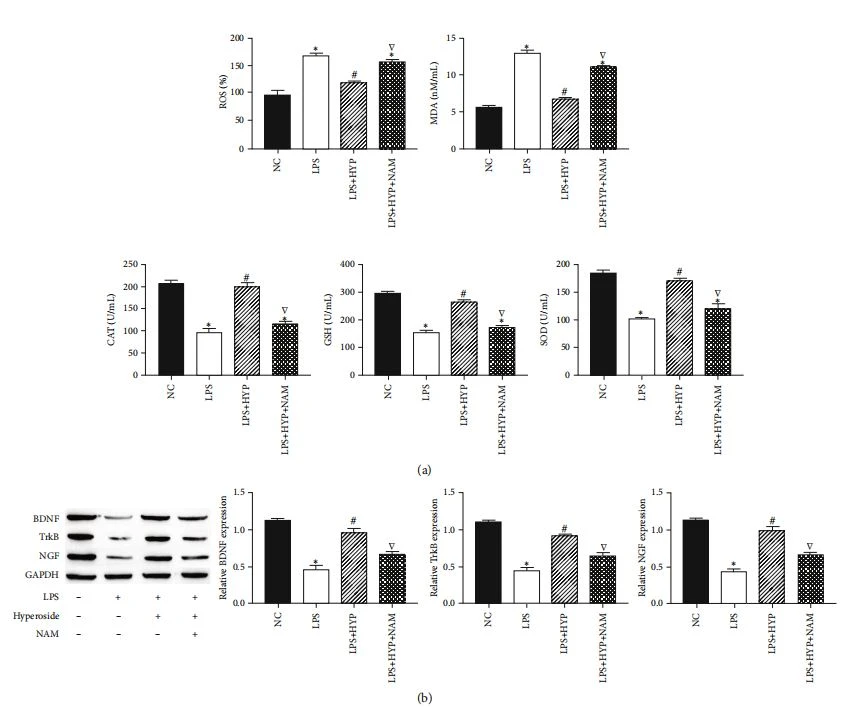
FIGuRE 4:Hyperoside inhibits LPS-induced HT22 cell oxidative stress and reduction of neurotrophic factor through SIRT1.The level of SOD, GSH, CAT,ROS,and MDA were measured by corresponding assay kits(a).The expression of BDNF, TrkB, and NGF in HT22 cells was measured by western blotting (b).* was considered significant compared to control (*P<0.05);# was considered significant compared to LPS (*P<0.05);V was considered significant compared to LPS+HYP (YP<0.05).
treatment(Figure 3(d)). These results indicated that hypero-side inhibits LPS-induced HT22 cell apoptosis and inflammation by upregulating SIRT1.

Cistanche can improve immunity
3.4.Hyperoside Inhibits LPS-Induced HT22 Cell Oxidative Stress and Reduction of Neurotrophic Factor through SIRT1.
We studied the effect of SIRTl on oxidative stress and neuro-trophic factors in LPS-induced HT22 cells. The results showed that NAM treatment decreased the levels of SOD, GSH, CAT, and neurotrophic factors BDNF and NGF and increased the levels of ROS and MDA in HT22 cells, com-pared with the treatment of cells with LPS+hyperoside treat-ment(Figures 4(a) and 4(b)). These results indicated that hyperoside inhibits oxidative stress and neurotrophic factor reduction in LPS-induced HT22 cells by upregulating SIRT.

Cistanche can improve immunity
3.5.Hyperoside Activates Wnt/β-Catenin and Sonic Hedgehog Pathways by Upregulating SIRT1.
To clarify whether SIRT1 alleviates HT22 damage through the Wnt/β-catenin and sonic hedgehog pathways, we measured the expression levels of signaling molecules in the Wnt/β-catenin and sonic hedgehog pathways. The results showed that LPS significantly decreased the expression levels of Wntl,β-catenin, Shh, and Patch;hyperoside treatment significantly increased the expression levels of Wntl,β-catenin, Shh, and patch; in addition, NAM treatment decreased the expression levels of Wntl, β-catenin, Shh, and patch. In addition, in HT22 cells, compared with NAM-treated cells, Wnt/β-catenin agonist(LiCl) and sonic hedgehog agonist(SAg) inhibited the inhibitory effect of NAM(Figures 5(a)and 5(b)). These results indicated that hyperoside activates Wnt/β-catenin and sonic hedgehog pathways by upregulating SIRT1.

FIGURE 5: Hyperoside activates Wnt/β-catenin and sonic hedgehog pathways by upregulating SIRT1.The expression of Wntl and β-catenin in HT22 cells was measured by western blotting (a). The expression of Shh and patch in HT22 cells was measured by western blotting (b),*was considered significant compared to control(*P<0.05);# was considered significant compared to LPS("P<0.05); V was considered significant compared to LPS+HYP(P<0.05);& was considered significant compared to LPS+HYP+LiCl or LPS+HYP+SAg ("P<0.05).
This article is extracted from Hindawi Neural Plasticity Volume 2021, Article ID 8706400, 10 pages https://doi.org/10.1155/2021/8706400



