

Hypoglycemic And Hypolipidemic Effects Of Blueberry Anthocyanins By AMPK Activation: In Vitro And in Vivo Studies Part 2
Mar 27, 2022
Please contact oscar.xiao@wecistanche.com for more information
3. Results
3.1.In Vitro studies
3.1.1. Effects of My, Mv-3-glc, Mv-3-gal, and BAE on cell viability in high glucose-stimulated HepG2 cells
High glucose causes hepatic oxidative stress and contributes to cellular cytotoxicity [28]. According to our study, high glucose (HG, 30 mM:35.67±1.90%)stimulation for 24 h significantly lowered the cell vitality to 35.67% in comparison with normal glucose(NG, 5.5 mM:100%)stimulated cells. The decrease of cell viability explained that high glucose disturbed the hepatic cellular homeostasis and led to apoptosis that caused degradation of glucose consumption and uptake, thus aggravating insulin resistance. Pretreatment with 5 μg/mL malvidin (Mv), malvidin-3-glucoside (Mv-3-glc), malvidin-3-galactoside (Mv-3-ga), and blueberry anthocyanin extract (BAE) ameliorated the glucose consumption and uptake after 24 h of high glucose incubation by significantly enhancing the cell vitalities at 99.04%, 97.76%, 96.47%,and 110.72%,respectively.Blueberry anthocyanin extract (BAE:110.72±14.00%)improved HepG2 cell vitality more than malvidin and malvidin derivatives. Malvidin(Mv:99.04 ± 1.49%) could protect the cell vitality slightly more than its derivatives. Anti-diabetic effect of malvidin-3-glucoside (Mv-3-glc: 97.76± 1.82%)was slightly but not significantly higher than malvidin-3-galactoside (Mv-3-gal:96.47±1.71%)(Fig. 1).

Please click here to know more
3.1.2. Antioxidant effects of Mvy, Mv-3-glc, Mv-3-gal, and BAE on ROS levels in high glucose-stimulated HepG2 cells
The intensity of fluorescence of ROS in HepG2 cells treated with normal glucose was low. After stimulation with high glucose, the fluorescence intensity of ROS was significantly enhanced(Fig.2).ROS level was significantly increased 6-fold when exposed to high glucose for 24 h (HG:3309.62± 23.82 A U.)in comparison with normal glucose (NG:560.41 ±26.38 A U.). This indicated that the level of reactive oxygen


had increased and hepatic oxidative stress had occurred. Fluorescence intensity of ROS in HepG2 cells was decreased after pretreatment with 5 ug/mL of Mv, Mv-3-glc, Mv-3-gal, and BAE for 24 h(Fig.2A). Pre-treatment with Mv,Mv-3-glc, Mv-3-gal, and BAE significantly inhibited ROS formations about 89.48%,81.54%,84.57%,and 92.38%, respectively(Fig.2B).Blueberry anthocyanin extract (BAE:769.83±20.41 A U.)significantly decreased ROS levels more than malvidin and its derivatives. Malvidin (Mv:849.53± 31.43 A U.)significantly exhibited a stronger antioxidant effect than its derivatives. Antioxidant effect of malvidin-3-galactoside(Mv-3-gal:984.66 ± 18.77 A U.)was significantly higher than malvidin-3-glucoside (Mv-3-glc: 1067.83± 20.85 A U.
3.1.3.Effects of My, Mv-3-glc, Mv-3-gal, and BAE on glucose metabolism in high glucose-stimulated HepG2 cells and supernatants
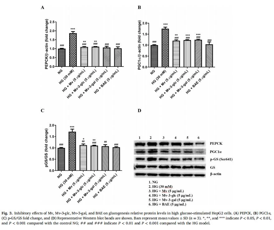
Gluconeogenesis is stimulated by the activation of transcription factor FOXO1, in which the interaction with its co-activator, PGClα, could increase the expression levels of gluconeogenesis (PEPCK and G6Pase)[29]. The hypoglycemic effects of Mv, Mv-3-glc, Mv-3-gal and BAE on PEPCK and PGCl-α in the high glucose-stimulated HepG2 cells were evaluated using Western Blot (Fig, 3), whereas on FOXO1 and G6Pase in the high glucose-stimulated HepG2 supernatant were evaluated using ELISA (Table 1). PEPCK level in HepG2 cells was increased 1.87-fold in the diabetic group compared to the control. Our results which can be seen in Fig.3A showed that BAE, malvidin, and its derivatives had a similar ability to decrease the up-regulation of PEPCK expression level in high glucose-induced HepG2 cells by 89.02%87.36%,92.59%,and 97.84%, respectively. The expression levels of PGC-1αin HepG2 cells increased 1.75-fold in the diabetic(high-glucose)group compared to the control (non-glucose), while Mv, Mv-3-glc, Mv-3-gal, and BAE significantly reversed this change. Pretreatment with BAE showed the strongest capacity to decrease PGC-1α(93.22%decrease) level in high glucose-induced HepG2 cells more so than malvidin(63.49% decrease)and its derivatives mv-3-glc (59.21% reduction) and mv-3-gal(55.57% decrease)which showed similar ability to decrease PGC-1α level in high glucose-induced HepG2 cells (Fig.3B).

Cistanche can improve immunity
Table1shows that with normal cells, FOXO1 expression was higher by 2.12-fold in high-glucose treatment compared with non-glucose treatment. FOXO1 over-activation stimulated by high glucose conditions could induce pro-inflammatory factors in diabetics and promote lipid accumulation [30]. FOXO1 up-regulation during high-glucose conditions was inhibited by pretreatment with Mv, Mv-3-glc, Mv-3-gal, and BAE, thus the inhibitory effect on FOXO1 in high glucose-induced HepG2 cells were 89.73%,89.94%,72.71%, and 76.64%, respectively (Table 1). The expression of G6Pase was strongly up-regulated when exposed to high glucose compared with a normal glucose group but these effects were reduced by BAE, malvidin, and its derivatives. These compounds decreased the up-regulation of G6Pase expression level in high glucose-induced HepG2 cells. The inhibitory effect of Mv, Mv-3-glc, Mv-3-gal and BAE on G6Pase in high glucose-stimulated HepG2 supernatant was about 74.12%,85.89%, 72.33%, and 77.58%, respectively (Table 1).

ELISA was also used to evaluate the hypoglycemic effects of Mv, Mv-3-glc, Mv-3-gal, and BAE on the phosphorylation of GSK3β at Ser9 inactive form in high glucose-stimulated HepG2 supernatant, whereas Western Blot was used to evaluate the phosphorylation of GS at Ser641 inactive form in high glucose-stimulated HepG2 cells. The expression level of the phosphorylation of GSK3β at Ser9 inactive form was high in unstimulated cells whereas the expression level of the phosphorylation of GS at Ser641 inactive form was low. When exposed to high glucose, the expression level of the phosphorylation of GSK3β at Ser9 inactive form was strongly down-regulated to 1.3-fold(Table 1), and the expression level of the phosphorylation of GS at Ser641 inactive form was strongly up-regulated to 2.6-fold. This experiment showed that BAE, malvidin, and its derivatives had the same ability to increase the phosphorylation of glycogenolysis enzyme GSK3β at Ser9 inactive form in high glucose-induced HepG2 cells. Pretreatment with Mv, Mv-3-glc, Mv-3-gal and BAE significantly increased the phosphorylation of GSK3β at Ser9 inactive form by about 1.54-fold, 1.78-fold, 2.08-fold, and 2.12-fold, respectively(Table 1). The results show a dramatic enhancement of GS activity. Pretreatment of BAE, malvidin, and its derivatives showed the same ability to increase GS synthesis by decreasing the phosphorylation of GS at Ser641 inactive form in high glucose-induced HepG2 cells. Pretreatment with Mv,Mv-3-glc, Mv-3-gal, and BAE significantly decreased p-GS/GS expression levels to 82.94%, 85.99%, 89.83%, and 96.22% in HepG2, respectively (Fig.3C). In addition, pretreatment of BAE, malvidin, and its derivatives showed the ability to inhibit the up-regulation of glucose transporter GLUT2 expression level in high glucose-induced HepG2 cells in this experiment. The inhibitory effects of Mv, Mv-3-glc, Mv-3-gal and BAE on GLUT2 in high glucose-stimulated HepG2 supernatant were 74.59%, 80.38%,54.68%, and 58.51%, respectively (Table 1).
3.1.4.Effects of Mv, Mv-3-glc, Mv-3-gal, and BAE on lipid metabolism in high glucose-stimulated HepG2 cells and supernatants
The effects of high glucose-induced hepatic fat accumulation on ACC and HMGCR in HepG2 supernatant were evaluated using ELISA (Table 1), whereas the phosphorylation of ACCat Ser79and SREBP-1cin

HepG2 cells were evaluated using Western Blot(Fig.4). Pretreatment of high glucose-induced HepG2 cells with Mv, Mv-3-glc, Mv-3-gal, and BAE inhibited ACC activity by significantly increasing the phosphorylation of ACC at Ser79 inactive form by 1.46-fold,1.87-fold,2.18-fold, and 1.97-fold, respectively (Fig。4B). High-glucose treatment significantly enhanced the expression level of ACC co-activator, SREBP-1c, in HepG2 cells.BAE had the strongest ability to inhibit ACC activity by decreasing the up-regulation of SREBP-1c expression level, nearly the same levels as the control. Malvidin-3-galactoside also significantly decreased the up-regulation of SREBP-1c expression level to nearly the same levels as control. Malvidin (78.59%) and malvidin-3-glucoside(77.53%) also showed the ability to decrease SREBP-1c expression level in high glucose-induced HepG2 cells but not to the same extent as malvidin-3-galactoside (92.72%)or BAE(95.58%)(Fig。4A). High-glucose treatment significantly enhanced ACC and HMGCR expressions in HepG2 supernatant. Pretreatment with BAE, malvidin, and its derivatives resulted in inhibited cholesterol synthesis to similar extents by inhibiting the up-regulation of ACC and HMGCR expression levels.

Fig. 4.Regulatory effects of Mv,Mv-3-glc, Mv-3-gal, and BAE on lipogenesis relative protein levels in high glucose-stimulated HepG2 cells.(A)SREBP-1c,(B)p-ACC/ACC fold change, and(C)Representative Western blot bands are shown.Bars represent mean values±SD (n=3).*,**,and*** indicateP<0.05,P<0.01,and P<0.001 compared with the control NG;## and ### indicate P<0.01 and P<0.001 compared with the HG model.
Pretreatment with Mv, Mv-3-glc,Mv-3-gal, and BAE decreased ACC expression levels by 67.93%,70.71%,62.12%, and 68.43%,respec-tively, and decreased HMGCR expression levels by 68.72%,68.18%, 77.55%, and 72.94%, respectively (Table 1).
On the other hand, pretreatment with BAE showed the strongest ability to inhibit the downregulation of lipolysis enzyme HSL expression levels in HepG2 supernatant caused by high-glucose treatment, followed by malvidin-3-glucoside, malvidin, and malvidin-3-galactoside. Pre-treatment with Mv, Mv-3-glc, Mv-3-gal, and BAE increased HSL expression levels by 89.69%,89.59%,76.07%,and 68.68%,respectively (Table 1).

3.1.5.Effects of My, Mv-3-GLC, Mv-3-gal, and BAE on AMPK activation in high glucose-stimulated HepG2 cells
AMPK is a major regulator of the liver and whole-body glucolipid homeostasis [31].AMPKis activated via increasing the phosphorylation of Thr172[32]. The phosphorylation of AMPK at Thr172 active form in HepG2 cells was significantly decreased in the high-glucose compared to the non-glucose group. BAE showed the strongest ability to activate AMPK in high-glucose-induced HepG2 cells compared to malvidin and its derivatives. Pretreatment with Mv, Mv-3-GLC, Mv-3-gal and BAE significantly increased p-AMPK/AMPK expression levels by about 1.32-fold,1.26-fold,1.39-fold, and 1.91-fold, respectively(Fig.5A and B)which indicated they have similar abilities to activate AMPK in high glucose-induced HepG2 cells.
3.2.In Vivo studies
3.2.1. Effect of BAE on body and tissue weight in induced diabetic mice
Oral administration of blueberry anthocyanin for five weeks to C57BL/6J mice did have a significant (P<0.0001)effect on the body weight, since the control had a significant high body weight compared to the diabetic model group, while BAE treatments alleviated the bodyweight decrease of diabetic mice (Fig. 6A). In blueberry anthocyanin extracts -low concentration (BAE-L)and blueberry anthocyanin ex-tracts-high concentration (BAE-H) treatments, daily administration of BAE had no significant differences on body weight indicating its dose-independent manner (P> 0.05). Furthermore, Table 2 shows the weight of selected internal mice organs. Results showed that weights were slightly similar to the control group. However, compared with the control group, the liver, spleen, and thymus weights were significantly different (P<0.05)in BAE-L. Moreover, there was no statistical significance in the liver length and body width among treatments (P>0.05).
3.2.2. Effect of BAE on blood glucose and urine glucose levels in induced diabetic mice
There was a significant (P <0.0001)elevation in fasting blood glucose in model mice as compared to the control group, implying that the diabetic model of mice was successful. However, supplementation of BAE to model mice, BAE-L and BAE-H for5 weeks resulted in significant recovery of fasting blood glucose levels(Fig. 6B) and ameliorated glucose tolerance (Fig.6C) in diabetic mice. The control group

Fig.5.Activation effects of BAE on glucolipid metabolism center the AMPK signaling pathway in vitro and in vivo.(A)p-AMPK/AMPK fold change by Mv, Mv-3-GLC, Mv-3-gal, and BAE in high glucose-stimulated HepG2 cells and (B)their representative Western blot bands are shown. (C)p-AMPK/AMPK fold change by BAE-Land BAE-H in induced diabetic mice and (D) their representative Western blot bands are shown. Bars represent mean values±SD(n=3 for cells in repeated experiment, n=4-6 for mice).*,**,and***indicate P<0.05,P<0.01,and P<0.001 compared with the group/the control;#,##,and### indicate P<0.05,P<0.01,and < 0.001 compared with the model HG group/the induced diabetic mice. maintained a constant glucose level during that period. Compared with the control, the BAE-L group notably reduced the blood glucose level in diabetic mice from week 2. Similarly, high-dose BAE obviously reduced the blood glucose levels from week 2 with continued gradual reduction until week 4. BAE at two doses(100 mg/kg and 400 mg/kg per day)significantly decreased blood glucose levels in diabetic mice at 30, 60, and 120 min after glucose load during glucose tolerance test. These results indicate that dietary inclusion of BAE for 5 weeks can effectively regulate glucose metabolism in diabetic mice. The diabetic model of mice drank more water and produced more urine than the control group. The supplementation of BAE to the model group could lessen the symptoms of polydipsia and polyuria in diabetic mice. Moreover, BAE-L and BAE-H treatments significantly decreased urine glucose levels(P<0.01 and P<0.001, respectively, Fig.7D). The levels of excreted urine showed significant increases in the model group compared to control, indicating that the former group of mice suffered from diabetes.
3.2.3.Effect of BAE on triglyceride, total cholesterol, and insulin levels in serum of induced diabetic mice
Administration of BAE (100 mg/kg and 400 mg/kg per day)significantly affected the lipid levels in induced diabetic mice. Total cholesterol and triglycerides were significantly elevated (P<0.01)in model mice in comparison to the control(Fig.7A and B). BAE-Treatment only induced a slight decrease in blood lipid indexes but there was no significant difference with the model diabetic mice(P>0.05). Treatment BAE-H resulted in a significant diminution of total cholesterol and triglycerides (P<0.01)and the levels of these parameters were similar to the control (P>0.05).On the other hand, Fig.7C shows the insulin levels in mice serum after five weeks of treatment. Both BAE-L and BAE-H significantly decreased (P<0.001) the insulin levels in serum of induced diabetic mice. These results indicate that the inclusion of dietary BAE for 5 weeks can effectively ameliorate insulin resistance in diabetic mice.
3.2.4. Effect of BAE on liver antioxidant and glucose transporter levels in induced diabetic mice
In this study, the effect of BAE on antioxidant enzyme activity of SOD and GSH-PX levels in diabetic mice was evaluated in order to elucidate whether the hypolipidemic and hypoglycemic effects of BAE were associated with the protection of the antioxidant defense systems. As shown in Fig.8A and B, the model group had a significant decrease in the activity of SOD and GSH-PX compared with the control group(P<0.05). After BAE treatment, the activity of SOD was significantly increased (P <0.01)in BAE-L and BAE-H when compared with the model group. Moreover, the GSH-PX activity of the BAE-H group(400 mg/kg BAE)was significantly higher than the model one(P<0.05). GLUT2 is the main glucose transporter in the plasma membranes of hepatocytes, therefore,

Fig. 6. Effects of BAE on body weight and blood glucose levels in induced diabetic mice. (A)Bodyweight and (B)Blood glucose change of mice from the control, model, BAE-L, and BAE-H groups during gavage for five weeks, and(C)Glucose tolerance of mice from the control, model, BAE-L, and BAE-H groups after gavage for five weeks. Bars represent mean values±SD (n=6).*,**,and *** indicate P<0.05,P<0.01, and P<0.001 compared with the NG group/the control;#,##,and ### indicate P<0.05,P<0.01,and P<0.001 compared with the model HG group/the induced diabetic mice. the effects of BAE on glucose GLUT2 content in induced diabetic mice were studied (Fig.8C). GLUT2 levels in mice liver were not statistically affected by BAE treatments but were significantly higher in diabetic mice treated with BAE, compared to the model group.
3.2.5. Effects of BAE on AMPK activation in induced diabetic mice
An AMPK signaling pathway is one of the principal factors for cellular energy homeostasis, which can be recognized as the crucial target in the prevention and treatment of obesity and diabetes. The effects of BAE on

the AMPK signaling pathway in induced diabetic mice are shown in Fig.5C and D.The level of AMPK significantly decreased (P<0.001)in the model group in comparison with the control. Groups with BAE showed the strongest ability to activate AMPK compared to the control. Moreover, BAE-L and BAE-H showed similar ability to activate AMPK in samples from diabetic mice, and also significantly increased p-AMPK/AMPK expression levels by about 0.54-fold and 0.45-fold, respectively (Fig.5C.
4. Discussion
Diabetes is a chronic metabolic disorder affecting a great proportion of the population worldwide. Blueberries are rich in anthocyanins which have been studied for their numerous beneficial effects on human health. Anthocyanins are able to ameliorate dysfunction in lipid and glucose metabolism, which are fundamental risk factors for obesity and diabetes. The objectives of the present study were to investigate the hypoglycemic and hypolipidemic effects of a blueberry extract rich in anthocyanins on HepG2 cells and induced diabetic mice, as well as to explore the underlying mechanisms. BAE intake in vivo for 5 weeks significantly attenuated the hyperglycemia and hyperlipidemia syndromes in diabetic mice. In this study, we found that administration of BAE, both 100 mg/kg and 400 mg/kg per day could reduce body weight loss. However, BAE treatments significantly decreased serum triglycerides and cholesterol in diabetic mice. The presence of hypocholesterolemic compounds that can act as inhibitors of some enzymes such as hydroxyl methyl glutaryl CoA reductase, which participates in the synthesis of cholesterol or reduces the absorption of cholesterol in the intestine, could be a factor that is attributable to the cholesterol-lowering property of BAE [33]. Furthermore, BAE resulted in a significantly reduced peak of glucose within 2 h, which is supported by other authors [34-36] that reported the potential antidiabetic effect of anthocyanins. Excessive oxidative stress in body cells plays an important role in the development of diabetes, and the persistent hyperglycemia of diabetes could also lead to increased oxidative stress in the body [37], therefore, the inhibition or reduction of oxidative stress could be an additional therapy for preventing or delaying the incidence of diabetes. It has been reported that anthocyanins could potentially exert antioxidant capacity under high oxidative stress conditions such as obesity and hypercholesteremia [38]. Our results showed a significantly increased activity of SOD levels in BAE-H treatment. These data suggested that BAE could suppress oxidant stress by improving the activity of SOD, which might be attributed to the hyperglycemia and hyperlipidemia alleviation effect of BAE. Similar findings of positive effects of BAE on antioxidant defensiveness were also established in the liver and hippocampus tissues, which was revealed by the strengthened antioxidant enzyme activities including SOD [39]. Anthocyanins could ameliorate dysfunction in lipid and glucose metabolism, which are important risk factors for

diabetes and obesity. One of the crucial factors for cellular energy homeostasis is the AMPK signaling pathway, which can be recognized as the key target in the prevention/treatment of diabetes and obesity [40]. Pancreatic β-cells in the liver are affected by glycoxidative stress because of the low quantities of ROS detoxifying enzymes there [41]. Hepatic glycoxidative stress is an essential contributor to diabetic progression as it decreases the insulin stimulation of the insulin signaling proteins and causes insulin resistance[41]. The increase in ROS after24 h of high glucose stimulation showed that high glucose disrupted the hepatic cellular homeostasis and led to apoptosis. The decrease in cell viability after 24 h of high glucose stimulation resulted in insulin resistance and caused dysfunction in glucose consumption and uptake. Pretreatment with BAE, malvidin, and its derivatives decreased ROS generation and ameliorated cell viability in this study.BAE showed the strongest antioxidant ability to keep β-cells from hepatoxicity triggered by hepatic glycoxidative stress, followed by malvidin, malvidin-3-galactoside, and malvidin-3-glucoside. The hydroxylation and methoxylation patterns of malvidin-3-glucoside and malvidin-3-galactoside on 3,5-dimethoxy substituents in the B-ring conferred these antioxidant properties [42]. Pretreatment with BAE resulted in the greatest ability to protect hepatic β-cells from hepato-toxicity triggered by oxidative stress. However, malvidin also showed the capacity to inhibit ROS generation. Therefore, blueberry anthocyanin extract has the potential to be developed as an anti-diabetic nutraceutical to inhibit insulin resistance induced by hepatic oxidative stress in diabetes prevention. Fig.9shows proposed mechanisms for improving hyperglycemia and hyperlipidemia through AMPK activation using BAE. AMPK is a molecule that has actions in the liver, skeletal muscle tissue, adipose tissue, and hypothalamus. Typically activated by nutrient deficiencies, it restores the energy balance in two main ways through a complex system of downstream activated molecules [43]. First, AMPK stimulates glucose uptake and lipid oxidation to increase the production of ATP, and second, it turns off energy consumption processes. AMPK is involved in pathways that regulate lipid and glucose metabolism. Upregulation of AMPK through a reduction in reactive oxygen species by BAE could lead to positive effects in reducing hyperglycemia and hyperlipidemia in diabetics. As AMPKactivity increases, it inhibits multiple factors involved in glucose synthesis including PGClα, FOXO1, PEPCK, G6Pase, and GS which in turn downregulates glucose synthesis [43,44]. According to this study, it inhibits the activity of GLUT2 which decreases glucose transport. It also increases the activity of GSK3β which is involved in glycogenolysis, thereby increasing the glycogenolytic activity of the cell. All these lead to a reduction in blood glucose. AMPK also inhibits factors that play a role in lipid synthesis including HMGCR which is involved in sterol and isoprenoid synthesis, SREBP-1 which promotes fatty acid synthesis, and ACC which is active in lipogenesis and in addition, AMPK promotes the activity of HSL which is involved in lipolysis [43,45]. These activities can then lead to a reduction in blood lipids. BAE, malvidin and its derivatives showed their strong capacity to enhance AMPK activation via phosphorylation at Thrl172 active form in the insulin-independent pathway. Therefore, BAE, malvidin, and its derivatives can be considered as potential nutraceuticals to improve insulin sensitivity in hepatic cells and increase glucose homeostasis.
Gluconeogenesis catalyzes the final release of glucose into the blood circulation, in which its over-expression in the liver would lead to insulin resistance and the rise of hepatic glucose production [46]. Insulin directly inhibits gluconeogenesis and its activators, thus consequently reducing the levels of circulating blood glucose [29]. Our results showed that BAE, malvidin, and its derivatives decrease the formation of hepatic glucose by suppressing gluconeogenesis (PEPCK and G6Pase)and its co-activators (PGC-1α and FOXO1)in the insulin-independent pathway. Therefore, BAE, malvidin, and its derivatives could improve insulin sensitivity and response to the post-prandial increases in blood glucose, thus inhibiting gluconeogenesis and its activators. The superfluous glucose which is not utilized as an instantaneous fuel for energy is initially stored as glycogen [47]. Glycogen synthase (GS)forms glycogen from glucose, but glycogen synthase kinase 3β(GSK3p) inhibits GS activity by converting glycogen to glucose. Insulin inhibits glycogenolysis by inactivating GSK3β, consequently promoting

glycogenesis by enhancing GS activity in hepatocytes[48].In this study, BAE, malvidin, and its derivatives could improve insulin sensitivity by acting as an effective GSK3β inhibitor and GS stimulator in the insulin-independent pathway.
GLUT2 is a bidirectional transporter that takes up glucose during the absorptive (glycolysis) phase and discharges it into the bloodstream during gluconeogenesis and glycogenolysis [49]. When the extracellular glucose concentration increases, more glucose enters pancreatic β-cell via the low-affinity glucose transporter GLUT2 [50].GLUT2 overexpression in the liver impairs glucose-stimulated insulin secretion and increases the risk of fasting hyperglycemia and T2DM [4]. Malvidin combined with sugars in blueberry extract prevented the sudden rise of glucose by decreasing GLUT2 expression level. Flavonoid-containing sugar could reduce glucose transport and absorption by decreasing the absorption rate, whereas anthocyanin itself is not carried away by GLUT2 [51]. The former would be beneficial in reducing the sudden increase of glucose. Anthocyanin and GLUT2 coalition occurred via hydrophilic moieties, i.e. the 3-glucosyl moiety and the B ring for the monoglucoside, the 5-glucosyl moiety, the A ring for the glucoside, and the A or B ring for the aglycones [52].In this study, BAE, malvidin, and its derivatives reduced GLUT2 over-expression levels in hepatic cells, thus keeping the balance of glucose-stimulated insulin secretion in pancreaticβ-cells and also reducing glucose transport and absorption into blood circulation.
The over-expression of lipogenesis, that is, ACC, SREBP-1c, HMGCR, and HSL in obese patients has been strongly associated with diabetes and fatty liver diseases [53]. Pretreatments with BAE, malvidin and its derivatives could have inactivated ACC by causing an increase in the phosphorylation of ACC at Ser79 inactive form.ACC inactivation could stimulate long-chain fatty acids to enter the mitochondria for oxidation, thus increasing fatty acid uptake [54]. These pretreatments also inhibited fatty acids synthesis by decreasing the expression of ACC cO-activator, SREBP-1c. 3-Hydroxy-3-methylglutaryl coenzyme A reductase (HMGCR)plays an important role in cholesterol synthesis [55]. Hormone-sensitive lipase (HSL) mediates the release of free fatty acids into vasculature [56]. BAE, malvidin and its derivatives effectively decreased cholesterol synthesis and free fatty acid circulation into vasculature by significantly decreasing HMGCR and HSL expression levels in hepatocytes. It was also reported that a high dose of BAE could suppress the accumulation of serum ceramides, diacylglycerols, triacylglycerol, and cholesterol, and thus prevent lipid metabolic dysfunction [57]. Therefore, BAE plays a role as a hypolipidemic nutraceutical that effectively reduces fatty acid and cholesterol synthesis, and increases fatty acid oxidation, thus consequently promoting lipid homeostasis. From our findings, BAE, malvidin, and its derivatives have the potential hypoglycemic and hypolipidemic effects of treating diabetes via AMPK activation that increase insulin sensitivity by inhibiting gluconeogenesis and lipogenesis in human hepatocarcinoma cells, and by decreasing glucose transport into the bloodstream.
5. Conclusion
The hypoglycemic and hypolipidemic effects of anthocyanin extract from rabbiteye blueberry cultivar in vitro and in vivo have been investigated in this study. The results demonstrated that hepatic oxidative stress was significantly increased by high glucose, which increased ROS by up to 6-fold and decreased cell viability. Pretreatment with Mv, Mv-3-glc, Mv-3-gal, and blueberry anthocyanin extract (BAE)significantly reduced this damage by lowering the ROS generation and increasing the cell viability. These findings show that BAE, malvidin, and its derivatives have a powerful antioxidant capacity to protect hepatic cells from oxidative deterioration by decreasing the formation of ROS and increasing cell viability. Therefore, these pretreatments could effectively decrease fatty acid and cholesterol synthesis and reduce free fatty acid circulation in blood vessels. BAE could significantly ameliorate hyperglycemia and hyperlipidemia in induced diabetic mice via a significant reduction in body weight loss, glucose and lipid levels. The antioxidant activity of SOD and AMPK activation was notably improved by BAE. BAE, malvidin, and its derivatives proved to be efficacious anti-diabetic nutraceuticals with hypoglycemic and hypolipidemic activities. They effectively activated AMPK which, in turn, improved insulin sensitivity and protected the body from the over-expression of gluconeogenesis and lipogenesis; therefore, they could effectively maintain glucolipid homeostasis and prevent diabetes. These findings suggest that BAE could exhibit great anti-diabetic effects in vitro and in vivo, and anthocyanins being one of the major constituents of the blueberry extract, play an important role in exerting hypoglycemic and hypolipidemic activities.
This article is extracted from https://doi.org/10.1016/j.redox.2021.102100 Received 21 July 2021; Received in revised form 9 August 2021; Accepted 11 August 2021





