

Kidney Pericyte Hypoxia-inducible Factor Regulates Erythropoiesis But Not Kidney Fibrosis
Mar 13, 2022
Contact: joanna.jia@wecistanche.com / WhatsApp: 008618081934791
Szu-Yu Pan1,2,3 et al
Prolyl hydroxylase domain enzyme (PHD) inhibitors are effective in the treatment of chronic kidney disease (CKD)- associated anemia by stabilizing hypoxia-inducible factor (HIF), thereby increasing erythropoietin and consequently erythropoiesis. However, concern for CKD progression needs to be addressed in clinical trials. Although preclinical studies showed an anti-inflammatory effect in kidney disease models, the effect of PHD inhibitors on kidney fibrosis was inconsistent probably because the effects of HIF are cell type and context-dependent. The major kidney erythropoietin-producing cells are pericytes that produce erythropoietin through HIF-2a-dependent gene transcription. The concern for the impact of HIF in pericytes on kidney fibrosis arises from the fact that pericytes are the major precursor cells of myofibroblasts in CKD. Since cells expressing Gli1 fulfill the morphologic and anatomic criteria for pericytes, we induced Gli1+ cell-specific HIF stabilization or knockout to study the impact of HIF in pericytes on kidney pathology of mice with or without fibrotic injury induced by unilateral ureteral obstruction. Compared with the littermate controls, mice with pericyte-specific HIF stabilization due to von Hippel-Lindau protein or PHD2 knockout showed increased serum erythropoietin and polycythemia rather than a discernible difference in kidney fibrosis. Compared with Gli1+ pericytes sorted from littermate controls, Gli1+ pericytes sorted from PHD2 knockout mice showed increased erythropoietin gene expression rather than discernible changes in Col1a1 or Acta2 expression. Furthermore, pericyte-specific knockout of HIF-1a or HIF-2a did not affect kidney fibrosis. Thus, our study supports the absence of negative effects of PHD inhibitors on kidney fibrosis of mice despite HIF stabilization in pericytes.
KEYWORDS: erythropoietin; fibrosis; hypoxia-inducible factor; pericyte

Cistanche is good for renal function
Translational Statement
Prolyl hydroxylase domain enzyme (PHD) inhibitors are effective in the treatment of anemia in chronic kidney disease (CKD) through stabilizing hypoxia-inducible factor (HIF), thereby increasing erythropoietin (EPO) and erythropoiesis. Renal pericytes produce EPO through HIF-2a in normal physiology, but they differentiate to scar-producing myofibroblasts in CKD. It is not clear whether HIF expression in renal pericytes impacts renal fibrosis. By preclinical murine models of HIF manipulation and renal fibrosis, we show that Gli1+ pericyte specific HIF manipulation affects EPO production and erythropoiesis but not renal fibrosis. Our study endorses the absence of negative effects of PHD inhibitors on renal fibrosis.
The mainstay of treatment for anemia in patients with chronic kidney disease (CKD) is erythropoiesis-stimulating agents and iron supplements. 1,2 Recently, the prolyl hydroxylase domain enzyme (PHD) inhibitor emerges as a promising therapeutic agent for CKD-associated anemia principally through stabilizing the hypoxia-inducible factor (HIF) and thereby increasing erythropoietin (EPO) production.3–5 The clinical application of PHD inhibitors or HIF stabilizers in patients with CKD is imminent in the foreseeable future if the concerns regarding possible angiogenesis, tumor growth, abnormal glucose metabolism, and accelerated decline of kidney function are to be addressed in the clinical trials.
HIF is the most important known cellular oxygen–sensing machinery.6,7 The functional HIF consists of a and b subunits. HIF-a is an oxygen labile subunit that is rapidly degraded at a normoxic condition in the presence of cofactors. HIF-b is not regulated by oxygen tension. The degradation of HIF-a is preceded by proline hydroxylation and polyubiquitination. The proline hydroxylation is catalyzed by PHD in the presence of oxygen, iron, and 2-oxoglutarate as cofactors. The polyubiquitination is then catalyzed by an E3 ubiquitin ligase composed of von Hippel Lindau protein (pVHL)-Elongin BC-CUL2 complex. The requirement of iron, PHD, and pVHL for HIF degradation makes it possible to stabilize HIF in the normoxic condition with the inhibition or decrease of any of these key components.
Renal pericytes are embedded within the microvascular basement membrane and in close contact with endothelial cells to support microvasculature and regulate blood flow.8–13 We have reported that renal pericytes are renal EPO-producing cells (REPCs) who produce EPO through HIF-2a–dependent gene transcription induced by anemia or hypoxia.14 Pericytes have been demonstrated as the major source of scar-producing myofibroblasts in CKD,8,10,11,15,16 a finding supported by the other independent reports.17,18 Although the transition to myofibroblasts leads to the repression of Epo transcription, overexpression of HIF-2a partially restores the EPO production in myofibroblasts.14,17
Our studies have shown that renal fibrosis can be attenuated by interruption of pericyte-myofibroblast transition through pharmacologic blockade of transforming growth factor-b, 19 platelet-derived growth factor,20 vascular endothelial growth factor,21 or WNT/b-catenin pathways.22
Regarding the concern for the accelerated decline of kidney function in patients under the treatment with PHD inhibitors, little is known about the effects of HIF activity in pericytes or myofibroblasts on renal fibrosis.23 In contrast, the effects of HIF overexpression or knockout on renal fibrosis have been extensively studied, either nonselectively24–27 or selectively in tubular cells,28,29 endothelial cells,30 and podocytes.31,32 However, the results were inconsistent probably because the effects of HIF are cell type and context-dependent.
In vitro studies using isolated human renal fifibroblasts33 and rat renal medullary interstitial cells34 suggested a profibrotic role of HIF-1a. However, Souma et al.17 reported that concomitant knockout of PHD1, PHD2, and PHD3 in REPCs of Tg(Epo-Cre) mice who express Cre recombinase under the control of Epo promoter does not affect renal fibrosis in the model of unilateral ureteral obstruction (UUO). However, renal Epo expression is low and the pool size of REPCs is small in a steady state without hypoxia or anemia,35 suggesting that Epo-Cre transgene only defines a small fraction of total REPCs. Recently, renal Gli1-expressing cells are shown to be pericytes and differentiate to myofibroblasts on kidney injury.11,36 Ablation of Gli1+ cells ameliorates UUO-induced renal fibrosis.11 Here we used mice with Cre recombinase under the control of Gli1 promoter/enhancer to study the effects of pericyte-specific HIF stabilization or knockout on renal fibrosis.

cistanche is good for kidney
METHODS
Mice
Gli1CreERT2/+, Tg(UBC-CreERT2), VhlF/F, ROSA26fstdTomato/fstdTomat, Hif1aF/F, and Hif2aF/F mice were purchased from the Jackson Laboratory (Bar Harbor, ME). Egln1F/F mice were kindly provided by Professor Guo-Hua Fong (University of Connecticut Health Center, Farmington, CT). All studies were carried out under a protocol approved by the Institutional Animal Care and Use Committee, National Taiwan University College of Medicine (IACUC 20160499).
Unilateral ureteral obstruction
The procedure had been detailed in a previous study.14 In short, in adult mice 7 to 9 weeks old under anesthesia with ketamine/xylazine (ketamine 100 mg/kg, xylazine 10 mg/kg), the left ureters of the mice were exposed and ligated with nylon suture through an incision at the left flank. The date of UUO surgery was identified as day 0 of the experiment.
Statistical analyses
Data were expressed as mean+ SD. Statistical analyses were carried out using GraphPad Prism (GraphPad Software, San Diego, CA). The statistical significance was determined by the Student t-test or Mann-Whitney U test as shown in the captions.
Supplementary methods
Full methods including mice, UUO, tamoxifen administration, quantitative polymerase chain reaction, immunoblot, immunohistochemistry, RNA in situ hybridization, microscopy, flow cytometry, and isolation of renal Gli1+ cells are in the Supplementary Methods.
RESULTS
Increased EPO production and erythropoiesis in mice with Gli1+ pericyte-specific PHD2 knockout
To demonstrate that Gli1+ pericytes produced EPO before we headed to study the effect of pericyte-specific HIF manipulation on renal fibrosis, we generated Gli1CreERT2/+; Egln1F/F mice to induce PHD2 knockout in Gli1+ pericytes conditionally (Figure 1a). Compared with littermate Egln1F/F mice, Gli1CreERT2/+; Egln1F/F mice showed a higher level of serum EPO (Figure 1b), heavier splenic weight (Figure 1c), more TER-119+ erythroid lineage in splenocytes by day 14 after tamoxifen (Figure 1d), but similar body weight change after tamoxifen administration (Supplementary Figure S1A). Hematocrit (Hct) was higher in Gli1CreERT2/+; Egln1F/F mice at day 21 (Figure 1e). We analyzed renal Epo mRNA and peripheral blood count 14 days after tamoxifen. Compared with littermate Egln1F/F mice, Gli1CreERT2/+; Egln1F/F mice had increased renal Epo expression (Figure 1f) and peripheral reticulocytes (Figure 1g), whereas peripheral white blood cell count and platelet count were not different (Supplementary Figure S1B and C). To identify renal cells responsible for Epo expression, we performed in situ hybridization of Epo on kidney sections from the Egln1F/F and Gli1CreERT2/+; Egln1F/Fmice (Supplementary Figure S2A–F). Increased Epo-expressing interstitial cells could be identified mainly in the corticomedullary junction, and to a lesser extent in the inner medulla and cortex in the Gli1CreERT2/+; Egln1F/F mice. These results suggested that Gli1+ pericyte-specific PHD2 knockout induced renal EPO production and erythropoiesis.


To determine whether PHD2 regulates EPO production in cell types and organs other than pericytes in the kidney, we generated Tg(UBC-CreERT2); Egln1F/F mice to induce global PHD2 knockout (Supplementary Figure S3A). After tamoxifen administration, knockout of the Egln1 gene in the kidney, liver, spleen, heart, and lung could be detected (Supplementary Figure S4). In the kidney, the expression of Egln1 mRNA reduced to 4%, indicating high efficiency of PHD2 knockout (Supplementary Figure S3B). As in Gli1CreERT2/+; Egln1F/F mice, Tg(UBC-CreERT2); Egln1F/F mice showed a progressive increase of Hct compared with littermate Egln1F/F mice(Supplementary Figure S3C). Notably, Tg(UBC-CreERT2); Egln1F/F mice showed a decrease in body weight (Supplementary Figure S3D). Remarkably high level of serum EPO, around 130,000 pg/ml, was found in Tg(UBC-CreERT2); Egln1F/F mice, which was much higher than that achieved by phlebotomy (less than 5,000 pg/ml) in wild-type mice (Supplementary Figure S3E). To identify the source of serum EPO, we checked Epo mRNA levels in different organs from Tg(UBC-CreERT2); Egln1F/F, and littermate control mice at day 15. In littermate control mice, Epo mRNA was hardly detected in any organ. In 5 major organs (kidney, liver, lung, heart, spleen) of Tg(UBC-CreERT2); Egln1F/F mice, only the kidney showed a remarkable increase of Epo mRNA, implying the kidney as a major source of serum EPO (Supplementary Figure S3F and G). In situ hybridization of Epo mRNA on kidney sections revealed that renal cells with Epo expression were interstitial cells with pericyte morphology in Tg(UBC-CreERT2); Egln1F/F mice (Supplementary Figure S5). Therefore, these data confirmed that renal Gli1+ pericytes produced EPO on PHD2 knockout, thereby increasing erythropoiesis and Hct.



Gli1+ pericyte-specific pVHL knockout leads to polycythemia but has no effect on renal fibrosis
Consistent with previous reports,11,36 increased Gli1+ pericytes were shown in the corticomedullary junction and medulla after UUO injury in Gli1CreERT2/+; ROSA26fstdTomato/ fstdTomato reporter mice (Supplementary Figure S6), suggesting that Gli1+ pericytes proliferate and contribute considerably to myofibroblasts during renal fibrosis. We hence studied the effect of HIF stabilization or knockout in Gli1+ pericytes on renal fibrosis.


We used mice with Gli1+ pericyte-specific knockout of pVHL, not PHD2 because of possible redundancy in 3 different PHDs. To investigate the long-term effect of pericyte-specific HIF stabilization on kidneys in mice without CKD, Gli1CreERT2/+; VhlF/F mice, and VhlF/F littermates were administered tamoxifen at 6 weeks of age and then observed until 30 weeks of age (Supplementary Figure S7A). One unexpected death in Gli1CreERT2/+; VhlF/F mice took place at 25 weeks of age (14.3% mortality rate), but there were no deaths in VhlF/F littermates. Weakness and decreased body weight gain were found in Gli1CreERT2/+; VhlF/F mice as early as 16 weeks of age (Supplementary Figure S7B). Because of emaciation and unexpected death, some mice were euthanized before 30 weeks of age. Pathology revealed diffuse severe erythropoiesis in the spleen and congestion of red blood cells in liver, lung, and kidney at 18 weeks of age (Supplementary Figure S8). Except for red blood cell congestion, no discernible fibrosis was observed in major organs including kidneys. The levels of Hct and serum EPO increased to 87+-2.6% and 1604 +-141 pg/ml at 30 weeks of age (i.e., 24 weeks after tamoxifen) (Supplementary Figure S9A and B)


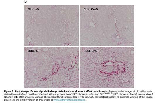
Because of no discernible renal fibrosis in mice with Gli1+ pericyte-specific pVHL knockout, we induced UUO in mice and assessed the severity of renal fibrosis 7 or 14 days later (Figure 2a). Deleted Vhl alleles in Gli1CreERT2/þ; VhlF/F mice were detected in kidney genomic DNA by polymerase chain reaction, indicating knockout of pVHL in pericytes (Supplementary Figure S10). Hct was increased in Gli1CreERT2/+; VhlF/F mice even after UUO surgery (Figure 2b). Notably, the expression of EPO mRNA was increased in both contralateral and UUO kidneys of Gli1CreERT2/+; VhlF/F mice (Figure 2c), suggesting HIF stabilization could activate Epo transcription in both pericytes and myofibroblasts. However, no difference in renal fibrosis between Gli1CreERT2/+; VhlF/Fand littermate VhlF/F mice could be detected, as measured by picrosirius red-stained area (Figures 2d and 3a and b) and expression of Col1a1 mRNA (Figure 2e) in the kidney. Besides, mRNA and protein levels of a-smooth muscle actin were not different in kidneys (Figure 2f and g and Supplementary Figure S11). To confirm that myofibroblasts retained Epo expression ability after pVHL knockout, we performed in situ hybridization of Epo and Acta2 on kidney sections (Supplementary Figures S12 and S13). Renal Acta2 expression increased drastically and comparably after UUO injury in both Gli1CreERT2/+; VhlF/F and VhlF/F mice. More interstitial Epo-expressing cells could be observed in Gli1CreERT2/+; VhlF/F mice than in VhlF/F mice, regardless of the presence of UUO injury. In addition, cells with concomitant expression of Acta2 and Epo could be identified in UUO kidney from Gli1CreERT2/+; VhlF/F mice but not VhlF/F mice. In fact, we could hardly identify any Epo-expressing cells in the UUO kidney from VhlF/F mice. These results suggested that Gli1+pericyte-restricted pVHL knockout promoted Epo expression but not myofibroblast differentiation or collagen production even after UUO injury.
Because UUO injury results in damage of proximal tubular cells and recruitment of macrophages,37 mRNA levels of Havcr1 (encoding kidney injury molecule-1) (Figure 4a) and Adgre1 (encoding endothelial growth factor-like module-containing mucin-like hormone receptor-like 1, also known as F4/80) (Figure 4b) increased in UUO kidneys, but no difference could be detected between Gli1CreERT2/+; VhlF/F and littermate VhlF/F mice. The mRNA levels of Vegfa, Hmox1, Egln1, and Egln3 (encoding vascular endothelial cell growth factor-A, heme oxygenase-1, PHD2, and PHD3, respectively) in kidneys were not changed (Figure 4c–f), probably reflecting that these transcripts were not dominantly produced by Gli1+ pericytes or not changed after Gli1+ pericyte-restricted VHL knockout. We assessed tubulointerstitial injury in periodic acid–Schiff–stained kidney sections, and no difference was detected between Gli1- CreERT2/+; VhlF/F, and littermate VhlF/F mice (Figures 4g and 5a and b). Because tamoxifen is reported to attenuate fibrosis,38,39 we extended the washout period between the last tamoxifen administration and UUO surgery from 2 weeks to 4 weeks (Supplementary Figure S14A). Again, no difference was detected between Gli1CreERT2/+; VhlF/F and littermate VhlF/F mice (Supplementary Figure S14B–E). These findings indicated that Gli1+ pericyte–specific pVHL knockout leads to EPO production and polycythemia but has no effect on renal fibrosis.

cistanche is good for kidney
Gli1+ pericyte-specific PHD2 knockout increases EPO production but not collagen production or myofibroblast differentiation
To confirm the effects of HIF stabilization in Gli1+ pericytes at the cellular level, we sorted tdTomato+ cells from the kidneys of Gli1CreERT2/+; ROSA26fstdTomato/fstdTomato and Gli1CreERT2/+; Egln1F/F; ROSA26fstdTomato/fstdTomato mice after UUO injury (Supplementary Figures S15 and S16). Under microscopic examination, the sorted cells emitted orange-red fluorescence on excitation (Supplementary Figure S17A–C). Compared with those from Gli1CreERT2/+; ROSA26fstdTomato/fstdTomato mice, the Egln1 mRNA level in tdTomato+ cells sorted from contralateral and UUO kidneys of Gli1CreERT2/+; Egln1F/F; ROSA26fstdTomato/fstdTomato mice 7 days after UUO injury reduced to 46% and 33%, respectively (Supplementary Figure S17D and E).
We analyzed the mRNA levels of Epo, Col1a1, Col3a1, and Acta2 in the sorted tdTomato+ Gli1+ pericytes from Gli1CreERT2/+;ROSA26fstdTomato/fstdTomato and Gli1CreERT2/+; Egln1F/F; ROSA26fstdTomato/fstdTomato mice. Consistent with the results obtained from mice with Gli1+ pericyte-specific pVHL knockout, PHD2 knockout in Gli1+ pericytes resulted in increased Epo expression, but had no effect on the expression of Col1a1, Col3a1, and Acta2 at the cellular level (Figure 6), confirming the neutral effect of HIF stabilization on the profibrotic properties of pericytes/myofibroblasts.
Gli1þ pericyte-specific HIF knockout does not affect renal fibrosis
Because these experiments confirmed the neutral effect of HIF stabilization on profibrotic properties of pericytes/myofibroblasts, we generated Gli1CreERT2/+; Hif1aF/F; Hif2aF/F mice to study the effect of HIF knockout in pericytes/myofibroblasts on renal fibrosis (Figure 7a). Deletion of Hif1a and Hif2a in the kidney could be detected in kidney genomic DNA after tamoxifen administration (Supplementary Figure S18). Compared with the littermate control, Gli1CreERT2/+; Hif1aF/F; Hif2aF/F mice had similar body weight change and Hct level 7 days after UUO (Figure 7b and c). In terms of renal fibrosis, no difference could be detected in the picrosirius red-stained area, mRNA levels of Col1a1 and Acta2 in the kidney (Figure 7d–f). The mRNA levels of Havcr1 and Adgre1 were also not different (Figure 7g and h). We changed the dose and washout period of tamoxifen (Supplementary Figures S19A and S20A), and the results for collagen deposition and gene expression were similar (Supplementary Figures S19B–E, S20B–E). We repeated the experiments in Gli1CreERT2/+; Hif1aF/F (Figure 8a–c) and Gli1- CreERT2/+; Hif2aF/F (Figure 8d–f) mice and did not demonstrate the difference in UUO-induced renal fibrosis when compared with the littermate control.


DISCUSSION
The main findings of this study included the following: (i) renal Gli1+ pericyte-specific HIF stabilization led to EPO production and erythropoiesis but no effect on the pathology of kidneys with or without UUO injury; and (ii) renal Gli1+ pericyte-specific HIF-1a and HIF-2a knockout did not affect renal fibrosis either.
We previously reported that renal Foxd1+ progenitor-derived pericytes are REPCs and regulated by HIF-2a. 14 In this study, we showed that renal Gli1+ cells are also REPCs. Although no direct evidence explains the relationship between FOXD1 and GLI1 in pericytes, prior studies have shown that both renal Foxd1+progenitor-derived cells and Gli1+ cells are pericytes and are able to differentiate to myofibroblasts on the injury.10,11,36 Our data showed that knockout of either PHD2 or pVHL specifically in Gli1+ pericytes resulted in increased renal EPO production, erythropoiesis, and polycythemia, which is in line with a recent report by Greenwald et al.40 In addition, compared with a marked decrease of Epo expression in the UUO kidney of wild-type littermates, Epo expression in UUO kidney of mice with Gli1-specific pVHL knockout was comparable to the level shown in the control contralateral kidney, endorsing HIF stabilization could overcome the mechanism of Epo repression in fibrotic kidney myofibroblasts.14,17 In line with our finding, Souma et al.17 reported that EPO synthesis is restored in UUO-kidney myofibroblasts in mice with PHD2 knockout. In correspondence with our findings, a very recent study41 reported that renal Gli1+ cells are a subpopulation of platelet-derived growth factor receptor-b+ REPCs and retain the ability of EPO production in UUO kidneys.
Clinical trials have proven the efficacy of PHD inhibitors in the treatment of CKD-associated anemia.3–5,42–45 Importantly, pooled analyses of phase III trials of roxadustat demonstrated non-inferior or even better cardiovascular outcome.46 However, no formal report regarding the effect of PHD inhibitors on CKD progression has been released so far. Experimentally, the role of hypoxia and HIF in renal fibrosis is controversial.47,48 In mice subjected to acute kidney injury (AKI) induced by ischemia-reperfusion injury, Kapitsinou et al.24 demonstrated that PHD inhibition before AKI ameliorates fibrosis, while inhibition in the early recovery phase of AKI does not. Moreover, Uchida et al.26 demonstrated that PHD inhibitor can ameliorate renal fibrosis in association with reduction proinflammatory cytokines and restoration of capillary density in the kidney of a rat CKD model induced by 5/6 subtotal nephrectomy, though no effect on proteinuria and serum creatinine level is shown. In contrast, a recent report showed that PHD inhibitor contributes to polycythemia and attenuation of tubulointerstitial nephritis, but it has no effect on renal fibrosis in a model of adenine-induced chronic tubulointerstitial nephritis.25 The anti-inflammatory effect of PHD inhibitors was also demonstrated in the kidney of an obese type 2 diabetes murine model.27 Whether PHD inhibitors ameliorate renal fibrosis through attenuation of interstitial inflammation warrants further study. Although these studies did not report the effects of PHD inhibitors on renal myofibroblast activation, the absence of a negative effect on renal fibrosis might support the ignorable effect of HIF expression on collagen production in renal pericytes. Evaluating the effect of HIF on the kidney is sophisticated.49 First, different cell types may express different HIF isoforms.50 Second, even the same HIF isoform may regulate different target genes in different cell types.51 Third, the expression and regulation of HIF in the physiologic condition may be different from the diseased kidney.50 Furthermore, the rapid degradation of HIF at normoxic conditions increases the difficulty of inaccurate analyses of HIF protein levels in tissue.52 Therefore, previous inconsistent results regarding the effect on renal fibrosis are probably because the effects of HIF are cell type and context-dependent. For example, evidence has shown that PHD inhibitor before ischemia-reperfusion injury-induced AKI ameliorates post-AKI fibrosis through an endothelial HIF-2a-dependent mechanism,24,30 while HIF-1a-dependent inflammation, fibrosis, and renal cell dysplasia have been found in Tg(Hoxb7-Cre); VhlF/F mice who have pVHL knockout in the collecting ducts and a subset of distal tubules.29 In our study focusing on whether HIF activity in pericytes modulated renal fibrosis, we demonstrated that EPO production was substantially increased in renal pericytes in mice with Gli1+ pericyte-specific HIF stabilization by the cell-specific pVHL or PHD2 knockout, and notably, the Epo expression was maintained in the UUO-kidney myofibroblasts. However, we did not find that pericyte-specific HIF stabilization affected the pericyte-myofibroblast transition, collagen production in the myofibroblasts, or UUO-induced kidney fibrosis. Moreover, the expressions of Havcr1 andAdgre1 were not affected in the UUO kidney, implying that the severity of the renal tubular injury and renal inflammation was not affected in mice with pericyte-specific HIF stabilization. Because the myofibroblast is the scar-producing cell type during renal fibrosis, our results may provide experimental evidence for the absence of negative effect of HIF stabilization by PHD inhibitors on renal fibrosis. In line with our study, Souma et al.17 also reported that renal fibrosis is not affected by concomitant knockout of PHD1, PHD2, and PHD3 in REPCs of Tg(Epo-Cre) mice.

cistanche is good for renal
In our study, Gli1CreERT2/+; VhlF/F mice were characterized by both increased endogenous EPO production and polycythemia. Whether increased serum EPO level of polycythemia affects renal fibrosis is controversial. There are extensive studies on the effects of exogenous recombinant EPO on AKI or CKD.53–57 In earlier studies, high-dose recombinant EPO is reported to cause polycythemia, hypertension, and deterioration of renal function.56,58 Interestingly, some studies revealed that low-dose exogenous EPO analog, as low as not to induce erythropoiesis, results in renoprotective effects in the remnant kidney and db/db mice models.59,60 In addition, phlebotomy to normalize polycythemia was shown to ameliorate the increased albuminuria in db/db mice subjected to high-dose exogenous EPO analog.60 In a clinical cohort of patients with polycythemia vera, a disease characterized with polycythemia without increased endogenous EPO,61 the frequency of CKD (defined as estimated glomerular filtration rate <60 ml/min per 1.73 m2 ) was 27%, which was higher than in the general population.62,63 However, the annual decline rate of estimated glomerular filtration rate was slower in patients with polycythemia vera than with other myeloproliferative diseases. In the study of Gli1CreERT2/+; VhlF/F mice subjected to UUO injury, our data showed increased renal Epo expression and polycythemia, but no alteration in renal fibrosis, inflammation, and tubular injury. Hence, the overall effects of pericytespecific HIF stabilization on renal injury, inflammation, and fibrosis were neutral, supporting the safety of PHD inhibitors in CKD progression. Renal pericytes may be defined as a heterogeneous population of fibroblasts in close contact with endothelial cells in the kidney.8–13 Because of the developmental complexity,64,65 heterogeneous biological function,8,11–13,23 and the differential PHD and HIF regulation,17,41,66 the optimal genetic marker for renal pericytes is yet to be defined. Gli1+ cells fulfill the morphologic and anatomic criteria for pericytes. After UUO injury, renal Gli1+ cells proliferate and differentiate to myofibroblasts. Renal Gli1+ cells accounted for around 45% of the total renal myofibroblast pool and ablation of renal Gli1+ cells reduced renal fibrosis.11,36 We were aware that renal Gli1+ cells are a subpopulation of platelet-derived growth factor receptor–b+ cells11,36,41 and that the effects of Gli1+ pericyte-restricted genetic manipulation may be masked by other fibroblasts. However, our experiments on sorted renal Gli1+ cells proved that HIF stabilization was able to increase Epo but not collagen or Acta2 expression at the cellular level. We cannot exclude the possibility that HIF stabilization driven by other pericyte markers may have different results. Mice with Cre recombinase is driven by Col1a1, Foxd1, or Pdgfrb promoter were not used to define pericytes in this study because their Cre recombinase is also active in glomerular podocytes,8 glomerular mesangial cells,67 vascular smooth muscle cells,68 and probably some tubular epithelial cells.69 pVHL knockout in podocytes and tubular cells were reported to induce renal pathologies such as crescentic glomerulonephritis31 and renal cysts.70 In addition, we used inducible Cre to knockout PHD2 or pVHL after 5 weeks of age because Foxd1 is expressed in the embryonic stage and knockout of either pVHL or PHD2 in Foxd1-expressing pericytes has been shown to impair nephrogenesis.71,72
In sorted renal Gli1+ cells with PHD2 knockout using Gli1CreERT2/+; Egln1F/F; ROSA26fstdTomato/fstdTomato mice, we observed a significant increase of cellular Epo mRNA expression but not Col1a1 or Acta2. The 50-fold increase of Epo mRNA confirmed HIF overexpression in sorted renal Gli1+ pericytes with PHD2 knockout. In addition, in vivo pVHL or PHD2 knockout in Gli1+ pericytes led to increased renal Epo expression and polycythemia but not renal fibrosis. We believe that if HIF downstream molecules in pericytes affected renal fibrosis, it would be observed in our study. The effects of PHD2 inactivation on HIF stabilization may be masked by the biological redundancy of PHD1 and PHD3. However, we observed consistent effects on EPO and collagen production in Gli1+ pericytes with either pVHL or PHD2 knockout, further supporting HIF stabilization in pericytes promotes erythropoiesis but not fibrosis. We were unable to generate Gli1CreERT2/+;VhlF/F; ROSA26fstdTomato/fstdTomato mice through the cross of Gli1CreERT2/þ; VhlF/F and ROSA26fstdTomato/fstdTomato mice because both Vhl and ROSA26 are located on chromosome 6.
We used UUO as a kidney injury model to investigate the effects of pericyte-specific manipulation of HIF on renal fibrosis because in the UUO model most Gli1+ pericytes differentiated to myofibroblasts and ablation of Gli1+ cells ameliorated renal fibrosis.11,36 UUO model has also been successfully used to demonstrate changes of collagen and EPO production during the pericyte-myofibroblast transition by our group and others.14,17,41 However, UUO has its own limitations as a CKD model and our findings in this study may not be generalized to other CKD models. Future studies are warranted to test the effects of pericyte-specific HIF manipulation in different CKD models.
In conclusion, we demonstrated that Gli1+ pericytespecific HIF stabilization increases EPO, erythropoiesis, but did not impact fibrosis in models with or without UUO injury. At the cellular level, Gli1+ pericyte-specific HIF stabilization increases Epo expression, but not an expression of Col1a1, Col3a1, and Acta2. Pericyte-specific HIF knockout did not affect renal fibrosis, either. These results provide experimental evidence for the absence of negative effects of PHD inhibitors on renal fibrosis and CKD progression.

Cistanche for improving kidney function
DISCLOSURE
All the authors declared no competing interests.
ACKNOWLEDGMENTS
SYP is supported by the Ministry of Science and Technology (grants 106-2314-B-418-006, 107-2314-B-418-001, and 108-2314-B-418-004) and Far Eastern Memorial Hospital (grants 2017-C-037 and 2020-C- 037). SLL is supported by the Ministry of Science and Technology (grants 108-2314-B-002-078-MY3 and 109-2314-B-002-260), National Health Research Institutes (grant EX108-10633SI), National Taiwan University Hospital (NTUH; grants 108-T16 and 109-T16), NTUH and NTU College of Medicine (grant NSCCMOH-131-43), Mrs. Hsiu-Chin Lee Kidney Research Foundation, and Taiwan Health Foundation.
We thank Professor Guo-Hua Fong (University of Connecticut Health Center) for Egln1F/F mice; Dr. Yi-Ting Tsai (NTU College of Medicine) for the pathological examination of Gli1CreERT2/+; VhlF/F and VhlF/F mice; and the Department of Medical Research of NTUH, the Cell Sorting and Imaging Core Facility of the First Core Laboratory, the Transgenic Mouse Model Core Facility and the Gene Knockout Mouse Core Laboratory of NTU College of Medicine for equipment support and technical assistance.
AUTHOR CONTRIBUTIONS
SYP, PZT, YHC, YTC, FCC, YLC, and WCC carried out experiments and analyzed data. SYP, YLC, TH, YMC, and TSC participated in experiment design and data analysis. SLL designed and directed the project. SYP and SLL wrote the manuscript.
SUPPLEMENTARY MATERIAL
Supplementary File (PDF)
Supplementary Methods.
Figure S1. Bodyweight, peripheral white blood cell count, and platelet count did not change in mice with pericyte-specific PHD2 knockout.
Figure S2. Representative images of Epo mRNA detected by RNA in situ hybridization in the kidney of Egln1F/F and Gli1CreERT2/þ; Egln1F/F mice. Figure S3. Global PHD2 knockout promotes EPO production and
erythropoiesis.
Figure S4. PCR analysis of genomic DNA extracted from the kidney, liver, spleen, heart, and lung in Tg(UBC-CreERT2); Egln1F/F and littermate control mice.
Figure S5. Representative images of Epo mRNA detected by RNA in situ hybridization in the kidney of Egln1F/F mice.
Figure S6. Representative images of Gli1þ pericytes in the CLK and UUO kidneys after UUO injury.
Figure S7. Long-term effects of pericyte-specific HIF stabilization in
Gli1CreERT2/þ;VhlF/F mice.
Figure S8. Pathology report of Gli1CreERT2/+; VhlF/F mice.
Figure S9. Increased hematocrit and serum EPO levels in Gli1CreERT2/ +; VhlF/F mice.
Figure S10. PCR analysis of kidney genomic DNA after tamoxifen induction.
Figure S11. Full gel pictures for immunoblotting of a-SMA and a[1]tubulin.
Figure S12. Renal Epo and Acta2 expression in the VhlF/F and Gli1CreERT2/+; VhlF/F mice 7 days after UUO injury.
Figure S13. Renal Epo and Acta2 expression in the VhlF/F and Gli1CreERT2/+; VhlF/F mice 14 days after UUO injury.
Figure S14. Renal fibrosis in the extended tamoxifen washout model.
Figure S15. Experimental schema for labeling renal Gli1þ cells with tdTomato.
Figure S16. Gating strategy for sorting of renal tdTomatoþ cells.
Figure S17. Validation of tdTomato expression and Egln1 deletion in the sorted renal tdTomatoþ cells.
Figure S18. PCR analysis of kidney genomic DNA from Gli1CreERT2/+; Hif1aF/F; Hif2aF/F and littermate Hif1aF/F; Hif2aF/F control mice after tamoxifen administration.
Figure S19. UUO induced renal fibrosis in Gli1CreERT2/+; Hif1aF/F; Hif2aF/F mice after high-dose and a short washout period of tamoxifen.
Figure S20. UUO induced renal fibrosis in Gli1CreERT2/+; Hif1aF/F; Hif2aF/F mice after half dose and an extended washout period of tamoxifen.
Table S1. Primers used for genotyping.
Table S2. Primers used for quantitative PCR.
Supplementary Macro. Macro for automatic quantification of picrosirius red-stained kidney area by ImageJ.

Cistanche products are good for renal
REFERENCES
1. Kidney Disease: Improving Global Outcomes (KDIGO) Anemia Work Group. KDIGO clinical practice guideline for anemia in chronic kidney disease. Kidney Int Suppl. 2012;2:279–335.
2. Astor BC, Muntner P, Levin A, et al. Association of kidney function with anemia: the Third National Health and Nutrition Examination Survey (1988-1994). Arch Intern Med. 2002;162:1401–1408.
3. Maxwell PH, Eckardt KU. HIF prolyl hydroxylase inhibitors for the treatment of renal anemia and beyond. Nat Rev Nephrol. 2016;12:157–168.
4. Chen N, Hao C, Peng X, et al. Roxadustat for anemia in patients with kidney disease not receiving dialysis. N Engl J Med. 2019;381:1001–1010.
5. Chen N, Hao C, Liu BC, et al. Roxadustat treatment for anemia in patients undergoing long-term dialysis. N Engl J Med. 2019;381:1011–1022.
6. Schodel J, Ratcliffe PJ. Mechanisms of hypoxia signaling: new implications for nephrology. Nat Rev Nephrol. 2019;15:641–659.
7. Pan SY, Chiang WC, Chen YM. The journey from erythropoietin to 2019 Nobel Prize: focus on hypoxia-inducible factors in the kidney. J Formos Med Assoc. 2021;120:60–67.
8. Lin SL, Kisseleva T, Brenner DA, et al. Pericytes and perivascular fibroblasts are the primary source of collagen-producing cells in obstructive fibrosis of the kidney. Am J Pathol. 2008;173:1617–1627.
9. Pan SY, Chang YT, Lin SL. Microvascular pericytes in healthy and diseased kidneys. Int J Nephrol Renovasc Dis. 2014;7:39–48.
10. Humphreys BD, Lin SL, Kobayashi A, et al. Fate tracing reveals the pericyte and not epithelial origin of myofibroblasts in kidney fibrosis. Am J Pathol. 2010;176:85–97.
11. Kramann R, Schneider RK, DiRocco DP, et al. Perivascular Gli1þ progenitors are key contributors to injury-induced organ fibrosis. Cell Stem Cell. 2015;16:51–66.
12. Schrimpf C, Xin C, Campanholle G, et al. Pericyte TIMP3, and ADAMTS1 modulate vascular stability after kidney injury. J Am Soc Nephrol. 2012;23: 868–883.
13. Lemos DR, Marsh G, Huang A, et al. Maintenance of vascular integrity by pericytes is essential for normal kidney function. Am J Physiol Renal Physiol. 2016;311:F1230–F1242.
14. Chang YT, Yang CC, Pan SY, et al. DNA methyltransferase inhibition restores erythropoietin production in fibrotic murine kidneys. J Clin Invest. 2016;126:721–731.
15. Chou YH, Pan SY, Shao YH, et al. Methylation in pericytes after acute injury promotes chronic kidney disease. J Clin Invest. 2020;130:4845–4857.
16. Chang FC, Chou YH, Chen YT, et al. Novel insights into pericytemyofibroblast transition and therapeutic targets in renal fibrosis. J Formos Med Assoc. 2012;111:589–598.
17. Souma T, Nezu M, Nakano D, et al. Erythropoietin synthesis in renal myofibroblasts is restored by activation of hypoxia signaling. J Am Soc Nephrol. 2016;27:428–438.
18. Asada N, Takase M, Nakamura J, et al. Dysfunction of fibroblasts of extrarenal origin underlies renal fibrosis and renal anemia in mice. J Clin Invest. 2011;121:3981–3990.
19. Wu CF, Chiang WC, Lai CF, et al. Transforming growth factor beta-1 stimulates profibrotic epithelial signaling to activate pericyte-myofibroblast transition in obstructive kidney fibrosis. Am J Pathol. 2013;182:118–131.
20. Chen YT, Chang FC, Wu CF, et al. Platelet-derived growth factor receptor signaling activates pericyte-myofibroblast transition in obstructive and post-ischemic kidney fibrosis. Kidney Int. 2011;80:1170–1181.
21. Lin SL, Chang FC, Schrimpf C, et al. Targeting endothelium-pericyte cross-talk by inhibiting VEGF receptor signaling attenuates kidney microvascular rarefaction and fibrosis. Am J Pathol. 2011;178:911–923.
22. Ren S, Johnson BG, Kida Y, et al. LRP-6 is a coreceptor for multiple fibrogenic signaling pathways in pericytes and myofibroblasts that are inhibited by DKK-1. Proc Natl Acad Sci U S A. 2013;110:1440–1445.
23. Kawakami T, Mimura I, Shoji K, et al. Hypoxia and fibrosis in chronic kidney disease: crossing at pericytes. Kidney Int Suppl. 2014;4:107–112.
24. Kapitsinou PP, Jaffe J, Michael M, et al. Preischemic targeting of HIF prolyl hydroxylation inhibits fibrosis associated with acute kidney injury. Am J Physiol Renal Physiol. 2012;302:F1172–F1179.
25. Schley G, Klanke B, Kalucka J, et al. Mononuclear phagocytes orchestrate prolyl hydroxylase inhibition-mediated renoprotection in chronic tubulointerstitial nephritis. Kidney Int. 2019;96:378–396.
26. Uchida L, Tanaka T, Saito H, et al. Effects of a prolyl hydroxylase inhibitor on kidney and cardiovascular complications in a rat model of chronic kidney disease. Am J Physiol Renal Physiol. 2020;318:F388–F401.
27. Sugahara M, Tanaka S, Tanaka T, et al. Prolyl hydroxylase domain inhibitor protects against metabolic disorders and associated kidney disease in obese type 2 diabetic mice. J Am Soc Nephrol. 2020;31:560–577.
28. Higgins DF, Kimura K, Bernhardt WM, et al. Hypoxia promotes fibrogenesis in vivo via HIF-1 stimulation of epithelial-to-mesenchymal transition. J Clin Invest. 2007;117:3810–3820.
29. Pritchett TL, Bader HL, Henderson J, et al. Conditional inactivation of the mouse von Hippel–Lindau tumor suppressor gene results in wide-spread hyperplastic, inflammatory and fibrotic lesions in the kidney. Oncogene. 2014;34:2631.
30. Kapitsinou PP, Sano H, Michael M, et al. Endothelial HIF-2 mediates protection and recovery from ischemic kidney injury. J Clin Invest. 2014;124:2396–2409.
31. Ding M, Cui S, Li C, et al. Loss of the tumor suppressor Vhlh leads to upregulation of Cxcr4 and rapidly progressive glomerulonephritis in mice. Nat Med. 2006;12:1081–1087.
32. Ding M, Coward RJ, Jeansson M, et al. Regulation of hypoxia-inducible factor 2-a is essential for the integrity of the glomerular barrier. Am J Physiol Renal Physiol. 2013;304:F120–F126.
33. Norman JT, Clark IM, Garcia PL. Hypoxia promotes fibrogenesis in human renal fibroblasts. Kidney Int. 2000;58:2351–2366.
34. Wang Z, Tang L, Zhu Q, et al. Hypoxia-inducible factor-1alpha contributes to the profibrotic action of angiotensin II in renal medullary interstitial cells. Kidney Int. 2011;79:300–310.
35. Koury MJ, Haase VH. Anemia in kidney disease: harnessing hypoxia responses for therapy. Nat Rev Nephrol. 2015;11:394–410.
36. Fabian SL, Penchev RR, St-Jacques B, et al. Hedgehog-Gli pathway activation during kidney fibrosis. Am J Pathol. 2012;180:1441–1453.
37. Lin SL, Castano AP, Nowlin BT, et al. Bone marrow Ly6Chigh monocytes are selectively recruited to injured kidneys and differentiate into functionally distinct populations. J Immunol. 2009;183:6733–6743.
38. Delle H, Rocha JR, Cavaglieri RC, et al. Antifibrotic effect of tamoxifen in a model of progressive renal disease. J Am Soc Nephrol. 2012;23:37–48.
39. Falke LL, Broekhuizen R, Huitema A, et al. Tamoxifen for induction of Crerecombination may confound fibrosis studies in female mice. J Cell Commun Signal. 2017;11:205–211.
40. Greenwald AC, Licht T, Kumar S, et al. VEGF expands erythropoiesis via hypoxia-independent induction of erythropoietin in noncanonical perivascular stromal cells. J Exp Med. 2019;216:215–230.
41. Broeker KAE, Fuchs MAA, Schrankl J, et al. Different subpopulations of kidney interstitial cells produce erythropoietin and factors supporting tissue oxygenation in response to hypoxia in vivo. Kidney Int. 2020;98:918–931.





