

Part 2:Calpastatin Prevents Angiotensin Il-mediated Podocyte Injury Through Maintenance Of Autophagy
Mar 11, 2022
Contact: joanna.jia@wecistanche.com / WhatsApp: 008618081934791
Pls click here to Part 1
Pls click here to Part 3
Angel + HSD activates calpain activity in podocytes that contributes to autophagy blockade
As calpain activity was found(i)to be activated by Angel in several cell types and(ii)to cleave several autophagy-related proteins, we wondered if AngII + HSD-mediated autophagy blockade could be attributable to increased Angl-induced calpain activity. We first found that primary podocytes expressed the 3 ubiquitous forms of calpains (Figure 3a). We then showed that AngII stimulated calpain activity in primary podocytes. This rise in calpain activity was blocked by a selective calpain inhibitor (Figure 3b). We used a knock-in mouse with additional calpastatin transgene expression (leading to decreased calpain activity)to assess the role of calpains in AngII + HSD-mediated kidney injury and autophagy blockade. Primary podocytes from CST'g mice showed decreased calpain activity in response to AngII when compared with podocytes from control mice (Figure 3c). Thus, calpastatin overexpression in podocytes decreased AngII-mediated calpain activation.

Cistanche tubulosa has many effects, click here to know more
We next assessed autophagy levels in podocytes from CSTTg mice. We generated CST mice with the GFP-LC3 transgene. LC3-GFP reporter allowed the counting of autophagosomes as GFP+/P62+ dots. P62 will accumulate in aggregates in cells when the autophagic flux is blocked. At the basal state, we counted fewer GFP+ P62+ dots(Figure 4a, b, and e)and less P62 accumulation(Figure4c,d, and f)in podocytes of CST1g GFP-LC3 mice than in podocytes of normal
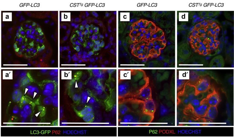

Figure 4|Evaluation of autophagic flux in podocytes of calpastatin transgenic (CST9) mice. (a,b)Representative immunofluorescence images of the expression of green fluorescent protein (GFP)(green) and P62 (red) in glomeruli from 12-week-old GFP-LC3 and CST'GFP-LC3 mice. The arrowheads indicate GFP+ P62+ autophagosomes. (c,d) Representative immunofluorescence images of the expression of P62 (green) and Podocalyxin (PODXL; red) in glomeruli from 12-week-old GFP-LC3 and CST'9 GFP-LC3 mice. Figure subparts with prime indicate higher magnification. Nuclei were stained with Hoechst (blue).Bars = 50 μm. (e) Quantification of the number of LC3+ P62+ dots per podocyte. Mann-Whitney test:**P=0.0065. (f)Quantification of P62+ area per glomerular section. Mann-Whitney test:*P= 0.0420.In(e,f), n =5 GFP-LC3 mice and n =8 CST'9 GFP-LC3 mice.Values are presented as individual plots and mean± SEM. To optimize viewing of this image, please see the online version of this article at GFP-LC3 mice, suggesting increased autophagic flux in podocytes of mice with high calpastatin abundance.
Autophagic flux can be monitored by the measurement of the conversion of the cytoplasmic form of LC3, LC3-I, to the autophagosomal cleaved and phosphatidylethanolamine-coupled form of LC3, LC3-II, on the Western blot. Podocytes from CST mice showed increased LC3-I to LC3-II conversion and decreased P62 expression, both in the presence and in the absence of bafilomycin A,(Figure 5a and b), indicating increased autophagic flux in podocytes with calpastatin overexpression. Finally, the accumulation of GFP+P62+ dots in podocytes after chloroquine administration was more important in CST'g GFP-LC3 mice than in GFP-LC3 mice, thus confirming that calpastatin overexpression in-duces autophagic flux in podocytes in vivo(Figure 5c-e). Altogether, our data suggest that AngII stimulates calpain activity in podocytes, that autophagy is inhibited by calpain in podocytes, and that inhibition of the endogenous calpain activity by calpastatin overexpression is sufficient to stimulate autophagic flux in podocytes.
CSTT9 mice are protected from Angel + HSD-induced podocyte injury
CSTT: mice did not show any kidney alteration until at least 12 months of age(Supplementary Figure S5). We analyzed podocyte injury in CST'号 mice during AngII + HSD treatment. Although WT mice developed mild glomerulosclerosis and podocyte injury after 4 weeks of hypertension, as shown by abnormal expression of podocalyxin and nephrin, CSTTg mice presented fewer sclerotic glomerular lesions(Figure 6a
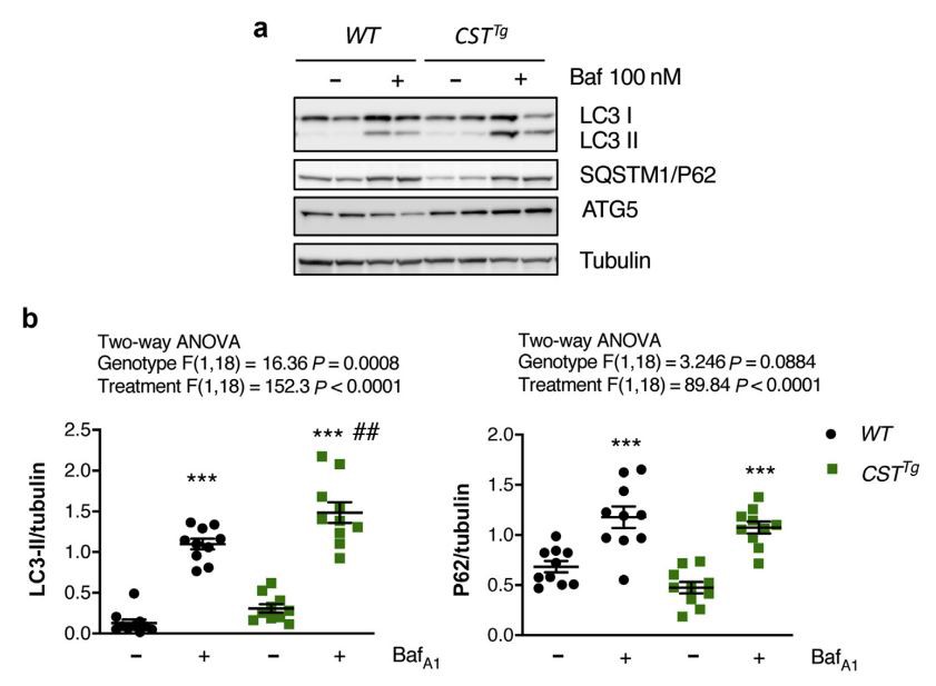
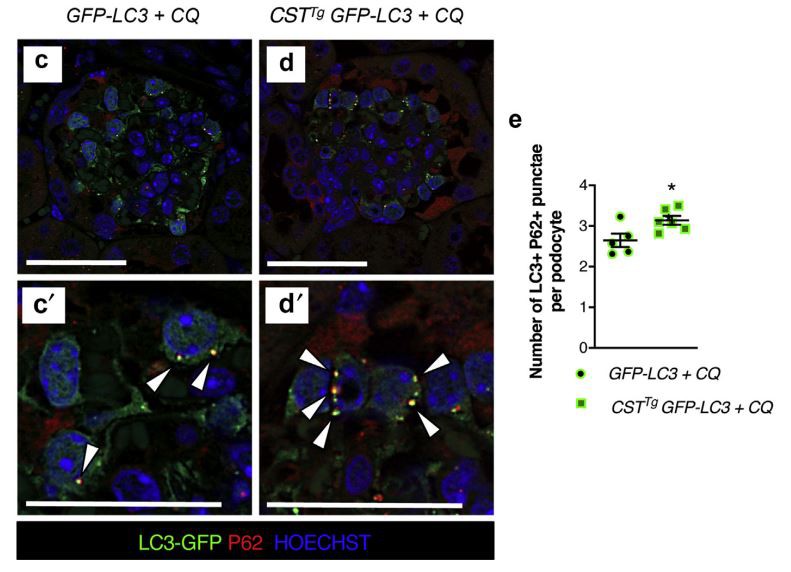
Figure 5|Blocking autophagosomal degradation confirmed increased podocyte autophagic flux in calpastatin transgenic (CSTT)mice.(a) Western blot analysis of the expression of LC3. Sequestosome 1(SOSTM1)/P62, and ATG5 in primary podocytes from wild-type(W or CST'9mice. Tubulin expression serves as normalization. Podocytes were treated or not treated with bafilomycin A1(BafA1; 100 nM) for 4 hours before culture arrest. (b) Quantification of the LC3-ll/'tubulin and P62/tubulin ratios.n = 10 WT mice and n =8 CST'9 mice. Values are presented as individual plots and mean ± SEM. Two-way analysis of variance paired for treatment: for LC3-ll/tubulin: genotype, P= 0.0008;treatment, P<0.0001; for P62/tubulin: genotype, P= 0.0884; treatment, P< 0.0001.Sidak's multiple comparison test: for LC3-II/tubulin:***P<0.0001 for WT-BafA1 versus WT+ BafA1,***P<0.0001 for CST'9-BafA1 versus CST'9+BafA1,"*P=0.0028 for WT+BafA1 versus CSTT9 +BafA1:for P62/tubulin:***P<0.0001 for WT-BafA1 versus WT+BafA1,***P<0.0001 for CST'9-BafA1 versus CST'9+ BafA1.(c,d)Representative immunofluorescence images of the expression of green fluorescent protein (GFP; green) and P62 (red) in glomeruli from 12-week-old GFP-LC3 and CST'9 GFP-LC3 mice. Figure subparts with prime indicate higher magnification. Nuclei were stained with Hoechst (blue). Bars = 50 μm. Mice were treated with chloroquine(CQ; 80 mg/kg) 4 hours before killing. The arrowheads indicate GFP+ P62+autophagosomes. (e) Quantification of the number of LC3+ P62+ dots per podocyte. n =5 mice per genotype. Values are presented as individual plots and mean ± SEM. Unpaired t-test with equal SD:*P= 0.0302. To optimize viewing of this image, please see the online version of this article at www.kidney-international.org.
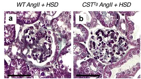


Figure 6| Calpastatin overexpression prevents angiotensin Il (Angel)+ high-salt diet (HSD)-mediated podocyte injury. (a,b)

Cistanche can relieve kidney disease
Representative images of Masson's trichrome-stained sections of glomeruli from wild-type (WT and calpastatin transgenic(CST'9) mice after 6 weeks of Angll+ HSD.Bars=50 um.(c,d,f,g)Representative immunofluorescence images of the expression of (c,d) Podocalyxin (PODXL) and (f,g)nephrin (NPHS1)in WT and CST'9mice after 6 weeks of Angel +HSD and (e,h) associated quantifications. Bars =50 um. (ii)

cistanche can treat acute renal failure
Representative immunofluorescence images of the expression of Wilm's Tumor 1 (WT1; green) and PODXL (red) in glomeruli from WT and CST'mice after 6 weeks of Angel +HSD and (k) associated quantification of the number of WT1+ cells per glomerular section. Nuclei were stained with Hoechst(blue).Bars =50 um. In (e,h,k), values are presented as individual plots and mean ± SEM,n =9 WT mice, and n=7 CSTTg mice.Unpaired t-test with equal SD:**P=0.0095 in (e),*P=0.0249 in (h), P=0.7891 in (k).
and b)and preserved podocalyxin and nephrin expression (Figure 6c-h). Such differences in podocyte phenotype were observed at a stage where the density of podocyte nuclei was not different between WT and hypertensive CST'g mice (Figure 6i-k).
Similarly, after 6 weeks of hypertension, GFP-LC3 and CST'1 GFP-LC3 mice presented podocyte injury but lesions were more prominent in GFP-LC3 mice (Figure 7). The urinary albumin-to-creatinine ratio was higher in GFP-LC3 mice after 4 weeks of AnglI + HSD(Figure 7a). Podocyte number was not different between the 2 genotypes, but nephrin expression was significantly more decreased in GFP-LC3 (control) mice(Figure 7b-e). Correlating with calpastatin-mediated protection, the ultrastructural analysis showed mild and focal podocyte foot process effacement in GFP-LC3 mice treated with Angl+ HSD with better preservation of the foot process effacement in CST'g mice, although the global quantification of the number of foot processes per glomerular basement membrane(GBM)length was not statistically different, most likely because podocyte injury is focal and segmental in our model(Figure 7f-h). Of note, macrophages and T-lymphocyte infiltration in kidneys were not different between the groups (Supplementary Figure S6). Taken together, these results demonstrated that calpastatin prevented AngII+ HSD-induced podocyte injury.
Calpastatin overexpression restores autophagic flux in podocytes from mice after Angel + HSD treatment
Finally, we wondered whether calpastatin-mediated glomerular protection in the AngII+ HSD model implicated autophagy maintenance in podocytes. Interestingly, AnglI +HSD-induced P62 accumulation in podocytes was prevented in CSTTg and GFP-LC3 CST'g mice(Figure 8a-d), indicating that calpastatin prevented blockade of podocyte autophagy. Of note, the number of GFP+ P62+ dots labeling of auto-phagosomes was similar in podocytes from hypertensive GFP-LC3 and GFP-LC3 CST mice, thus suggesting that AnglI + HSD induced a slowdown of podocyte autophagic flux rather than a complete arrest(Figure 8e-g).
In silico prediction of the calpain cleavage site in podocyte proteins
We used several in silico tools to predict potential calpain targets in podocytes(Supplementary Tables S1 and S2). Three online databases were compared: GPS-CCD,4CaMPDB,5, and DeepCalpain.° In silico analysis identified several podocyte proteins, nephrin and podocin among them, that could be cleaved by calpains. Thus, calpastatin-mediated glomerular protection could be linked to a reduction of calpain enzymatic activity, leading to reduced degradation of some podocyte proteins.
Furthermore, at least 3 autophagy-related proteins are direct targets of calpains. ATG5 is cleaved by calpains, leading to a disturbance in the ATG12-ATG5 complex formation.57 Administration of calpain inhibitors in vivo also prevented cleavage of the autophagy protein Beclin-1.5"In silico analysis of the putative cleavage site on autophagy-related proteins supports the hypothesis that calpain could regulate autophagy through the enzymatic cleavage of autophagy proteins.
mRNA expression of endoplasmic reticulum (ER) and oxidative stress markers in glomeruli from mice treated with Angll+ HSD
We evaluated endoplasmic reticulum(ER)stress and oxidative stress by quantitative PCR in glomeruli during AngII +HSD treatment(Table 1). At baseline, we did not observe any change in mRNA expression of analyzed genes in glomeruli5loxlox mice(Supplementary from WT and Nphs2.cre Atg5 Figure S7). After 6 weeks of hypertension, glomeruli from Nphs2. cre Atg5loxlox mice showed different mRNA profiles of genes of the ER stress and oxidative stress pathways with increased expression of Sod1, Prdxl, Atf4, Gpxl, and Hsp90b1 as compared with WT glomeruli, thus suggesting that autophagy depletion in podocytes favored AngII + HSD-induced ER stress and oxidative stress. Conversely, glomeruli from CST mice presented downregulation of several genes of the ER stress and oxidative stress pathways as well as decreased expression of some pro-apoptotic genes(Figure 9).
Taken together, these results indicate that calpastatin overexpression could prevent glomerular injury by reducing AngII + HSD-induced ER and oxidative stress.
DISCUSSION
In the present study, we demonstrated that in AngII+HSD-induced hypertension, podocyte autophagy is markedly downregulated. Furthermore, mice with podocyte-specific deletion of Atg5 were more prone to AngI+ HSD-induced glomerulosclerosis and podocyte loss, thus showing that autophagy in podocytes prevents the development of hypertensive nephropathy, highlighting the critical role of autophagy in Angl + HSD-induced podocyte injury. Little is known about the extracellular stimuli that regulate cellular autophagy, and these results shed light on the pathophysiological regulation of podocyte autophagy by AngII.
In most human glomerulopathies, podocyte foot process effacement is a hallmark of glomerular injury leading to proteinuria. Autophagy is likely to play an essential role in maintaining podocyte function because these terminally differentiated cells display high rates of autophagy even in the absence of stress. A previous study showed that Angi pro-motes autophagy through the generation of reactive oxygen species in a conditionally immortalized murine podocyte cell line.5 Reactive oxygen species production is indeed a general inducer of autophagy in many cell types and the reasons for such discrepancy with our findings are unclear. Unlike this latter study, we used murine primary cultured podocytes retaining podocin and nephrin expression and in vivo approaches and not murine cell lines. We confirm previous reports that postmitotic podocytes exhibit an unusually high level of constitutive autophagy. Measurement of the increased amount of LC3-II after AngII stimulation in the presence or absence of lysosomal inhibitors is necessary to determine


whether autophagic flux is increased or blocked. Previous studies have neglected this phenomenon.
Here, we used a hypertensive model based on AnglI perfusion and HSD. Podocytes exhibit ATI receptors and are exposed to free filtered peptides such as AngII1,6,26,61-6 It is assumed that the observed renoprotective effects of RAAS inhibition could be—-at least partially—due to a blockade of this podocyte-specific RAAS. Likewise, in vivo studies

Cistanche can relieve kidney disease





