

Research On The Antidepressant Effects And Mechanisms Of Cistanche Phenylethanol Glycosides in Mice Ⅱ
Dec 17, 2024
2 Results and analysis
2.1 The effects of CPhGs on SPT and 5-HT and DA in serum of depression model mice are shown in Table 1.
Compared with Con, Mod's sugar water preference rate, serum and hippocampal tissue neurotransmitter 5-HT and DA contents were significantly higher. Significantly decreased (P<0.01 or P<0.05). It shows that Mod mice show depression-like behavior and the model is successfully established. Compared with Mod, CPhGs-H, CPhGs-M, CPhGs-L and FLX significantly increased the sugar water preference rate, neurotransmitter 5-HT and DA contents in serum and hippocampal tissue of mice (P<0.01 or P< 0.05), among which the sugar water preference rates increased by 72.91%, 46.59%, 23.43% and 87.93% respectively; the serum 5-HT levels increased by 31.77%, 25.24%, 20.44% and 37.59% respectively; the serum DA levels increased by 47.07% respectively. , 35.57%, 27.77% and 52.93%; the 5-HT content in hippocampal tissue increased by 31.77%, 25.24%, 20.44% and 37.59% respectively, and the DA content in hippocampal tissue increased by 47.07%, 35.57%, 27.77% and 52.93% respectively. This shows that CPhGs has an alleviating effect on the anhedonia of CUMS mice. CPhGs may restore the anhedonic ability of mice by regulating neurotransmitter transmission in the central nervous system, especially pathways related to reward and emotion regulation such as 5-HT and DA.

Herbal Cistanche Raw Materials For Depression
2.2 Effect of CPhGs on OFT in depression model mice
According to the physiological characteristics of rodents and some special behaviors in unfamiliar environments, it can be used to evaluate the exploratory behavior and anxiety and depression state of mice in new environments. The open field experiment mainly examines the autonomous activities of mice, and then reflects In its depressive state, the longer the total distance of movement of the mouse per unit time, the longer the activity time in the central area, and the longer the distance of movement in the central area, the lighter the degree of depression of the mouse [11]. The experimental results are shown in Figure 1. Compared with Con, the above three indicators of Mod were significantly reduced (P<0.01); compared with Mod, the above three indicators of CPhGs-H, CPhGs-M, CPhGs-L and FLX were significantly increased (P<0.01 or P<0.05), among which the total movement distance CPhGs-H, CPhGs-M, CPhGs-L and FLX increased by 46.63%, 29.86%, 23.92% and 52.65% respectively, and the central area activity time respectively The increases were 127.92%, 64.65%, 43.81% and 119.81%, and the exercise distance in the central area increased by 122.44%, 53.21%, 33.97% and 102.56% respectively. Experimental results show that CPhGs can regulate brain neural pathways and improve functions related to emotion regulation and behavioral drive, affecting the behavioral performance of OFT in depression model mice.
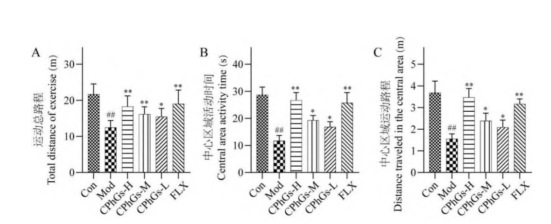
Fig. 1 Results of OFT in each group of mice (x ± s, n = 10)
2.3 Effects of CPhGs on TST and FST in depression model mice
The results of TST and FST are shown in Figure 2. Compared with Con, the immobility time of Mod's TST test and the floating immobility time of FST test were both significantly longer (P<0.01); compared with Mod, CPhGs- The total immobility time and floating immobility time of H, CPhGs-M, CPhGs-L and FLX were significantly shortened (P<0.01 or P<0.05). Among them, the immobility time of forced swimming was reduced by 34.67%, 30.71%, 20.78% and 44.97% respectively, and the immobility time of tail suspension was reduced by 35.91%, 27.88%, 23.30% and 42.14% respectively. This suggests that CPhGs improves the function of the central nervous system and restores the normal activity of neuronal functions and neural circuits. In desperate stress environments, the negative emotions of mice are significantly reduced, and the desire to survive becomes positive, thus improving the TST of depressed mice. and behavioral manifestations of FST.

2.4 Effect of CPhGs on inflammatory factors in depression model mice
To evaluate the anti-inflammatory effects of CPhGs, this study measured the expression levels of key inflammatory factors in the hippocampus, including TNFα, IL-1β, IL-18, IL-6, and IFN-γ (see Figure 3 ). Compared with Con, the levels of TNF-α, IL-1β, IL-18, IL-6 and IFN-γ in Mod were significantly increased by 132.92%, 107.32%, 69.13%, 62.32% and 76.55% (P<0.01) . Compared with Mod, TNF-α in CPhGs-H, CPhGs-M, CPhGs-L and FLX mice decreased by 42.93%, 29.95%, 11.08% and 23.00% respectively (P<0.01 or P<0.05); IL-1β respectively decreased by 45.72%, 21.91%, 12.32% and 48.65% (P<0.01 or P<0.05); IL-18 decreased by 35.17%, 18.22%, 9.13% and 22.06% respectively (P<0.01 or P<0.05); IL- 6 were reduced by 34.67%, 11.47%, 9.06% and 19.47% respectively (P<0.01 or P<0.05); IFN-γ levels were reduced by 34.97%, 14.06%, 9.89% and 26.25% respectively (P<0.01 or P<0.05) .

Fig. 3 Changes in the levels of inflammatory factors in the hippocampal tissue of each group of mice (x ± s, n = 10)
2.5 Effect of CPhGs on inflammatory mediators in depression model mice
To evaluate the effect of CPhGs on inflammatory mediators, this study measured the expression levels of key inflammatory mediators (iNOS, COX-2, and PGE2) in the hippocampus (see Figure 4 ). Compared with Con, iNOS, COX-2 and PGE2 levels were significantly increased in Mod by 278.34%, 147.45% and 124.03% (P<0.01). Compared with Mod, the iNOS levels of CPhGs-H, CPhGs-M, CPhGsL and FLX were reduced by 49.55%, 40.52%, 11.99% and 68.57% respectively (P<0.01 or P<0.05); the COX-2 levels were reduced by 39.58% respectively. , 26.87%, 17.96% and 48.39% (P<0.01 or P<0.05); PGE2 levels were reduced by 27.89%, 14.28%, 10.45% and 37.51% respectively (P<0.01 or P<0.05), among which the PGE2 of CPhGs-L The level is not statistically significant compared with Mod. The above results indicate that CUMS successfully induced the mouse depression model and caused significant inflammatory response and oxidative stress state. CPhGs can effectively reduce the levels of these inflammatory mediators after administration, showing good anti-inflammatory and antioxidant effects, and its effects are dose-dependent.

Fig. 4 Results of the content of inflammatory mediators in the hippocampal tissue of each group of mice (x ± s, n = 10)
2.6 Effect of CPhGs on hippocampal tissue structure in depression model mice
HE staining was used to analyze pathological changes in mouse hippocampal tissue (see Figure 5). Compared with Con, Mod mice showed obvious pathological changes in the dentate gyrus region of the hippocampus, including disordered cell arrangement, nuclear pyknosis, and cytoplasmic vacuolation, showing significant neuronal damage. Compared with Mod, CPhGs-L has no obvious improvement effect on the above damage phenomena. CPhGsM and CPhGs-H significantly improved these pathological changes. The effect of CPhGs-M was close to FLX, and the effect of CPhGs-H was even better than FLX. The morphological improvement of hippocampal neurons was dose-dependent, suggesting that CPhGs have a protective effect on the hippocampus.

Equine neurons, improving depressive symptoms. This result is consistent with previously observed improvements in behavioral and biochemical markers and further supports the potential value of CPhGs in the treatment of depression.
Nissl bodies in Nissl staining (see Figure 5) are cytoplasmic structures unique to neurons and are primarily responsible for protein synthesis. It is abundant in healthy, metabolically active neurons but is reduced or disassembled when damaged. Therefore, the state of Nissl bodies directly reflects the function and health of neurons and is an important indicator for evaluating nerve cell activity. Compared with Con, the neuronal arrangement in the dentate gyrus region of the hippocampus of Mod mice was disordered and Nissl corpuscles were significantly reduced, suggesting reduced neuronal activity. CPhGs-L, CPhGs-M, and CPhGs-H all increased the number of Nissl bodies compared with Mod. Among them, CPhGs-H has the most significant effect, with the neuron arrangement tending to be regular and the number of Nissl bodies approaching the Con level. FLX also showed significant improvement to a comparable extent as CPhGs-H. These results indicate that CPhGs can dose-dependently ameliorate CUMS-induced hippocampal neuronal damage and increase neuronal viability.
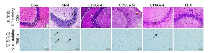
Fig. 5 Results of HE staining and Nissl staining in the hippocampal tissue of each group of mice
2.7 Effect of CPhGs on glial cells in the hippocampus of depression model mice
Astrocytes are the most abundant glial cell type in the brain and play a crucial role in maintaining neurotransmitter transmission and establishing the blood-brain barrier [12]. To analyze the density of astrocytes in mouse hippocampal tissue, IHC was used to detect the expression of the astrocyte marker GFAP in hippocampal tissue (see Figure 6A and Figure 7A ). Compared with Con, GFAP expression in the hippocampus of Mod mice was significantly reduced by 47.47% (P<0.01), showing significant astrocyte damage. Compared with Mod, CPhGs-L increased by 18.48%, but did not reach statistical significance; the GFAP expression of CPhGs-H, CPhGs-M and FLX significantly increased by 64.23%, 40.07% and 57.45% respectively (P< 0.01 or P<0.05), among which the effect of CPhGs-M is close to FLX, and the effect of CPhGs-H is even better than FLX. Changes in the number of astrocytes in the hippocampus of the CPhGs administration group
The effect was dose-dependent, suggesting that CPhGs can protect astrocytes and improve depressive symptoms.
Microglia are immune cells of the central nervous system, which play an important role in brain immune surveillance, neurogenesis, synaptic pruning and other processes [13]. Clinical studies have found that microglia are overactivated in the brains of depressed patients. Animal studies have also found that chronic stress induces depressive-like behaviors in animals and can also cause structural and functional abnormalities and excess of microglia in the brain (prefrontal lobe, hippocampus and other brain areas). Activate[14]. To analyze the density of astrocytes in mouse hippocampal tissue, IHC was used to detect the expression of the astrocyte marker Iba-1 in hippocampal tissue (see Figure 6B and Figure 7B). Iba-1 expression was significantly increased in Mod by 72.34% compared to Con (P<0.01). Compared with Mod, Iba-1 expression of CPhGs-H, CPhGs-M, CPhGs-L, and FLX was significantly reduced by 23.73%, 18.87%, 14.89%, and 29.67%, respectively (P<0.05 or P<0.01). Description CPhGs
It can reduce the release of pro-inflammatory cytokines and inflammatory mediators caused by microglial activation, thereby CUMS-induced central nervous system inflammation.
2.8 Effect of CPhGs on neuroinflammation in hippocampal tissue of depression model mice
The NLRP3 inflammasome pathway plays a key role in the inflammatory response in the central nervous system and is closely related to the pathological process of depression. NLRP3 and Caspase-1, as the core of the inflammasome, are activated when exposed to external stimuli, catalyzing precursor inflammatory factors and triggering a series of inflammatory responses [15]. In order to analyze the severity of neuroinflammation in mouse hippocampal tissue, IHC was used to detect the expression of NLRP3 inflammasome key proteins NLRP3 and Caspase-1 in hippocampal tissue (see Figures 6C, 6D and Figures 7C, 7D). Compared with Con, the NLRP3 level of Mod was significantly increased by 121.62% (P<0.05), and the Caspase1 level was significantly increased by 172.65% (P<0.01). Compared with Mod, the NLRP3 levels of CPhGs-H, CPhGs-M, CPhGs-L and FLX were reduced by 50.00%, 40.24%, 32.93% and 71.95% respectively (P<0.05), and the Caspase-1 levels were reduced by 57.65% and 37.99 respectively. %, 31.91% and 60.59% (P<0.05 or P<0.01). This shows that CPhGs can effectively inhibit the activation of NLRP3 inflammasome, thereby reducing neuroinflammatory response.

Fig. 6 IHC results of GFAP, Iba-1, NLRP3, and Caspase-1 in the hippocampal tissue of each group of mice (200

Fig. 7 Quantitative analysis results of IHC for GFAP, Iba-1, NLRP3, and Caspase-1 in the hippocampal tissue of mice from each group (x ± s, n = 10)
2.9 Effect of CPhGs on the expression of TLR4 and p-NFκB p65 in mouse hippocampus
TLR4/NF-κB signaling is a key mediator in inducing the activation of NLRP3 inflammasome and initiating downstream inflammatory responses. The expression of NLRP3 is driven by NF-κB activation and its nuclear translocation of p65 subunit, and the entire inflammatory signaling cascade is driven by Mediated by the TLR4 signaling pathway [16]. In order to analyze the changes in the expression of NLRP3 inflammasome upstream proteins in mouse hippocampal tissue, immunofluorescence double staining was used to detect the expression of TLR4 and p-NF-κB p65 in hippocampal tissue (see Figures 8 and 9). Compared with Con, the TLR4 level of Mod was significantly increased by 111.17% (P<0.01), and the p-NF-κB p65 level was significantly increased by 184.51% (P<0.01).
Compared with Mod, the TLR4 levels of CPhGs-H, CPhGs-M, CPhGs-L and FLX were reduced by 15.24%, 27.12%, 39.40% and 33.51% respectively (P<0.01 or P<0.05), p-NF-κB p65 The levels were reduced by 19.46%, 33.61%, 48.82% and 41.54% respectively (P<0.01 or P<0.05). The above results, combined with pathological examination and inflammatory indicators, indicate that CPhGs can effectively inhibit the activation of the TLR4/NF-κB signaling pathway, thereby reducing the activation of the NLRP3 inflammasome and the resulting inflammatory response.

Fig. 8 Immunofluorescence staining results of TLR4 and p-NF-κB p65 in the hippocampal tissue of each group of mice
Green fluorescence represents p-NF-κB staining results; Red fluorescence represents TLR4 staining results; Blue fluorescence represents DAPI nuclear staining.

Fig. 9 Average fluorescence intensity of TLR4 and p-NF-κB p65 in the hippocampal tissue of each group of mice (x ± s, n = 10)
3 Discussion and conclusion
This study systematically evaluated the therapeutic effects and underlying mechanisms of CPhGs on CUMS-induced depression mouse model.
Research results show that CPhGs can significantly improve CUMS-induced depressive-like behaviors, and this improvement effect is closely related to regulating neurotransmitter systems, inhibiting neuroinflammatory responses, and affecting NLRP3 inflammasome-related pathways. At the behavioral level, CPhGs effectively improved various behavioral indicators of CUMS model mice. CPhGs significantly increased the sugar water preference rate, shortened the immobility time in the forced swimming and tail suspension tests, and reduced anxiety behaviors in the open field experiment. These results confirm that CPhGs has significant antidepressant and anxiolytic effects, and its effects are comparable to the positive control drug fluoxetine. in neurobiochemical water On average, CPhGs significantly increased the levels of 5-HT and DA in the serum of model mice. It shows that CPhGs may improve depressive symptoms by protecting nerve cells and regulating neurotransmitter systems.

Inflammatory factors play an important role in the occurrence and development of depression, which has been confirmed in animal models of depression and clinical studies [17]. This study found that CUMS significantly increased the expression of multiple inflammatory factors in mouse hippocampal tissue, including TNFα, IL-1β, IL-18, IL-6, and IFN-γ. CPhGs treatment can significantly reduce the levels of these inflammatory factors, thereby improving depressive-like behavior in CUMS mice. Studies have found that the levels of pathogen-related molecular patterns such as iNOS and NO and risk-related molecular patterns such as IL-1β and IL-18 are increased in the hippocampus of patients with depression. After these molecules enter the cytoplasm of microglia, they are detected by pattern recognition receptors. NLRP3 recognizes and stimulates immune responses [18]. The NLRP3 inflammasome is a key protein complex that regulates the innate immune inflammatory response and includes LRP3, ASC, and the effector protein Caspase-1. When activated, NLRP3 recruits ASC and Pro-Caspase-1 to complete the assembly of the inactive NLRP3 inflammasome [19]. At the same time, PAMPs and DAMPs can also cause mitochondrial damage and lysosome rupture, leading to the release or exposure of mitochondrial contents, cathepsin B release from lysosomes, promoting the binding of NEK7 to the NLRP3 inflammasome and activating it, and inducing IL-1β and IL-18 matures and is released extracellularly, thereby inducing neuroinflammation [20]. CPhGs can reduce the levels of iNOS, COX-2 and PGE2, reduce the activation of NLRP3 inflammasome by inhibiting the production of PAMPs and DAMPs, thereby reducing the maturation and release of inflammatory factors such as IL-1β and IL-18 in hippocampal tissue, thereby Improved depressive-like behavior in CUMS mice.
Studies have shown that the NLRP3 inflammasome is involved in the pathogenesis of depression and anxiety and is a key mediator connecting chronic stress and depressive states. Activation of the NLRP3 inflammasome is a complex process that requires the receipt of initiating signals, such as the activation of TLR4/NF-κB signaling. Activated NF-κB further upregulates the mRNA transcription levels of NLRP3 and pro-IL-1β, activates inflammasomes, and initiates downstream inflammatory responses [21]. The results of this study showed that TLR4/NF-κB signaling and NLRP3 showed consistent high expression after CUMS stimulation, which is consistent with previous findings in depression mouse models [22]. CPhGs treatment can significantly reverse this high expression state, suggesting that CPhGs may affect the activation of NLRP3 inflammasome by regulating TLR4/NF-κB signaling. By inhibiting the TLR4/NF-κB signaling pathway, CPhGs block the initiation signal of NLRP3 inflammasome activation, thereby reducing the production and release of downstream inflammatory factors. The present study also observed the regulatory effects of CPhGs on glial cells. CPhGs significantly increased the number of GFAP-positive astrocytes while reducing the number of Iba-1-positive microglia. This finding may be related to inhibition of the TLR4/NF-κB signaling pathway. Studies have shown that activation of the TLR4/NF-κB signaling pathway can promote the formation of neurotoxic A1 astrocytes [23], and CPhGs may help maintain astrocytes by inhibiting this pathway. Normal functions and reduces neuroinflammation and neuronal damage.
In summary, this study systematically elucidates the effect of CPhGs in ameliorating CUMS-induced depressive-like behaviors and its underlying mechanisms. CPhGs exhibit potential as antidepressants by modulating neurotransmitter systems, inhibiting neuroinflammatory responses, and affecting NLRP3 inflammasome-related pathways.
References
1 Ren L, Shao Y T, Qin SH, et al. Study on target identification and effect of saponin A on depressive disorder[J]. Nat Prod Res Dev(Natural Product Research and Development), 2024, 36: 44-51.
2 Report on nutrition and chronic diseases status of Chinese residents (2020)[J]. Acta Nutr Sin( Nutrition Science Journal), 2020, 42, 521-521.
3 Qin TT, Hu JW, Ceng JS, et al. Antidepressant effect of essential oil from Schizonepeta tenuifolia Briq. on lipopolysaccharide-induced depression model mice[J]. Nat Prod Res Dev(Natural Product Research and Development), 2023, 35: 1480-1485.
4 Yin YS, Ju T, Zeng DY, et al. "Inflamed" depression: a review of the interactions between depression and inflammation and current anti-inflammatory strategies for depression[J].Pharmacol Res,2024,207:107322. 5 Xiong TW, Zhang J, Shi JS. Research progress on the echanism of antidepressant effects of traditional Chinese medicine [J]. Trad Chin Med J (Chinese Medicine Bulletin), 2024, 23: 65-69. 6 Fan L, Peng Y, Chen XN, et al. Integrated analysis of hytochemical composition, pharmacokinetics, and network pharmacology to probe distinctions between the stems of Cistanche deserticola and C. tubulosa based onantidepressant activity[J].Food Funct,2022,13:8542-8557





