

Serum Neurofilament Light Chain Is Not A Useful Biomarker Of Central Nervous System Involvement in Women With Fabry Disease
Jan 05, 2024
Neurofilament Light Chain (NfL) serum concentration is a new noninvasive marker of neurodegenerative disorders. Fabry disease (FD) leads to accumulation of glycosphingolipids in tissues leading to progressive damage of critical body systems and organs, including peripheral and central nervous system. There are no established serum markers of neurodegeneration in FD. Our cross-sectional single-center study was designed to prove the concept that serum NfL levels could reflect the severity of cognitive impairment and indirectly, the level of central nervous system involvement in women at earlier stages of FD. Twelve women with a diagnosis of FD confirmed by genetic tests and 12 matched healthy subjects were included. Serum concentrations of NfL were measured in all subjects together with neuropsychological tests that included the Mini-Mental State Examination (MMSE) and Montreal Cognitive Assessment Scale (MoCA). Quality of life was assessed with the Short Form Survey (SF-36). FD patients and healthy subjects did not differ concerning serum NfL concentration, results of neuropsychological tests, and quality of life. There was a significant positive correlation between NfL and globotriaosylosphingosine (lyso-Gb3) concentration in women with FD (R = 0,69, p = 0.01). There was also a correlation between NfL concentration and MoCA score but not MMSE score. Receiver operating characteristic (ROC) analysis showed that the best predictor for Mild Cognitive Impairment in both groups was eGFR. Serum NfL concentration does not appear to predict the degree of nervous system involvement in women with FD.
biomarker, neurodegeneration, quality of life


CLICK HERE TO GET NATURAL ORGANIC CISTANCHE EXTRACT WITH 25% ECHINACOSIDE AND 9% ACTEOSIDE FOR KIDNEY FUNCTION
Supportive Service Of Wecistanche-The largest cistanche exporter in the China:
Email:wallence.suen@wecistanche.com
Whatsapp/Tel:+86 15292862950
Shop For More Specifications Details:
https://www.xjcistanche.com/cistanche-shop

1. Introduction
Fabry disease (FD), is an ultra-rare lysosomal storage disease inherited as an X-linked disorder. FD is caused by a deficiency of the lysosomal enzyme alpha-galactosidase A (α-Gal A; E.C. 3.2.1.22). GLA gene, located on X chromosome at Xq22, encodes a 429 amino acid precursor that is processed to a 398 amino acid glycoprotein functioning as a homodimer. The mutation of GLA leads to a deficiency or absence of the enzyme α-galactosidase A (α-Gal A), which catalyzes the hydrolysis of globotriaosylceramide.
Alpha-Gal A deficiency leads to progressive accumulation of glycolipids, and globotriaosylosphingosine (lyso-Gb3) in different body cells, leading to damage and dysfunction of affected organs. The most affected cells and tissues in FD include glomerular podocytes, cardiomyocytes, endothelial cells, vascular muscles and peripheral and central nerves. It all leads to dysfunction and failure of vital organs including the heart, kidneys, and nervous system. The severity of the disease depends on the gender, age, and type of mutation. Males with classic phenotypes have the highest risk of complications and early symptoms, while younger women mostly become affected by FD later in life (1).

Neurofilaments are the main component of the neuronal cytoskeleton. Light (NfL), intermediate (NfM) and heavy (NfH) chains have been distinguished on the basis of their molecular mass. The serum concentration of NfL and its importance as a marker of central nervous system diseases have been demonstrated in several recent studies (2,3). Neuronal damage and physiological changes of the central nervous system (CNS) cause the release of neurofilaments. This translates into elevated levels of NfL in the cerebrospinal fluid and ultimately in the blood, where its concentration reflects the rate at which NfL is released from the neurons (3). Several studies have shown a strong positive correlation between NfL in the blood and in the cerebrospinal fluid (4). Serum NfL concentration positively correlated with the severity of various diseases of the central nervous system including multiple sclerosis, amyotrophic lateral sclerosis, frontotemporal dementia, Alzheimer's disease, traumatic brain injuries and degeneration of the nervous system associated with HIV infection (2).
The main physiologic factor influencing the NfL concentration is the age of the patients. With aging NfL concentration in healthy subjects increased by 2.2% annually. After the age of 60, a further significant increase in NfL concentration is observed (5). These changes could be attributed both to aging itself and the aggregation of comorbidities. It has been well-proven that patients with FD are characterized by much faster brain aging compared to the healthy population (6). Patients with FD are at increased risk of developing cognitive dysfunction and most of them also have symptoms of depression (7). Patients diagnosed with severe depression have more cognitive impairment compared to the general population (8). It also was shown that in major depressive disorder, higher levels of NfL were observed (9). However, it has not been confirmed in FD that the cognitive impairment is due to depressive symptoms but its risk factors include male gender, patients with classic disease phenotype, lower intelligence quotient (IQ), and a history of stroke (10,11).
Many clinical tests have been developed to assess cognitive functions, each of which assesses specific domains of cognitive functioning, but in a different aspect. Screening tests play a key role in allowing each clinician to perform an initial assessment of cognitive impairment. The best-validated tests used for screening for cognitive impairment include the Mini-Mental State Examination (MMSE) and the Montreal Cognitive Assessment Scale (MoCA) (12,13). MMSE and MoCA were used and well-validated in recent studies assessing cognitive impairment in patients with FD (11,14).
The study aimed to assess whether the serum concentration of NfL could be a marker of early central nervous system involvement and cognitive impairment in women with FD.
2. Materials and Methods
The study was approved by the Local Ethics Committee and was conducted by the Declaration of Helsinki. All patients gave informed written consent to participate in the study.
Twenty-four subjects were enrolled, including 12 women with confirmed FD and 12 matched healthy controls. The study was conducted between March and October 2020. The characteristics of the study population is provided in Table 1. Diagnosis of FD was based on the blood concentration of α-Gal A, lyso-Gb3, and on genetic tests. The tests were performed using the Dry Blood Spot method (DBS). Individual results are presented in Table 2. Only one woman from the study group has been qualified for enzyme replacement therapy.
The patients with FD included in our study came from three different families. The degree of kinship and family trees of the patients are presented in Figure 1.
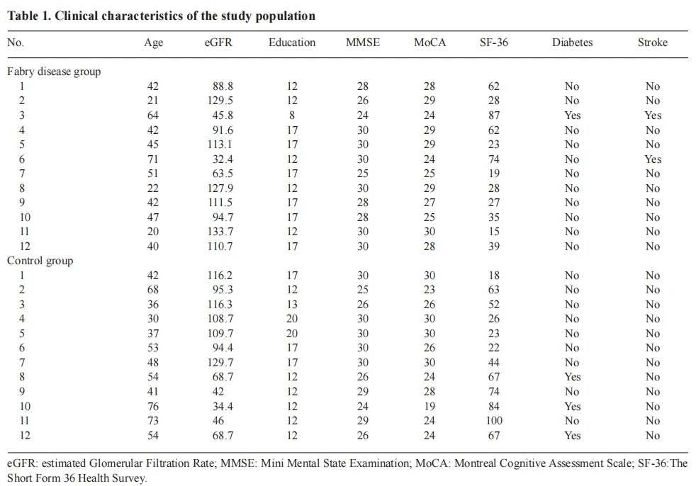
Table 2. Type of genetic variant, globotriaosylosphingosine and α-galactosidase A concentration in blood in women with Fabry disease

The control group consisted of 12 healthy women, matched to women with FD for age, education level, and kidney function. The exclusion criteria were the diagnosis of any disease of the central nervous system other than associated with FD, a disability that would hinder any of the study procedures such as hearing or vision loss, chronic kidney disease with eGFR < 30 mL/ min, dementia, acute functional psychiatric disorder and uncontrolled hypertension (systolic BP > 130 mmHg or diastolic BP > 80 mmHg). Every patient completed a quality-of-life questionnaire The Short Form 36 Health Survey (SF-36) and two screening tests assessing cognitive function, MoCA and MMSE.

The MoCA and MMSE are commonly used as the screening neuropsychological scales. In MMSE, the most commonly used cutoff point for the diagnosis of dementia is a score of 24 points or less. The maximum score for the MoCA test is 30 points; a result of 26 or more points is defined as normal. A score from 19 to 25 is considered as mild cognitive impairment (MCI) (13). During the same visit, blood was collected in a fasting state after an overnight rest from all participants to determine serum concentration of NfL, creatinine, urea, calcium, phosphate, parathyroid hormone (PTH), and blood hemoglobin. The concentration of NfL was assessed with a Neurofilament Light Polypeptide (NEFL) ELISA Kit (antibodies-online GmbH, Aachen, Germany). Other parameters were measured using routine automated laboratory methods in a local laboratory.
The mean value and standard deviation were calculated for each normally distributed variable. For non-normally distributed variables median value with the interquartile range (IQR) was calculated. Analysis of the normality of the distribution was performed with the Shapiro-Wilk test, and on the basis of its results, the parametric t-test or the non-parametric Mann-Whitney U test was used. Receiver operating characteristic (ROC) curves were drawn to assess the value of serum concentration of NfL, eGFR, lyso-Gb3 and α-Gal A indicating the presence of mild cognitive impairment in the MoCA test. Statistica 13.1 software was used to perform the statistical analysis.
Supportive Service Of Wecistanche-The largest cistanche exporter in the China:
Email:wallence.suen@wecistanche.com
Whatsapp/Tel:+86 15292862950
Shop For More Specifications Details:
https://www.xjcistanche.com/cistanche-shop
CLICK HERE TO GET NATURAL ORGANIC CISTANCHE EXTRACT WITH 25% ECHINACOSIDE AND 9% ACTEOSIDE FOR KIDNEY INFECTION





