

Aqueous Extracts From Cultivated Cistanche Deserticola Y.C. Ma As Polysaccharide Adjuvant Promote Immune Responses
Mar 23, 2022
Contact: joanna.jia@wecistanche.com / WhatsApp: 008618081934791
Shuangshuang Feng a,1 , Xiumei Yang a , Xiang Weng a,1 , Bin Wang b , Ailian Zhang a,*
ABSTRACT
Ethnopharmacological relevance: Herbal polysaccharides have exhibited great immune-enhancing potential. Adjuvants are a key tool for developing efficacious vaccines. In our previous study, a water-soluble polysaccharide extracted from wild Cistanche deserticola Y.C. Ma showed potent immunostimulatory activity.
Aim of the study: In this study, the immune profiles and efficacy of aqueous extracts of cultivated Cistanche deserticola Y.C. Ma (AECCD) on ICR mice against ovalbumin (OVA) were investigated. In in vitro experiments, the possible DC activation mechanism by AECCD was evaluated.
Materials and methods: AECCD were extracted using hot water after which the crude polysaccharides were precipitated by ethanol. Mice were firstly immunized subcutaneously with OVA (10 μg per mouse) alone or OVA (10 μg per mouse) respectively containing different doses of AECCD (200, 400, and 800 μg per mouse) on Days 1 and 14 and the magnitude and kinetics of antibodies and cell-mediated responses were then assessed.
Results: AECCD elicited vigorous and long-term IgG responses with mixed Th1/Th2 responses and up-regulated levels of Th-associated cytokines (CD4+IL-4, CD4+IFN-γ, and CD8+IFN-γ). Moreover, AECCD induced the strong cellular immune response characterized by increased splenocyte proliferation as well as the activated T cell response. Notably, AECCD significantly enhanced the maturation of dendritic cells (DCs) and inhibited Tregs. In vitro experiments, Preliminary tests indicated that AECCD induced DC activation by promoting phenotypic maturation, cytokine section, and allostimulatory activity. Toll-like receptor 4 (TLR4) was an essential receptor for DCs to directly bind AECCD. The inhibitors of NF-κB decreased the expression levels of CD40, CD80, CD86, and MHC-II and the production of IFN-γ, TNF-α, and IL-6 through DCs.
Conclusions: Finally, these findings suggested that AECCD could elicit potent and durable antigen-specific immune responses through DC activation, which was involved in the regulation of maturation markers and cytokine expression via the TLR4-related NF-κB pathway. The study indicates that AECCD is a potential immunomodulator.

Cistanche herba improves immunity.
1. Introduction
Many studies have suggested that adjuvants can enhance the efficacy of vaccines against various infectious diseases such as HIV, COVID-19, influenza, and foot-and-mouth disease (Cao et al., 2020; Giuseppe et al., 2018). Adjuvants can induce different types and magnitudes of immune responses, so an appropriately selected adjuvant may induce the immune response required for a given pathogen or antigen (Reed et al., 2013). Natural herbaceous extracts have long been widely used in daily life or healthcare. In many cases, traditional Chinese medicine (TCM) and its active components, especially multiple TCM polysaccharides-derived adjuvants, significantly improved the efficacy of vaccines. TCM polysaccharides (Advax™, Astragalus polysaccharide, Ginseng polysaccharide, Lycium barbarum polysaccharide, and Ganoderma lucidum polysaccharide) had several advantages over other adjuvants, such as better safety, biocompatibility, strong immune enhancement, and low reactogenicity (Li and Wang, 2015; Schijns et al., 2020). TCM polysaccharide-based compounds and formulations, therefore, have the potential as adjuvant candidates.
Cistanche deserticola Y.C. Ma (Roucongrong in Chinese) is a traditional Chinese medicine recorded in Chinese Pharmacopeia and has been used as a tonic food for hundreds of years in China (Chinese Pharmacopoeia Commission, 1992). Modern pharmacological studies have shown its multiple pharmacological and immunomodulatory applications (Dong et al., 2007; Fu et al., 2017). In our previous study, the crude polysaccharides derived from wild Cistanche deserticola Y.C. Ma promoted the antigen-specific humoral and cellular responses and regulated the maturation of dendritic cells (DCs) (Zhang et al., 2018; Zhao et al., 2019). However, wild Cistanche deserticola Y.C. Ma has been excessively explored and listed as endangered species. Cistanche deserticola Y.C. Ma has been planted in a large scale and is widely used in the development and application of TCM resources. However, the adjuvant potential of cultivated Cistanche deserticola Y.C. Ma is still unclear.
DCs are essential regulators against infectious diseases. After DCs are activated, surface molecules and pro-inflammatory cytokines on DCs interact with corresponding receptors to activate T cells and elicit antigen-specific responses and the production of cytokines directing adaptive immune responses (Banchereau and Steinman, 1998). It has been reported that toll-like receptor 4 (TLR4) on DCs functions as an important polysaccharide receptor (Park et al., 2014; Qi et al., 2016). Indeed, many polysaccharides can interact directly or indirectly with TLR4 on DCs to activate the downstream nuclear factor-kappa B (NF-κB), which can induce the expression of various proinflammatory genes such as IFN-γ, TNF-α, and IL-6 and the inflammatory response to eliminate pathogens (Tian et al., 2019; Zhou et al., 2017; Zhu et al., 2013). Therefore, DCs and NF-κB have been extensively studied as potential targets for disease therapy.
In the study, with the in vivo (ICR mice) strategy, we first evaluated the immunostimulatory effects of aqueous extracts of cultivated Cistanche deserticola Y.C. Ma (AECCD) with different doses (low, medium, and high) to elicit an immune response against OVA and explain the in vitro way to activate DCs and downstream receptor-related pathways. With a mouse model, we revealed that AECCD promoted the long-term antibody response with increased IgG titers and cellular immune response. Importantly, AECCD induced Th-associated cytokines, especially IFN-γ production, which led to Th1 response against infections. The induction effect was closely correlated with an enhanced DC maturation and decreased Tregs. Mechanistically, AECCD promoted DC activation via TLR4-related NF-κB pathway. In summary, the study confirmed the potent adjuvant activity of AECCD in promoting the adaptive response. The study laid a foundation for the development of AECCD polysaccharide adjuvants.
2. Materials and methods
2.1. Reagents
Ovalbumin (OVA), 3-(4, 5-dimethylthiazol-2-yl)-2, 5-diphenyltetrazolium bromide (MTT), polymyxin B (PMB), concanavalin A (ConA), lipopolysaccharide (LPS), mitomycin C and 3,3′ ,5,5′ -Tetramethylbenzidine (TMB) were obtained from Sigma (USA). Fetal bovine serum (FBS) was purchased from Biological Industries. Cell Counting Kit-8 (CCK-8) was purchased from Biosharp (China). Goat anti-mouse IgG/IgG1/IgG2a-HRP conjugates were from Southern Biotech (USA). Inject Alum was from Thermo Scientific Pierce (USA). GM-CSF was from Peprotech (USA). Fluorescently-labeled antibodies (anti-CD3-PE, anti-CD4-APC, anti-CD8a-FITC, anti-CD44-PE, anti-CD62L-PE, anti-MHCII-FITC/Percp-cy5.5, anti-IL-4-PE, anti–IFN–γ-PE, anti-CD86-APC, anti-CD40-FITC/PE, anti-CD11c-PE/FITC, anti-CD80-APC), Golgistop, Cytofix/Cytoperm, and Perm/Wash buffer were all from BD Bioscience (USA). The regulatory T-cell staining kit was from eBiosciences (USA). TAK-242 was from MedchemExpress (Shanghai, China). PDTC was from Beyotime Biotechnology (China). The multi-analyte flow assay kit was from Biolegend (USA).
2.2. Preparation of AECCD
C. deserticola (A voucher specimen No. CD10041602) was identified by Professor Jiang He. AECCD was obtained by water extraction, ethanol precipitation, and the deproteinization method with some minor modifications (Zhang et al., 2018). Briefly, cultivated C. deserticola was firstly extracted with water many times and then crude polysaccharides were overnight precipitated by adding ethanol according to a volume ratio of 1:4. Then the obtained crude polysaccharides were deproteinized by the Sevag method (Liu et al., 2012). The total content of sugar was detected to be 76.82% with the anthrone-sulfuric acid method (Dubois et al., 1951).
cistanche male benefits in immunity
2.3. Infrared spectroscopic analysis
For the purpose of analyzing organic functional groups, AECCD with KBr was pressed into pellets for the infrared (IR) spectra measurement. The IR spectra of samples were determined within 4000–400 cm− 1.
2.4. In vivo experiments
2.4.1. Animal immunization
Female C57BL/6 mice and ICR mice (6 weeks old) from Xinjiang Medical University (Urumqi, China) were acclimatized for 3 days. All experimental procedures were conducted in compliance with the regulations of the Committee on the Ethics of Animal Experiments of Xinjiang Key Laboratory of Biological Resources and Genetic Engineering in Xinjiang University (BRGE-AE001).
Mice were subcutaneously immunized with OVA alone or OVA with different concentrations of AECCD and boosted on Day 14 according to different groups as follows (6 mice in each group): Control group (0.9% NaCl), OVA group (10 μg OVA), AECCD-H group (AECCD 800 μg), OVA/ AECCD-L group (10 μg OVA, AECCD 200 μg), OVA/AECCD-M group (10 μg OVA, AECCD 400 μg), OVA/AECCD-H group (10 μg OVA, AECCD 800 μg), and OVA/Alum group (10 μg OVA, Alum100 μg). Vaccine formulations in 100 μL of 0.9% NaCl were injected at two different sites (50 μL at each site) in the back skin of each mouse. After vaccination, blood samples were collected from the retro-orbital plexus, and sera were acquired and stored at − 80 ◦C for the further analysis of IgG detection. Splenocyte proliferation and the detection of cytokine, T cell subsets, DCs surface markers and Tregs were conducted after the same vaccination protocol.
2.4.2. Antibody analysis
IgG titres in serum against OVA were determined by ELISA on Days 14, 28, 42 and 56 after primary vaccination mentioned previously (Zhang et al., 2018). IgG1 and IgG2a were measured on Day 21. The absorbance at 450 nm/630 nm was obtained by using a reader (Bio-Rad, USA). The results of antibody response are expressed as IgG titers and IgG1 and IgG2a isotypes are expressed as corresponding absorbances.
2.4.3. Splenocyte proliferation assay
Splenocyte proliferation assay in mice was performed with a CCK-8 assay kit according to the manufacturer’s instructions. On Day 21 after the first immunization, the splenocytes were isolated from the spleen of immunized mice and the erythrocytes were lysed after grinding. Splenocytes (1 × 10⁶ cells/well) were stimulated with OVA (10 μg/mL), OVA323-339 (10 μg/mL), ConA (10 μg/mL), and LPS (10 μg/mL) for two days and blank cells were used as the control. The proliferation activity was expressed as stimulation index (SI), which was calculated the absorbance ratio of a stimulated cell to an unstimulated cell under the wavelength of 570/630 nm.
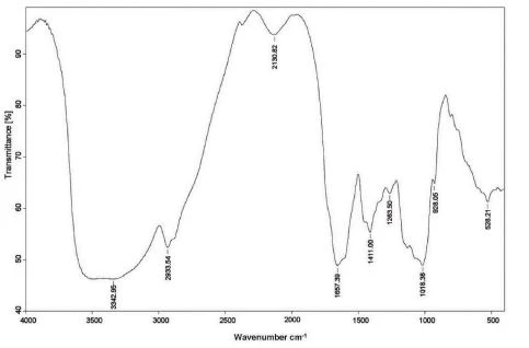
Fig. 1. FTIR spectra of AECCD.
2.5. In vitro experiments
2.5.1. Generation of DCs, cell viability, and endotoxin levels
DCs derived from the bone marrow of C57BL/6 mice were obtained according to the method described previously (Inaba et al., 1992). Briefly, cells were cultured with RPMI-1640 complete medium (20 ng/mL GM-CSF and 5 ng/mL IL-4). After six days, all suspended cells were collected as immature DCs for subsequent assays. The percentage of CD11c+ DCs in the non-adherent cells was determined to be 94% by flow cytometry.
The cell viability of DCs and splenocytes from mice upon AECCD were detected with the CCK-8 kit according to the manufacturer’s instructions. The cells from DCS and splenocytes were obtained and then stimulated with different concentrations of AECCD (0, 10, 100, 400, 800, and 1600 μg/mL). The cell viability was expressed as the ratio of the absorbance of the treated group to the absorbance of the control group) × 100%.
For the purpose of excluding the effect of endotoxin in AECCD, after AECCD (100, 200 and 400 μg/mL) and LPS (100 ng/mL) were pretreated with PMB (100 μg/mL) for 60 min, DCs (5 × 10⁵ cells/mL) were incubated with pretreated AECCD (100, 200 and 400 μg/mL) and LPS for 12 h. After the above treatment, the levels of CD40, CD86, CD80, and MHCII were evaluated.
2.5.2. Analysis of DC phenotype
The levels of costimulatory molecules were determined to evaluate DC maturation through in vitro experiments. On Day six, DCs were treated with AECCD (20, 100, 200 and 400 μg/mL) or LPS (100 ng/mL) for 12 h. Then, DCs were stained with surface molecules and analyzed by flow cytometry.
2.5.3. Cytokine production by DCs in vitro
The cell culture supernatant was collected to measure the level of cytokines by using an ELISA kit or multi-analyte flow assay kit with the CBA method. All experimental steps were performed according to the manufacturer’s instructions.
2.5.4. Allogeneic mixed lymphocyte reaction (MLR)
Splenocytes from BALB/c mice were aseptically obtained in RPMI-1640 medium (Zhang et al., 2018). Mature C57BL/6 DCs (matured in the presence of different concentrations of AECCD or LPS) were pretreated with 50 μg/mL Mitomycin C for 60 min at 37 ◦C. After washing, DCs were mixed with splenocytes according to a ratio of 1:5 or 1:10 for 2 days. Cell proliferation was determined by MTT assay.

Fig. 2. OVA-specific antibody titers and IgG isotypes in serum. Serum samples were collected after primary and booster immunizations for antibodies with ELISA
2.6. Statistical analysis
Data are expressed as mean ± SD. The significant level of statistical differences was determined by One-way ANOVA and Tukey’s multiple comparison test. P < 0.05 is considered to be statistically significant.
3. Results
3.1. FTIR spectroscopy
The main absorption peaks of AECCD in Fig. 1 were characteristic absorption peaks of glycosidic structures corresponding to the O–H stretching vibration at 3342.95 cm− 1. The band at 2933.54 cm− 1 had the characteristic weak absorption of C–H stretching vibrations. The peak at 1657.39 cm− 1 was attributed to O–H flexural vibration. The absorption at 1411.00 cm− 1 was ascribed to O–H or C–H and C–O stretching vibrations. The peaks at 1018.38 cm− 1 also indicated CO and C–O–C glycosidic band vibrations.
3.2. OVA-specific serum antibody response
Sera from the immunized mice were tested to check for the presence of generated antibodies (IgG, IgG1, and IgG2a) against OVA. As displayed in Fig. 2A, IgG titers in mice immunized with OVA/AECCD-M elicited higher total IgG (titers: 220,000, 250,000 and 250,000) relative to those of OVA group (titers: 100,000, 100,000 and 120,000), and was close to Alum group (titers: 250,000, 250,000 and 270,000) on Days 28, 42 and 56 after primary vaccination. OVA/AECCD-M/H significantly enhanced IgG1 levels compared to the OVA group (P < 0.05) and the difference between OVA/AECCD-M/H group and Alum group was not significant (P> 0.05). OVA/AECCD-M/H also markedly enhanced IgG2a levels compared to the OVA group and Alum group (Fig. 2B and C) (P < 0.05). The enhanced IgG2a levels might prevent a disease requiring IgG2a immune response.
3.3. Cellular immune responses
Spleen lymphocyte proliferation was investigated by MTT assay under restimulation with OVA or OVA323-339. Proliferation analysis revealed that OVA/AECCD-M both markedly increased splenocyte proliferation after the exposure to OVA, OVA323-339, Con A or LPS relative to the OVA group. OVA and OVA323-339 significantly improved the higher proliferation response than that in the Alum group (Fig. 3) (P < 0.01).
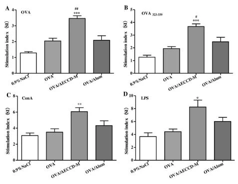
Fig. 3. Splenocyte proliferation upon AECCD treatment in vivo
3.4. Activation of T cell subsets in draining lymph nodes and spleen
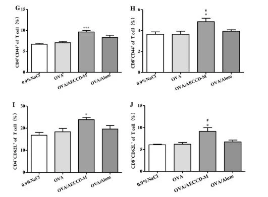
Lymph nodes and spleen from immunized mice were harvested to detect T cell subsets on Day 21 after primary vaccination. The percentage of T cell subsets (CD4+, CD8+, CD44+ T cells) from lymph nodes in the OVA/AECCD-M group was significantly higher than that of the OVA group and Alum group (Fig. 4A–D) (P < 0.05). The percentages of T cell subsets (CD4+, CD8+, CD44+, and CD62L+ T cells) in a spleen were significantly higher than those of the OVA group (Fig. 4E–J) (P < 0.05). These findings indicated that AECCD promoted cell-mediated immune response.
3.5. Cytokines secreted by restimulated splenocytes in vivo
Th1/Th2 cytokines from the spleen of immunized mice were determined by intracellular staining on Day 21 after the first vaccination. The levels of CD4+IL-4, CD4+IFN-γ, and CD8+IFN-γ in the OVA/AECCD-M group were significantly higher than that in the OVA group and Alum group (P < 0.05, Fig. 5). The differences indicated that AECCD could stimulate Th1/ Th2 responses, especially T-cell mediated immune response.
3.6. DC maturation and Tregs in vivo
DC activation can initiate adaptive immunity. The effects of AECCD on DC maturation in immunized mice were evaluated on Day 3 after the first immunization. OVA/AECCD-M induced the highest levels of CD40, CD80, CD86, and MHCII on DCs relative to the OVA group and the difference between OVA/AECCD-M group and Alum group was significant (Fig. 6A–D) (P < 0.05). The frequency of CD4+CD25+Foxp3+ Tregs in the OVA/AECCD-M group was dramatically lower than that in the OVA group (Fig. 6E) (P < 0.05). These data suggested that AECCD could activate DCs and lower Treg frequency.
3.7. Assays of cytotoxicity and endotoxin contamination
After the treatment with different concentrations of AECCD for 2 days, the cells from DCs and splenocytes of mice were used to determine the potential cytotoxicity with the CCK-8 kit. There was no significant inhibited proliferation response in the cell viability of DCs and splenocytes compared to that of the control groups (Fig. 7A–B) (P > 0.05). These results displayed that AECCD at the concentration of 10–1600 μg/mL showed no significant cytotoxicity to DCs and splenocytes compared to the control (P > 0.05). Thus, the concentration of AECCD in subsequent experiments was set to be under 1600 μg/mL. To exclude the interference of endotoxin contamination in the effects of AECCD on DC activation, PMB, an antibiotic widely recognized due to its LPS-neutralizing effect, was used to exclude endotoxin contamination (Morrison and Jacobs, 1976). As depicted in Fig. 7C–F, CD40, CD80, CD86 or MHC-II expression levels in AECCD-treated DCs in the presence of PMB showed no significant change (P > 0.05). However, a dramatic decline of CD40, CD80 CD86, and MHC-II expression levels was observed in LPS-treated DCs in the presence of PMB (P < 0.001). These results demonstrated that AECCD was free of endotoxin contamination.
3.8. Phenotypic maturation and functional activation of DCs in vitro
Based on immunization experiments above, the phenotypic maturation of DCs was further explored after the treatment with AECCD in vitro. On Day 6, immature DCs were stimulated with a given concentration of AECCD or LPS to analyze surface molecules. AECCD induced cell maturation characterized by a significant increase in the percentages of CD40, CD80 CD86, and MHC II in a dose-dependent manner (P < 0.001, Fig. 8A–D). A similar phenotypic maturation of DCs was also observed after the stimulation with 100 ng/mL LPS. The analysis data revealed that AECCD improved DC maturation. From the above results, AECCD enhanced the phenotypic maturation of DCs. In order to observe the effect of AECCD on DC function in vitro, the ability of AECCD-treated DCs to stimulate allogeneic T cell proliferation in an MLR at a DC-splenocyte ratio of 1:5 or 1:10 was investigated. DCs treated with different concentrations of AECCD induced significant T cell proliferation compared to untreated-DCs (Fig. 8E–F) (P < 0.001). DC maturation could induce the production of cytokines. Next, the supernatants of DCs treated with AECCD or LPS were detected by ELISA kit. The levels of IL-12 and TNF-α in the supernatants of DCs were dramatically increased in a dose-dependent manner (P < 0.001, Fig. 8G–H).
3.9. TLR4, a receptor for AECCD on DCs
DCs can initiate cellular signaling by membrane receptors. To determine the role of TLR4 in participating in AECCD-treated DC maturation, after the pre-treatment with TLR4 inhibitor TAK-242, DCs were stimulated with a given concentration of AECCD, and then the activation status of DCs was examined by flow cytometer. The expression levels of cytokines in culture supernatants were analyzed by a multi-analyte flow assay kit. The upregulation of CD40, CD80, CD86, and MHC-II induced by AECCD was significantly inhibited by blocking TLR4 pathways (P < 0.01, Fig. 9A–D). Moreover, compared to untreated control, blocking TLR4 with TAK-242 had a more significant inhibition effect on the production of IFN-γ, TNF-α, and IL-6 (P < 0.01, Fig. 9E–G).
3.10. DC activation by AECCD via TLR4-related NF-κB pathway
To investigate whether the expressions of surface molecules and cytokines of AECCD-treated DCs were regulated by NF-κB after immature DCs pretreated with NF-κB inhibitor PDTC were stimulated with AECCD for 12 h, the expression levels of costimulatory molecules and cytokines were measured by flow cytometer. The levels of CD40, CD80 CD86, and MHC-II induced by AECCD were significantly decreased compared to the untreated control by blocking NF-κB pathways with corresponding inhibitors (P < 0.001, Fig. 10A–D). Simultaneously, the expression of IFN-γ, TNF-α, and IL-6 was also significantly inhibited by PDTC (P < 0.05, Fig. 10E–G).
4. Discussion
Currently, various polysaccharides from TCM were investigated as the adjuvants for human and animal vaccines (Sun et al., 2018; Rey-Ladino et al., 2011). In this study, the immune-modulating effects of AECCD and the primary activation mechanism of DCs were investigated in vivo and in vitro by a series of experiments in order to evaluate the adaptive immune response against OVA and the activation status of DCs. In vivo experiments indicated that AECCD remarkably promoted the production of OVA-specific IgG and IgG isotypes, enhanced splenocyte proliferation, and induced T cell activation and cytokines (IL-4/IFN-γ). Besides, AECCD improved DC maturation and exhibited diminished Tregs. These findings suggested that the function of AECCD as an efficient adjuvant was realized by promoting DC maturation and inhibiting Tregs. In vitro experiments indicated that AECCD promoted phenotypic maturation of DCs, cytokine section, and allostimulatory activity via TLR4-related NF-κB pathway. These experimental results indicated that AECCD had significant potential as an immune-enhancing stimulator.
Each adjuvant has unique immune characteristics against different antigens, indicating that adjuvants have application potential in different diseases. Firstly, the adjuvant properties of co-administration of AECCD with OVA were tested by determining their effects on adaptive immune response in mice. Many studies showed that the immune-stimulatory activities were directly proportional to the dose within a certain range (Li et al., 2020; Reed et al., 2020; Zhang et al., 2017). Similarly, in the study, the analysis of antibody and isotype revealed that AECCD in a certain dose range enhanced OVA-specific IgG, IgG1, and IgG2a responses and that the medium dose of AECCD was the optimal dose. In subsequent experiments, we mainly investigated the stimulatory effect of AECCD at an optimal medium dose. The medium dose of AECCD significantly increased the splenocyte proliferation response in immunized mice, indicating that AECCD promoted cellular immune responses against OVA. These results suggested that AECCD enhanced both humoral and cellular immune responses, which were associated with adjuvant potential.
cistanche reviews about immunity
The elicited substantial T-cell activity may be conducive to disease eradication. During an immune response, activated T cells may reach secondary lymphoid tissues, including spleen and lymph nodes (Steinman, 2012). The medium dose of AECCD effectively improved vaccine efficacy as it could significantly enhance CD4, CD8, CD44, and CD62L T cell responses in draining lymph nodes and spleen. Activated CD8 T cells mediated pathogen eradication by secreting IFN-γ. Proliferated CD4 T cells were differentiated into Th1 cells and Th1 cells. Th1 cells secreted Th1 cytokine to stimulate lymphocyte proliferation and Th2 cells secreted Th2 cytokine to enhance antibody production (Seder et al., 2008). In the study, the levels of CD4+IL-4, CD4+IFN-γ, and CD8+IFN-γ were increased by AECCD at a medium dose. AECCD induced a balanced Th1/Th2 response and directed the immune response by altering the cytokine network.
An adjuvant should induce specific immunity and control the outcome of immune response by regulating DCs status at numerous points (Carrera Silva et al., 2013). The expression of surface molecules on DCs is a crucial indicator for evaluating DC maturation and is typically correlated with T lymphocyte differentiation (Steinman, 2012). In this study, AECCD enhanced the expression of costimulatory molecules and facilitated DC maturation when it was used as an adjuvant against OVA. This may be one of the ways that AECCD promoted humoral and cellular immune responses. Meanwhile, AECCD also decreased the frequency of CD4+CD25+Foxp3+ Tregs, which provided a balanced immunity after vaccination. Our results supported recent findings that various polysaccharides from TCM were the regulator of DC maturation (Bo et al., 2019; Li et al., 2015; Zhu et al., 2016).
Activation of adaptive immune responses by polysaccharide adjuvants is mediated primarily via their recognition by specific receptors of DCs (Akira, 2004). DCs activated through TLR4-related pathways can secrete various immunomodulators as the critical mediator against infections (Akira, 2007). Therefore, in subsequent experiments, we preliminary explored the molecular mechanism of the stimulatory effect of AECCD on DCs in vitro. In order to observe the cytotoxicity of AECCD, the cell viability of DCs and splenocytes from mice was firstly determined. AECCD at 10–1600 μg/mL displayed no cytotoxicity to DCs and splenocytes. To rule out LPS contamination, we pre-treated AECCD with PMB. The expression levels of CD40, CD80 CD86, and MHC-II in AECCD-treated DCs were not decreased, but PMB effectively inhibited the expression of costimulatory molecules in LPS-treated DCs. This demonstrated that AECCD was free of endotoxin contamination. Next, the activation status of DCs was further explored. Similar to LPS-treated DCs, in a dose-dependent manner, AECCD-treated DCs increased the levels of CD40, CD80 CD86, and MHC II as well as the production of the pro-inflammatory cytokine, which was a parameter of functional maturation of DCs. In an MLR, the proliferation ability of allogeneic T cells was evaluated in AECCD-treated DCs. AECCD could prime and activate allogeneic T cells. DCs were firstly pre-treated with Mitomycin C and then thoroughly washed to remove this reagent, so the effects of AECCD on DCs were not likely to be attributed to the mitogenic activity. Experimental data revealed that DCs phenotypic maturation and functional activation might be attributed to the stimulation from AECCD.
A number of TCM polysaccharides might stimulate DCs maturation via TLR4. NF-κB as a member of TLR4-related downstream pathway plays an important role in regulating phenotypic and functional maturation of DCs and inducing the expression of various genes involved in immune responses (Qi et al., 2016; Re and Strominger, 2001; Wei et al., 2016; Zhu et al., 2013). Through in vitro experiments, we found that AECCD promoted DC maturation and that TAK-242 (TLR4 inhibitor) and PDTC (NF-κB inhibitor) decreased the expression levels of CD40, CD80, CD86, and MHC-II and the secretion levels of IFN-γ, TNF-α, and IL-6 on AECCD-treated DCs, These findings indicated that TLR4 and NF-κB were involved in AECCD-induced DC activation and cytokine secretion. These results were consistent with previously reported studies because AECCD as an adjuvant improved antigen targeting onto DCs.
In summary, our results showed that AECCD induced durable and Th1/Th2 responses, especially T-cell mediated immune response against OVA. AECCD activated DCs through the TLR4-mediated NF-κB pathway, thus enhancing the production of immunomodulators and allogeneic T cell proliferation. AECCD may be used as an effective adjuvant in vaccine development. However, an accurate component analysis will be performed to determine which component in AECCD activated DCs. With a focus on the mechanisms regarding how AECCD boosts immunogenicity, including optimization of adjuvant formulations and route of vaccine administration, we will further explore the immunomodulatory components and the specific adjuvant properties of AECCD for the purpose of designing effective vaccine adjuvants against infectious diseases.

cistanche improve immunity




