Cistanche Phenylethanoid Glycosides And Autophagy: Reading The Mouse Study Behind The Headlines
Nov 13, 2026
The Word That Keeps Appearing in Longevity Research
If you have spent any time reading about aging, cellular health, or metabolic medicine in the past decade, you have run into the word autophagy. It gets used loosely - sometimes as a synonym for "cellular cleanup," sometimes as a buzzword attached to fasting protocols, sometimes as a reason to buy a supplement.
The loose usage hides something genuinely interesting. Autophagy is a real, well-characterized biological process with defined molecular machinery, measurable markers, and a documented role in diseases ranging from neurodegeneration to cancer. It is also one of the more counterintuitive processes in cell biology, because whether autophagy helps or harms depends entirely on context - the same mechanism that protects a starving cell can help a tumor survive chemotherapy.
That context problem is precisely why a 2022 study on Cistanche phenylethanoid glycosides (CPhGs) and autophagy in tumor-bearing mice is worth reading carefully rather than skimming. The study found something clean and measurable: at higher doses, the extract increased autophagosome formation and shifted two marker proteins in a direction consistent with autophagy activation. Those are real findings.
What they are not is a cancer treatment. This article walks through the biology, the experimental design, the actual numbers, and - most importantly - the boundaries of what the data can support. If you sell, buy, or simply read about Cistanche, this is the version of the story that will not mislead you.
Autophagy 101: The Cell's Recycling and Quality-Control System
Every cell you have is constantly producing damaged proteins, misfolded proteins, and worn-out organelles. Left alone, that debris accumulates, interferes with metabolism, and eventually kills the cell. Cells therefore run a continuous cleanup operation. Autophagy - literally "self-eating" - is the heavy-duty branch of that operation.
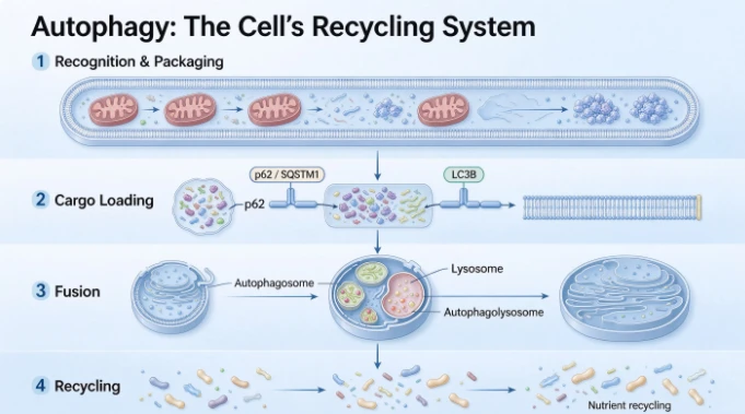
The sequence runs in four stages.
1. Recognition and packaging. A double-membrane structure begins to form around material destined for disposal: damaged mitochondria, fragments of endoplasmic reticulum, aggregated proteins, even invading pathogens. The forming structure is called an autophagosome.
2. Cargo loading. Specific receptors recognize specific cargo. One of the most important is p62 (also written SQSTM1), a protein that simultaneously binds ubiquitinated cargo and binds to the autophagosome membrane protein LC3B. p62 is, functionally, the loading dock supervisor - it physically links "this needs to be destroyed" to "here is the bag."
3. Fusion. The autophagosome merges with a lysosome - the cell's acid-filled digestion chamber. The combined structure is an autophagolysosome, and inside it, enzymes break the cargo down into amino acids and basic building blocks.
4. Recycling. The breakdown products are returned to the cytoplasm and reused for new protein synthesis.
The whole continuous sequence - from packaging to fusion to degradation to reuse - is called autophagic flux. This distinction between "autophagy happened" and "autophagy flowed" is one of the most commonly blurred concepts in popular writing, and it matters enormously for interpreting data. More autophagosomes can mean autophagy is running faster. It can equally mean autophagy is stuck - bags piling up because the fusion step failed. Only flux measurements can tell the difference.
Biologically, autophagy serves two roles that pull in opposite directions:
Survival support. When a cell faces starvation, hypoxia, or oxidative stress, autophagy digests non-essential components to keep essential functions running. It buys time.
Quality control and programmed death. When damage is severe or autophagy is over-activated, the digestion becomes so complete that the cell dies - a process sometimes called autophagic cell death.
Both roles operate in cancer, often simultaneously, in different cells of the same tumor.
The Two-Faced Role of Autophagy in Tumors
This is the conceptual heart of the whole topic, and it explains why "induces autophagy" is not automatically a good thing or a bad thing.
In early tumor development, autophagy usually protects. It maintains genomic stability, clears damaged organelles that would generate mutation-inducing oxidative stress, and supports the immune surveillance that eliminates abnormal cells. In this phase, autophagy is on the body's side.
In established tumors, autophagy often protects the tumor. A growing mass outgrows its blood supply. Cells in the interior face hypoxia, glucose deprivation, and metabolic stress. Autophagy lets them survive conditions that would kill a normal cell. In fact, tumor cells in the process of metastasis are frequently in a state of high stress with limited nutrient supply, which makes them heavily autophagy-dependent. In those settings, autophagy inhibition can slow tumor growth - which is why autophagy inhibitors are an active area of oncology research.
And in some contexts, autophagy kills tumor cells. Push autophagy hard enough, or block the machinery that would normally restrain it, and the cell digests itself past the point of viability. A meaningful number of anticancer agents - including several natural products - appear to work partly by hyper-activating autophagy in tumor cells.
So the phrase "compound X induces autophagy in tumor cells" needs a follow-up question: is the autophagy protective or destructive here? Studies that measure only autophagosome counts cannot answer that. Studies that measure flux markers together with cell viability and tumor growth start to.
What Exactly Are Cistanche Phenylethanoid Glycosides?
A brief recap for readers new to the plant.
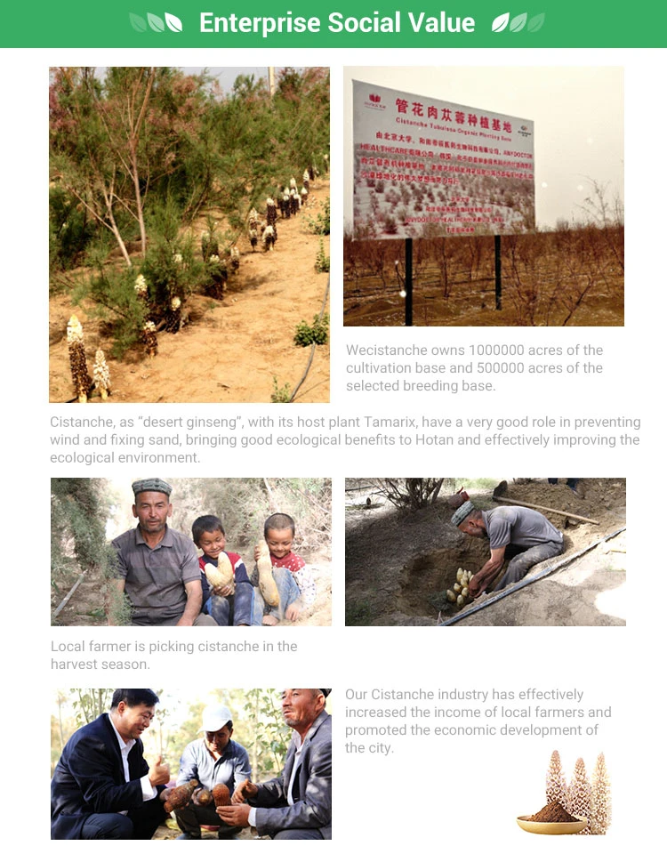
Cistanche is a genus of parasitic desert herbs in the family Orobanchaceae, native to the arid regions of northwest China and warmer dry zones of Eurasia. It attaches to the roots of desert shrubs - mainly saxaul and tamarisk - and lives off their water and nutrients. The medicinal material is the dried fleshy stem with its scaly leaves. It has a long history in Chinese pharmacy and is classified as both a food and a medicine.
Its dominant bioactive family is the phenylethanoid glycosides (PhGs) - small polyphenols built on a phenethyl or phenylpropenyl core with attached sugar chains. Echinacoside and acteoside (verbascoside) are the two most abundant and most studied. The Chinese Pharmacopoeia uses exactly those two compounds as quality markers for the herb, which is a useful practical detail: any serious extract is described by its content of them.
In this particular study, the material was a total phenylethanoid glycoside fraction (CPhGs) at 85% purity, containing at least 35% echinacoside and at least 16% acteoside. That is a highly standardized product - a meaningful contrast to the unlabeled "10:1 powder" that dominates the lower end of the market.
One more thing worth carrying forward from the metabolism literature: these glycosides absorb poorly intact. Oral bioavailability of echinacoside is under 1%, and acteoside is even lower. What reaches the tissues is largely a set of smaller metabolites produced by gut bacteria - caffeic acid, hydroxytyrosol, acteoside itself, and related products. Any honest discussion of how Cistanche "works" has to account for that conversion step, because the molecule in the capsule is rarely the molecule at the target.
The H22 Model: What Kind of Experiment Is This?
To read the study, you need to know what an H22 mouse model is.
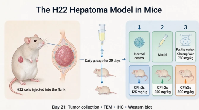
H22 is a mouse hepatocellular carcinoma cell line - a liver cancer line originally derived from mice and maintained in culture. Researchers inject a known number of these cells under the skin of a mouse's flank (in this study, 200 microliters of a suspension at 5 × 10⁶ cells per milliliter), and within days a measurable solid tumor grows at the injection site.
Why this model? Because it is fast, reproducible, and gives a subcutaneous tumor you can weigh. You can measure whether a treatment reduced tumor mass, and you can harvest the tumor tissue afterward for molecular analysis. It is a workhorse screening model used across pharmaceutical and natural-product research.
Why is it not a model of human cancer? Several reasons, and they all matter:
It is not a human tumor. H22 cells are mouse cells, with mouse genetics and mouse biology. Findings do not automatically transfer across species.
It is not orthotopic. The tumor grows under the skin, not inside the liver. Real hepatocellular carcinoma develops in a liver that is often already damaged by cirrhosis or viral hepatitis, in a microenvironment full of specific immune and metabolic pressures that a flank tumor does not replicate.
It is a transplant, not a spontaneous tumor. The immune system's relationship to a suddenly implanted mass differs from its relationship to a tumor that arose gradually from the host's own tissue.
Mouse dosing does not translate directly. Milligrams per kilogram in a mouse, scaled by body surface area or metabolic rate, produce very different human equivalent doses.
None of this makes the model worthless. It makes it a discovery tool: good for asking "does this compound do anything measurable to tumor tissue?" and "which molecular pathway does it touch?" It is a first chapter, not the final word.
How the Experiment Was Built
The design was straightforward and reasonably thorough for a screening study.
Sixty male Kunming mice, SPF-grade, acclimated for three days, were randomized into six groups of ten:
表格
| Group | Treatment |
|---|---|
| Normal control | Distilled water, no tumor |
| Model | Distilled water, tumor-bearing |
| Positive control | Xihuang Wan 780 mg/kg body weight |
| CPhGs low dose | 125 mg/kg body weight |
| CPhGs medium dose | 250 mg/kg body weight |
| CPhGs high dose | 500 mg/kg body weight |
All groups except the normal control received the H22 implant, then began daily gavage treatment 24–48 hours later - distilled water, Xihuang Wan, or CPhGs at the assigned dose. Treatment ran once per day for 20 consecutive days. On day 21, animals were fasted (water allowed), sacrificed by cervical dislocation, and tumors were photographed, weighed, and split for analysis: part fixed for microscopy and immunohistochemistry, part snap-frozen at −80 °C for protein work.
The choice of Xihuang Wan as the positive control is worth noting. It is a well-known traditional Chinese formula used as an adjunct in oncology, and it gives the researchers a benchmark: does CPhGs perform comparably to something with an established clinical reputation? That is a much more demanding comparison than saline alone.
Dose selection follows a standard toxicology logic - a low, a mid, and a high dose spanning a fourfold range, so that a dose-response relationship can be detected rather than a single data point.
Three Ways of Looking at the Same Question
The study used three complementary methods. Understanding why all three were needed tells you a lot about how autophagy research actually works.
Transmission electron microscopy (TEM)
TEM shoots a beam of electrons through an ultra-thin slice of tissue (60–80 nanometers) and produces images at a resolution far beyond any light microscope. At that magnification, autophagosomes are directly visible - you can literally see the double-membrane vesicles and, crucially, what is inside them.
This is the gold-standard structural evidence for autophagy. Protein markers can be misleading; a vesicle containing a half-digested mitochondrion cannot.
Immunohistochemistry (IHC)
IHC uses an antibody tagged with a visible marker to show where a protein is and how much of it is present in intact tissue. Brown or amber staining indicates positive signal. The researchers quantified it semi-quantitatively using ImageJ: ten random fields per slide, measuring integrated optical density divided by the protein's distribution area, yielding an average optical density that correlates with protein concentration per unit area.
This preserves spatial context - you can see whether the protein is in tumor cells versus stroma, and compare staining intensity across groups.
Western blot
Western blot homogenizes the tissue, separates proteins by molecular weight on a gel, transfers them to a membrane, and detects specific proteins with antibodies. It gives quantitative band intensities for a specific protein - in this case LC3B and p62 at 1:2000 antibody dilution.
The advantage is precision and quantification. The disadvantage is that you lose all spatial information: the band tells you how much protein was in the whole piece of tissue, not where it was.
Why all three? Because they triangulate. TEM proves the structures exist. IHC shows they are in the right cells and tracks intensity across a dose range. Western blot gives hard numbers for statistical testing. When all three point the same direction, the finding is solid. When they disagree, you have a problem.
What They Found, Part 1: The Structures
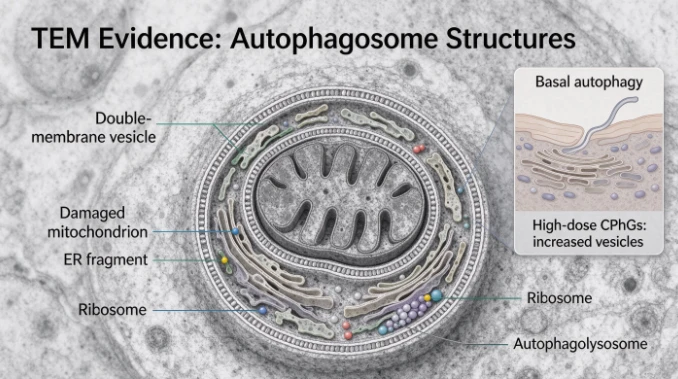
Under TEM, the model group and the low-dose CPhGs group showed tumor cells with intact structure - clearly visible irregular nuclei, intact nuclear membranes, mitochondria and endoplasmic reticulum with normal morphology. A small number of autophagosomes were present, which is expected: basal autophagy operates in every living cell.
As the CPhGs dose increased, the picture changed.
Autophagosome and autophagolysosome counts rose with dose.
The vesicles contained damaged mitochondria, endoplasmic reticulum fragments, and ribosomes - that is, recognizable cellular components awaiting degradation, not empty bags.
At the high dose, the tumor cells showed nuclear dissolution and fragmentation, loss of clear cell boundaries - structural signs of cell collapse.
The high-dose appearance was described as similar to the Xihuang Wan positive control group.
The detail about what is inside the vesicles is the most important part of this finding. Empty autophagosomes suggest a cell ramping up packaging. Vesicles stuffed with recognizable organelles suggest a cell actively cannibalizing its own machinery. The latter is what you see when autophagy is genuinely engaged under stress.
The nuclear dissolution and loss of cell boundaries add a second layer: this is not just a cell recycling more aggressively. It is a cell in the process of dying.
What They Found, Part 2: The Marker Proteins
Both LC3B-II and p62 were confirmed to be expressed in the cytoplasm. The staining patterns moved in opposite directions - and that opposition is the signature of autophagy activation.
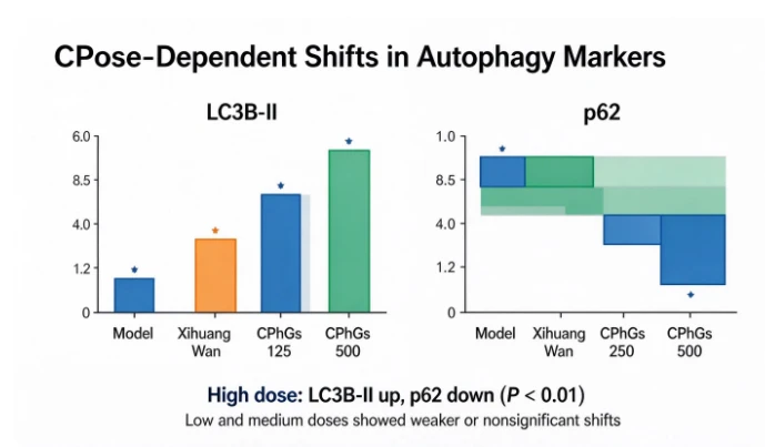
LC3B-II increased with dose. Staining intensity was proportional to CPhGs dose. In the model group and the low-dose group, cytoplasmic staining was relatively pale. In the medium- and high-dose groups, the number of positive cells rose markedly and staining intensified.
p62 decreased with dose. Staining intensity was inversely proportional to dose - as CPhGs went up, p62 staining faded.
The Western blot quantification confirmed both trends with hard numbers:
表格
| Group | LC3B-II (mean ± SD) | p62 (mean ± SD) |
|---|---|---|
| Model | 40.28 ± 3.34 | 56.74 ± 13.10 |
| Positive control (Xihuang Wan) | 59.50 ± 2.60 | 29.55 ± 5.56 |
| CPhGs low (125 mg/kg) | 41.93 ± 2.95 | 53.41 ± 6.00 |
| CPhGs medium (250 mg/kg) | 46.73 ± 5.85 | 33.19 ± 3.62 |
| CPhGs high (500 mg/kg) | 70.60 ± 11.98 | 31.50 ± 9.47 |
Compared with the model group, the high-dose CPhGs group showed LC3B-II up-regulation and p62 down-regulation at P < 0.01 - a strong statistical signal. The low and medium doses did not differ significantly from the model group on LC3B-II.
Compared with the positive control, the high-dose CPhGs group actually had higher LC3B-II (70.60 vs. 59.50, statistically significant at P < 0.05), while p62 levels were comparable. On these two markers, the highest dose of the extract moved the system at least as far as the reference formula.
That is a notable result. It is also where careful reading is required, because "moved further on a marker" and "better clinical outcome" are two different claims.
Why LC3B-II Up and p62 Down Means What It Means
These two proteins are not arbitrary choices. Together they constitute one of the most reliable marker pairs in autophagy research, and understanding the logic is genuinely useful for anyone reading this literature.
LC3B exists in two forms. LC3B-I floats free in the cytoplasm. During autophagy, it is conjugated to a lipid and becomes LC3B-II, which anchors to the autophagosome membrane. So LC3B-II is, quite directly, a count of autophagosome membranes. When autophagy activates, total LC3B barely changes but the LC3B-I to LC3B-II conversion increases - meaning LC3B-II content rises. The LC3B-II level and the LC3B-II/LC3B-I ratio are standard readouts for whether autophagy is switched on.
p62 is a selective autophagy receptor. It has two binding domains that matter: one grabs ubiquitinated cargo, the other grabs LC3B on the autophagosome. It is the physical bridge between "this protein is marked for destruction" and "here is the vehicle." Because p62 is itself destroyed during autophagy, p62 levels fall when autophagy is running and rise when autophagy is blocked.
That gives the diagnostic logic:
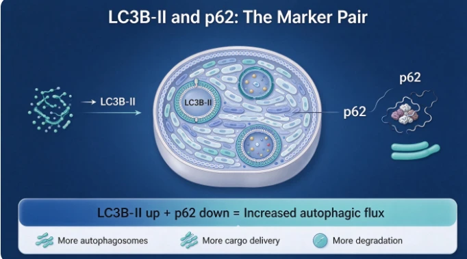
LC3B-II up + p62 down → autophagy is being activated and cargo is actually being delivered and degraded. Consistent with increased flux.
LC3B-II up + p62 up → autophagosomes are accumulating but degradation is failing. Autophagy is blocked, not activated. This is a trap that catches inexperienced researchers.
LC3B-II down + p62 up → autophagy is suppressed.
The study's pattern - LC3B-II significantly up, p62 significantly down, in both IHC and Western blot - fits the first scenario. That is why the authors concluded that CPhGs induces autophagy in tumor cells and promotes autophagic flux activation.
There is one methodological caveat the authors themselves flag, and it deserves emphasis: reading flux from two static markers is an inference, not a measurement. The rigorous version of this experiment adds autophagy modulators - an inhibitor like chloroquine or 3-methyladenine, applied alongside the treatment. If blocking lysosomal fusion prevents the LC3B-II rise, you have proven flux. If LC3B-II rises even with the inhibitor, you have proven blockade. The authors explicitly state that their group plans exactly this follow-up. It is an honest limitation, and it is the single most important next experiment.
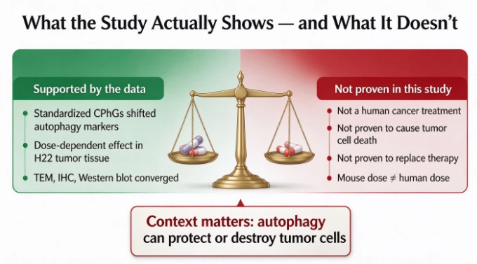
The Honest Boundaries: What This Study Does Not Show
This is the section that most articles about this paper would skip. It should not be skipped.
1. It does not show that CPhGs treats liver cancer in humans. The subjects were mice with a transplanted mouse tumor cell line. No human data exist in this paper. Nothing here establishes safety or efficacy in people.
2. It does not show that autophagy induction caused the tumor cells to die. Autophagy was measured. Tumor size was measured in a previous study by the same group, which reported that CPhGs inhibited subcutaneous H22 tumor growth. This paper examined the mechanism. But the causal chain - autophagy induction → tumor cell death → smaller tumor - was not directly demonstrated here. It is a supported hypothesis, not a proven pathway.
3. It does not distinguish protective from destructive autophagy. In established tumors, autophagy frequently helps tumor cells survive stress. It is entirely possible for a compound to increase autophagic flux in tumor tissue while the net effect on tumor viability is modest or even ambiguous. Determining the direction requires viability assays, apoptosis markers, and - critically - autophagy inhibitor controls.
4. The statistical signal is concentrated at the high dose. Low dose (125 mg/kg) was statistically indistinguishable from the model group on LC3B-II (41.93 vs. 40.28). Medium dose moved partway (46.73, significant vs. model at P < 0.05 for p62 but not for LC3B-II). The dramatic result is at 500 mg/kg. Dose matters, and extrapolating from a high-dose mouse experiment to a low-dose human supplement is not valid.
5. Sample size is ten animals per group. That is standard for this type of screening study, and adequate for detecting large effects. It is not adequate for subtle ones, and it does not support fine-grained dose-response claims.
6. One cell line, one model, one species. H22 is a single hepatocellular carcinoma line. Results in a different tumor type, a different genetic background, or an orthotopic model could differ substantially.
7. The endpoint is molecular, not clinical. Survival, metastasis, quality of life, and toxicity were not the focus here. Molecular marker shifts are mechanistic information, not outcome data.
Read those seven points and the finding becomes what it actually is: a clean, well-controlled piece of mechanistic evidence that a standardized Cistanche extract shifts autophagy markers in tumor tissue in a dose-dependent way, in a mouse model, consistent with flux activation. That is a real contribution to the literature. It is not a therapeutic claim.
Where This Fits in the Wider Cistanche Literature
This study is one thread in a larger body of preclinical work on Cistanche and cancer-adjacent biology. Summarizing it accurately requires the same restraint.
Osteosarcoma. Cistanche has been reported to induce apoptosis in human osteosarcoma MG-63 cells, and to reverse chemoresistance in that line. Cell-culture work.
Breast cancer bone metastasis. Cistanche has been reported to inhibit breast cancer bone metastasis in preclinical models - interesting because it connects to the bone-remodeling biology that Cistanche is better known for.
Renal cell carcinoma. Echinacoside has been reported to induce apoptosis in human renal carcinoma 786-O cells via a mitochondrial pathway. Cell-culture work.
Liver cancer. Acteoside (one of the two main PhGs) has been shown to promote autophagy and inhibit proliferation in liver cancer cells in vitro, with JNK signaling involvement. This is a useful corroboration: the autophagy effect seen with the total glycoside fraction in vivo has an in vitro counterpart with one of its principal constituents.
Bone. Cistanche extracts have shown anti-osteoporotic activity in ovariectomized rat models through the RANKL/RANK/OPG axis and several other pathways - a much deeper literature than the cancer work.
Two patterns are worth noting. First, these are overwhelmingly cell-culture and animal studies. Second, they involve different compounds, different doses, and different endpoints, which means they cannot be added together into a single "Cistanche fights cancer" conclusion. The correct synthesis is: multiple preclinical lines suggest Cistanche-derived phenylethanoid glycosides interact with apoptosis and autophagy machinery in tumor cells. Human relevance is undetermined.
Autophagy Beyond Cancer: Why This Mechanism Matters Elsewhere
Here is the part that makes autophagy research genuinely interesting for general readers, and it connects directly to why a bone-health audience might care about this paper at all.
Autophagy is not a cancer-specific process. It is a universal quality-control system, and its decline is one of the hallmarks of aging. As autophagy efficiency falls with age, damaged proteins and organelles accumulate, mitochondrial function degrades, and inflammatory signaling rises. That cascade is implicated in neurodegeneration, metabolic disease, sarcopenia, and - relevant here - bone loss.
In bone specifically, autophagy supports osteoblast differentiation and survival, and modulates osteoclast formation. Oxidative stress suppresses autophagy and accelerates bone cell death; maintaining autophagic capacity helps preserve the osteoblast population. This is why the autophagy literature and the bone literature keep intersecting, and why a compound that modulates autophagy is interesting well beyond oncology.
The same polyphenol chemistry shows up in both stories. Echinacoside and acteoside carry ortho-diphenolic hydroxyl groups that neutralize reactive oxygen species, and their metabolites - caffeic acid and hydroxytyrosol - are independently antioxidant. Oxidative stress and autophagy are tightly coupled: ROS both damages the machinery autophagy repairs and modulates the signaling that controls it. A compound that reduces ROS burden is, indirectly, protecting autophagic capacity.
So the honest big picture: Cistanche's PhGs appear to interact with cellular stress-response machinery - autophagy, oxidative stress, apoptosis - across multiple tissue contexts. Cancer is one lens on that. Bone and aging are others.
Translating Mouse Doses: The Arithmetic Nobody Does
A detail that separates serious analysis from marketing: 500 mg/kg in a mouse is not 500 mg/kg in a human.
Drug doses are typically converted between species using body surface area normalization rather than straight weight scaling, because metabolic rate per kilogram differs dramatically between a 25-gram mouse and a 70-kilogram human. The commonly used conversion factor for mouse-to-human (based on body surface area) is roughly 12.3 - meaning a mouse dose divided by about 12.3 approximates the human equivalent on a mg/kg basis.
Run the numbers on this study: 500 mg/kg in mice corresponds to roughly 40 mg/kg human equivalent. For a 70 kg adult, that is on the order of 2,800 mg per day of a material that is 85% total phenylethanoid glycosides.
That is a large amount. It is also - and this is the key point - a number derived from a mouse study with a molecular endpoint, not from a human trial with a clinical endpoint. It tells you the scale at which the observed effect appeared in animals. It does not tell you what dose a person should take, because no human dose-finding study exists for this endpoint.
Anyone who quotes "500 mg/kg was effective" as a reason to buy a product is doing arithmetic that stops halfway. Anyone selling 300 mg capsules and implying they replicate this experiment is not doing arithmetic at all.
What the Standardization Numbers Actually Tell You
The study material was described precisely: 85% total phenylethanoid glycosides, with echinacoside ≥35% and acteoside ≥16%.
Those three numbers do real work.
85% total PhGs means the fraction is highly enriched - 85 cents of every gram is the target chemistry, with the remainder being other plant constituents, sugars, and processing residues. Compare that to a 10:1 extract, where the ratio describes raw-material input, not output chemistry. A 10:1 powder can contain 1% PhGs or 40%, and the label will not tell you.
Echinacoside ≥35% anchors the product to the compound the Pharmacopoeia treats as the primary quality marker, and to the compound most of the mechanistic literature used.
Acteoside ≥16% matters because acteoside is both independently active and the compound echinacoside converts into in the colon. A product with high echinacoside but negligible acteoside is relying entirely on that conversion step.
If you are evaluating a Cistanche product and the certificate of analysis does not give you these three figures - total PhG percentage, echinacoside percentage, acteoside percentage - you are buying blind. Everything in this article, and everything in the research literature, is contingent on the chemistry actually being in the bottle.
Practical Takeaways for Readers
Distilled down, here is what a careful reader should carry away from this study.
1. The finding is real but narrow. A standardized Cistanche phenylethanoid glycoside fraction increased autophagosome formation and shifted LC3B-II up and p62 down in H22 tumor tissue in mice, in a dose-dependent manner, comparable to or exceeding a reference formula at the highest dose. That is a legitimate mechanistic observation.
2. "Induces autophagy" is not a benefit claim. In tumors, autophagy can protect or destroy depending on context. Without flux controls and viability data, the direction of the net effect on tumor cells remains to be established. The authors say so themselves.
3. This is not evidence for treating anything in humans. No human trial exists for this endpoint. Anyone converting a mouse marker study into a human health claim is skipping several required steps.
4. Dose scaling matters more than dose headlines. The effective dose in this experiment translates to a human equivalent far above typical supplement dosing. That gap is a reason for humility about expected effects, not a reason to megadose.
5. Standardization is the whole ballgame. 85% total PhGs with defined echinacoside and acteoside floors is a fundamentally different product from an unlabeled powder. Demand the numbers.
6. Autophagy is worth understanding on its own terms. It is one of the most important quality-control systems in your body, it declines with age, and it intersects with bone health, metabolic health, and neurodegeneration. That knowledge is useful regardless of what you decide to buy.
Safety and Cautions
Cistanche has a long history as a food and tonic, is classified as both food and medicine in China, and is generally well tolerated at customary doses. It is mildly laxative by tradition and by observation, so people with loose stools may need to start low.
Reasons for caution specific to this context:
Do not use it as a cancer treatment or as a substitute for oncology care. This is the single most important statement in this article. Delaying or replacing conventional treatment on the basis of a mouse study is dangerous.
If you are in active cancer treatment, talk to your oncologist before adding any botanical. Autophagy-modulating compounds are theoretically relevant to how some therapies work. That is a reason for supervision, not for self-experimentation.
Hormone-sensitive conditions. Echinacoside and acteoside show weak phytoestrogenic activity in cell assays. Discuss with your physician if you have a history of estrogen receptor–positive cancer.
Pregnancy and breastfeeding. Insufficient data; avoid.
Immunosuppressants and autoimmune conditions. Cistanche has immunomodulatory effects. Get advice first.
Quality and contamination. Desert-grown parasitic plants accumulate soil minerals, so heavy metal and pesticide testing is essential. Adulteration is a bigger real-world risk than the herb itself.

Frequently Asked Questions
Does this study prove Cistanche can treat liver cancer? No. It proves that a standardized extract changed autophagy markers in a mouse tumor model. That is mechanistic evidence, and it is several steps removed from a human clinical claim.
What is autophagy, in one sentence? It is the process by which a cell packages its damaged proteins and worn-out organelles into vesicles, delivers them to digestive chambers, breaks them down, and recycles the pieces.
Why did they measure both LC3B-II and p62? Because either marker alone can mislead. LC3B-II rising can mean autophagy activated or that degradation is blocked. p62 falling confirms that cargo is actually being delivered and destroyed. Together they indicate flux.
Was the effect dose-dependent? Yes, clearly. Low dose was statistically indistinguishable from the untreated tumor group; medium dose moved partway; high dose produced the largest and most statistically robust shift in both markers.
Is a mouse study meaningless for humans? No - it is how nearly every drug and supplement candidate gets discovered. But it is a first chapter. Species differences, dose scaling, and model limitations all mean findings must be confirmed in humans before they support human health claims.
Can autophagy be a bad thing in cancer? Yes. In established tumors, autophagy frequently helps cancer cells survive hypoxia, nutrient deprivation, and treatment stress. Whether inducing it helps or harms depends on the tumor, the stage, and the treatment context. This is why oncology autophagy research is so careful about directionality.
Does Cistanche have other preclinical evidence? Yes - apoptosis in osteosarcoma and renal carcinoma cell lines, autophagy and antiproliferative effects in liver cancer cells with acteoside, inhibition of breast cancer bone metastasis in animal models, and a much more developed anti-osteoporotic literature in ovariectomized rats. All preclinical.
What dose was used, in human terms? 500 mg/kg in mice, which by body-surface-area conversion is roughly 40 mg/kg human equivalent - about 2,800 mg/day of an 85% standardized fraction for a 70 kg adult. That is an animal-to-human arithmetic estimate, not a recommended dose.
Closing: The Value of Reading Carefully
The reason this study is worth an article is not that it produces a dramatic headline. It is that it is well-built - a defined standardized material, a fourfold dose range, a legitimate positive control, twenty days of dosing, and three independent methods that converge on the same conclusion. That is good science at the screening stage.
And the reason to write about it carefully is that its conclusions are easy to overstate. "Cistanche induces autophagy in tumor cells" is a true statement about a mouse experiment. "Cistanche fights cancer" is a claim the data does not support, that regulators do not permit, and that could genuinely harm someone who substituted it for treatment.
The productive middle ground - the one this article has tried to occupy - is to understand the mechanism honestly. Autophagy is a real system. Cistanche's phenylethanoid glycosides are real, well-characterized molecules with documented metabolic fates and measurable effects on cellular stress machinery. The research on how they interact with that machinery is early, interesting, and ongoing.
Knowing what a study measured, and knowing what it did not, is the difference between information and marketing. That distinction is worth more than any single finding.

Disclaimer
This article is educational and informational only and does not constitute medical advice. The research described was conducted in cell culture and animal models; it does not establish safety or efficacy in humans. Cistanche supplements are not intended to diagnose, treat, cure, or prevent any disease, including cancer. Nothing here should be interpreted as a recommendation to use Cistanche in place of conventional medical treatment. If you have a cancer diagnosis, are undergoing treatment, take prescription medication, are pregnant or breastfeeding, or have a hormone-sensitive condition, consult a qualified healthcare professional before using any botanical supplement. Statements about dietary supplements have not been evaluated by regulatory authorities.