Crosstalk Between Neuron And Glial Cells in Oxidative Injury And Neuroprotection Part 3
Mar 22, 2024
4. Microglia
4.1. Microglia in the Brain
Microglia, which have numerous fine and motile processes that survey the parenchymal environment, represent approximately 10% of CNS cells. Each microglial cell has its territory, which is approximately 50 µm in diameter [66].
Microglia are a type of nerve cells that play a vital role in our brains. They remove waste products from around neurons, maintain neuron health, and facilitate communication between neurons, all of which are necessary to maintain memory.
Among microglia, there is a type of cell called "astrocyte", which has a special shape and function. They monitor and regulate the connections between neurons, helping our brains process information more efficiently. This is similar to a computer room administrator who constantly monitors the connection status of network cables and cables to ensure the smooth flow of the entire network.
Research shows that microglia are also involved in the process of learning and memory. They release neurotransmitters, facilitate communication between neurons, and enhance memory consolidation and retrieval. At the same time, microglia can also promote the formation of new connections between neurons, thereby improving memory ability.
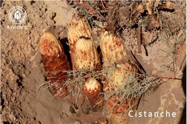
Therefore, maintaining the health and number of microglia in the brain is key to maintaining memory. We can promote the generation and maintenance of microglia by paying attention to diet, exercising appropriately, and maintaining a good mentality. Only by maintaining good microglia function can our brains stay young, healthy, and strong. Memory will naturally be improved. It can be seen that we need to improve memory, and Cistanche deserticola can significantly improve memory, because Cistanche deserticola can also regulate the balance of neurotransmitters, such as increasing the levels of acetylcholine and growth factors. These substances are very important for memory and learning. In addition, Cistanche deserticola can also improve blood flow and promote oxygen delivery, which can ensure that the brain receives sufficient nutrients and energy, thereby improving brain vitality and endurance.

Click know supplements to improve memory
Microglia, referred to as the resident macrophages in the CNS, are long-lived and self-renewing cells. In a healthy brain, microglia have a ramified morphology and are in a "quiescent" or "resting" state [67].
Microglial processes undergo continuous cycles of extension and withdrawal, scan their environment for disruptions in brain homeostasis, and systematically synapse to monitor and regulate neuronal activity via a specific signaling mechanism [68,69]. Microglia change their morphology from the resting state to the reactive amoeboid state during a pathological brain condition.
Reactive microglia, which evolve into phagocytic or amoeboid microglia, have an increased cell body size, fewer processes, reduced process length and branching, and increased numbers and proliferation, indicating an intimate link between morphology and function [70–73] (Figure 2).
Microglia are highly sensitive to environmental signals and respond to maintain their homeostatic phenotype in a disease-specific and brain-region-specific manner. White and gray matter microglia show different immune regulation; cortex-associated microglia play a role in neurodegeneration and white-matter-associated microglia play a role in de-/remyelination [74].
Usually, activation of the neurotransmitter receptors inhibits the inflammatory activation of microglia and inhibits the production of abnormal molecules and abnormal concentrations of physiological molecules.
Once activated upon brain injury or infection, microglia initiate immune responses and produce several cytokines, chemokines, and growth factors, and upregulate the expression of cell surface receptors, such as toll-like receptors (TLRs), phagocytic receptors, scavenger receptors, and various complement factors [75,76]. Microglia express several neurotransmitter receptors, including GABA, glutamate, dopamine, and noradrenaline [66,77].
4.2. Microglia in Oxidative Injury
During oxidative stress, activated microglia produce several inflammatory mediators, including NO and superoxide, which freely cross the cell membrane and act as signaling molecules.
NO and superoxide can form peroxynitrite, which causes DNA fragmentation, and lipid oxidation, and induces neuronal death [78,79]. In cultured microglia, superoxide production, which is catalyzed by nitrates/nitrites (NOx), is induced by phorbol ester, and NO production is stimulated by the induction of iNOS upon treatment with bacterial lipopolysaccharide (LPS) and interferon-γ (IFNγ) [80,81].

The expression of iNOS after intrahippocampal treatment with LPS was induced more rapidly in microglia than in astrocytes, and a lower concentration of LPS was required for iNOS induction in microglia than in astrocytes [82,83].
In addition, arginine is a well-known physiological substrate of NOS. Activated microglia with an insufficient amount of arginine lead to iNOS-mediated production of NO and superoxide, which form toxic peroxynitrite [84]. The induction of iNOS or activation of NOx alone does not cause substantial damage to microglia, but the simultaneous production of superoxide and NO by NOx and iNOS has the potential to harm microglia [85,86].
In activated microglia that generate superoxide upon NOx activation, the oxygen and H2O2 levels quickly become imbalanced and may affect microglial functions. ROS facilitates phagocytosis by amoeboid microglial cells and enhances vesicle formation, which was observed upon treatment of microglial cells with H2O2 [87]. Microglia-derived ROS can damage adjacent brain cells.
Therefore, microglial proliferation and ROS production are potential therapeutic targets that may protect the brain from oxidative damage and neurodegenerative disease [88].
4.3. Microglia-Mediated Antioxidant Defense
To prevent oxidative stress by ROS, microglia contain a high cellular GSH concentration and express and upregulate diverse antioxidant enzymes, including SOD, GPx, GR, and catalase.
Brain cell cultures labeled with fluorescence showed that microglia express a higher level of GSH than the other cell types in the rat brain [89]. This high concentration of intracellular GSH in microglia contributes to its antioxidant defense system against radical and peroxide-mediated damage. Microglial cultures stimulated with TNFα showed twice as much GSH as unstimulated microglial cultures [90].
However, the cellular GSH content was lower in microglia treated with LPS/IFNγ, which induces iNOS production, but the mitochondrial GSH content was unaffected [91]. Thus, the microglial GSH content shows a binary effect, in which it increases upon improvements in GSH synthesis and decreases upon accelerated GSH consumption, depending on the type of stimulation.
SOD, another antioxidant enzyme, was observed by immunocytochemical staining in activated microglia after quinolinic acid treatment but was not detected in microglia under basal conditions [92,93]. The specific activity of MnSOD is 20 and 4 times higher in cultured microglia than in cultured astrocytes and oligodendrocytes, respectively [94]. In microglia treated with LPS/IFNγ or TNFα to induce oxidative stress, mitochondrial MnSOD expression was upregulated, which improved the ability of cells to decompose mitochondrial superoxide [90,95].
Elevated SOD activity in activated microglia reduces the risk of cell damage by superoxide-derived hydroxyl radicals and peroxynitrite. The upregulation of GSH peroxidases (GPx) in microglia is also a crucial mechanism against oxidative stress. The specific activity of GPx and GSH reductase (GR) is significantly higher in microglia than in neurons [96–98].
However, the specific activity of catalase was similar and/or a little lower in microglia than in other brain cell types, including neurons, astrocytes, and oligodendrocytes [97,99]. Although microglial GSH disulfide (GSSG) increases to almost 30% of total cellular GSH after exposure to H2O2, microglial GSSG is barely detectable under basal conditions [98,100].
5. Neuron–Glia Crosstalk in the Antioxidant Defense Mechanism
Neurons depend on a continuous supply of glucose and oxygen from outside the brain via cerebral blood flow, even though they do not directly contact microvessels. However, 99% of the brain capillary surface is covered with astrocyte end-feet processes, indicating that neurons must interact with astrocytes to receive essential materials from the cerebral circulation [101].
Crosstalk between astrocytes and neurons is essential for neuronal defense against ROS. Activated astrocytes exhibit ambidextrous properties such as A1 and A2 astrocytes. A1 astrocytes lead to neuronal loss by promoting inflammation via the NF-kB pathway, which loses the ability to protect neurons and control synaptogenesis [102,103].
A2 astrocytes promote neuronal survival via the Janus kinase/signal transducer and activator of the transcription 3 (JAK-STAT3) signaling pathway by upregulating neurotrophic factors [104]. Neurons produce glutamate, which stimulates ascorbate release from astrocytes during glutamatergic synaptic activity, and then ascorbate enters neurons inhibits glucose consumption, and stimulates lactate transport.
The antioxidant and metabolic interplay
between neurons and astrocytes is described in Figure 3. Astrocytes are responsible for the
maintenance and support of neurons by regulating oxidative stress via GSH production and
glucose transformation into lactate, which ensures the energetic support of neurons [105].
The intrinsic antioxidant GSH, which is produced in both neurons and astrocytes, acts as an
independent ROS scavenger and as a substrate for an antioxidant. Neuronal cells depend
on astrocyte-derived GSH, for example, neurons depend on shuttling of the GSH precursor
from astrocytes to neurons. Cysteine is the rate-limiting substrate for GSH synthesis, and
extracellular cysteine is readily auto-oxidized to cystine [53].
Cystine uptake occurs via the
cystine/glutamate exchange transporter in astrocytes, and then astrocytes reduce cystine
back to cysteine for GSH synthesis. GSH directly reacts with ROS or acts as a substrate
for GSH S-transferase or GSH peroxidase [50]. For the efficient use of extracellular cystine
as a cysteine precursor, neurons depend on astrocytes to supply cysteine, even though
neurons can synthesize GSH [54,106].
It has been shown that neuronal GSH levels are significantly higher when co-cultured with astrocytes [107]. Upon H2O2-induced oxidative stress, noradrenaline treatment protects neurons by increasing the supply of GSH from astrocytes to neurons via stimulation of the beta3-adrenoreceptor in astrocytes [108]. The other interactions between neurons and astrocytes that are related to antioxidant activity include an astrocyte–neuron lactate shuttle and the recycling of ascorbate [55]. Astrocytes play a crucial role in coupling neuronal activity and brain glucose uptake through an astrocyte–neuron lactate shuttle [109].

Neuronal activity triggers glucose metabolism in astrocytes; glucose is converted to pyruvate by glycolysis and converted to lactate, which is released from astrocytes and taken up by neurons for oxidative phosphorylation. Ascorbate that is concentrated in the brain is released from glial reservoirs into the extracellular space and taken up by neurons. Highly activated neurons generate ROS, which oxidize ascorbate to dehydroascorbic acid (DHA), and scavenge ROS by taking up ascorbate [110,111].

Figure 3. This diagram represents neuron–glia crosstalk involved in neuroprotection and the antioxidant defense mechanism. Astrocyte-neuron: Astrocytes contain a variety of antioxidant molecules, including glutathione (GSH), ascorbate, vitamin E (VE), and ROS-detoxifying enzymes, such as GSH S-transferase, GSH peroxidase, thioredoxin reductase, and catalase.
Astrocytes project the end-feet processes onto the brain capillary surface so that astrocytes control the movement of molecules and cells between the vascular compartments and the brain. In the lactate shuttle, astrocytes support neurons by regulating glucose transformation into lactate, which ensures the energetic support of neurons. Neuronal activity triggers glucose metabolism in astrocytes. Glucose is converted to pyruvate by glycolysis and to lactate, which is released from astrocytes and taken up by neurons (blue arrow).
Astrocytes can synthesize GSH via activation of Nrf2 and can shuttle GSH precursors to neurons for GSH synthesis. Astrocytes release GSH into the extracellular space and neurons take up the GSH directly or use extracellular neuronal aminopeptidase N to form glycine and cysteine (black arrow). In glutamate uptake and recycling, glutamate from the synaptic space enters astrocytes through EAAT and is converted by glutamine synthetase (GS) to inactive glutamine. After its release and import into neurons, glutamine can be re-converted to glutamate (red arrow).
Recycled ascorbate can directly scavenge ROS and act as a cofactor for the recycling of oxidized vE and GSH. Astrocytes take up dehydroascorbic acid (DHA), an oxidation product of ascorbate, from the extracellular space and recycle it back to ascorbic acid. Astrocytes capture and transport excess extracellular K+ to the astrocytic syncytium through Na+/K+ ATPase. Nrf2 induction of glutamate cysteine ligase (GCL) increases GSH synthesis in astrocytes, and GSH is subsequently exported to the extracellular medium.
Astrocytes also participate in metal sequestration in the brain to prevent the generation of free radicals by redox-active metals. Microglia-neuron: Microglia contain a high cellular GSH concentration and express and upregulate diverse antioxidant enzymes. The expression of classical antioxidant proteins is controlled by Nrf2 in microglia. Heme oxygenase-1 (HO-1), an antioxidant enzyme upregulated by Nrf2, inhibits NOX2 activation.
Fractalkine (FKN) is predominantly expressed in neuronal cells, and microglia and neurons exclusively express the fractalkine receptor (CX3CR1); this is an interesting signaling axis for communication. Abbreviations: ARE, antioxidant response element; ASC, ascorbate; ApoE, apolipoprotein E; xCT, cysteine-glutamate exchanger; Cys, cysteine; DHA, dehydroascorbic acid; DMT1, divalent metal transporter; EAAT, excitatory amino acid transporter; mFKN, membrane-anchored fractalkine; sFKN, soluble fractalkine; CX3CR1, fractalkine receptor; Glc, glucose; GLUT, glucose transporter; Glu, glutamate; Gln, glutamine; GSH, glutathione; GCL, glutamate-cysteine ligase; GS, glutamine synthetase; GLAST, glutamate aspartate transporter; GLT1, glutamate transporter 1; Gly, glycine; HO-1, heme oxygenase-1; JNK, c-Jun amino-terminal kinase; LRP, lipoprotein receptor-related protein; MCT, monocarboxylate transporter; Nrf2, nuclear erythroid-related factor 2; Pyr, pyruvate; SVTC-2, sodium-dependent transporter; TRPC, transient receptor potential canonical.
In neurotransmitters, overstimulation with glutamate induces excitotoxicity, which is involved in the pathogenesis of many brain disorders. Astrocytes use two main transporters, excitatory amino acid transporter1 (EAAT1)/glutamate aspartate transporter (GLAST) and EAAT2/glutamate transporter-1 (GLT1), to take up glutamate and return glutamate to neurons via the well-established glutamate–glutamine cycle that involves the astrocyte-specific enzyme glutamine synthetase (GS), which converts glutamine into glutamate.
If failure to convert glutamine back to glutamate occurs, the glutamate pool in presynaptic terminals would rapidly be depleted and excitatory neurotransmission would be disrupted [112,113]. An insufficient supply of glutamine to GABAergic neurons induces GABAergic dysfunction [114,115]. Glutamine in astrocytes is critical for GABA replenishment by glutamate decarboxylase, known as the GABA–glutamine cycle, in GABAergic neurons [116].
Neuronal activity and action potentials increase extracellular K+ in restricted spaces and lead to hyper-excitable membrane potentials when tight regulatory mechanisms are absent [117]. Astrocytes have a high number of membrane K+ channels and high K+ permeability [118,119]. Astrocytes capture and transport excess extracellular K+ to the astrocytic syncytium through Na+/K+ ATPase.
Astrocytes also regulate the Ca2+ concentration within neurons via astrocytic calcium signaling and astrocyte-neuron crosstalk. Neuronal activation, which induces a reduction in extracellular Ca2+, evokes spatiotemporal changes via the Ca2+/Na+ exchanger in astrocytes and generates astrocytic Ca2+ waves that propagate from the cytoplasm into the extracellular space [120,121].

Astrocytes are also highly mechanosensitive, and a drop in extracellular Ca2+ due to synaptic activity leads to the release of ATP from astrocytes via the opening of connexin 43 hemichannels [122–124]. Neuronal activity can elicit metabolic changes in astrocytes via dual Na+ and Ca2+ signaling, which triggers glucose mobilization and glycolysis to support neuronal function. Astrocytic metabolism correlates with the high metabolic demands from neurons [125,126].
For more information:1950477648nn@gmail.com