

Expression Of Renal Vitamin D And Phosphatonin-Related Genes in A Sheep Model Of Osteoporosis
Jul 29, 2022
Please contact oscar.xiao@wecistanche.com for more information
Abstract: Osteoporosis is a significant public health issue around the world, with post-menopausal osteoporosis due to estrogen deficiency resulting in approximately 3/4 of cases. In this study,18 aged Merino ewes were ovariectomized, and 10 were controls. Three of the ovariectomized ewes were treated weekly with 400 mg of methylprednisolone for 5 months and three were treated weekly for 2 months, followed by a 3-month recovery period. At 2 months, five control animals and six ovariectomized animals were euthanized. At 5 months, all the remaining ewes were euthanized. Kidney samples were collected postmortem for qPCR analysis of NPT1, PTH1R, NPT2a, NPT2c, Klotho, FGFR1Illc, VDR, CYP24A1, CYP27B1, TRPV5, TRPV6, CalD9k, CalD28k, PMCA, and NCX1. Ovariectomized sheep had significantly greater VDR expression compared with other groups. Ovariectomized sheep treated with glucocorticoids for 2 months followed by euthanasia at 5 months showed significant differences in TRPV5, CYP24A1, and klotho gene expression compared to other groups. Differences in klotho expression were most marked after adjustment for repeated measures (p=0.1). Klotho is known as the"anti-aging" hormone and is involved in calcium and phosphorus metabolism. Klotho may be involved in the recovery of bone mineral density in ovariectomized sheep treated with glucocorticoids for 2 months followed by euthanasia at 5 months. Further research on the role of klotho is recommended.

Please click here to know more
Keywords: osteoporosis; sheep; vitamin D; phosphating; qPCR; calcium metabolism; phosphorus metabolism; klotho
1. Introduction
Osteoporosis is a major public health issue for the world's increasingly aging population, costing the United States of America nearly USD 17 billion in 205 and projected to grow to USD 25 billion in 2025 [1,2]. Approximately 70-75% of the osteoporosis burden is due to post-menopausal osteoporosis in women [1], primary osteoporosis whereby estrogen deficiency results in decreased bone formation and increased bone resorption, leading to decreased bone mass and bone fragility [3]. Osteoporosis may also occur secondary to specific causes, such as glucocorticoid treatment. The sheep is a well-established large animal model for different human skeletal diseases, including osteoporosis[4]. One of the advantages of a large animal model, such as the sheep, is the ability to test orthopedic implants, surgical techniques, and biomaterials [4]Additionally, the sheep is relatively low cost, easy to handle, and provides abundant material for testing[4,5].
The most common sheep models of osteoporosis are the 12month post-operatively ovariectomized sheep and the 6-month ovariectomized sheep on a calcium and vitamin D deficient diet given glucocorticoids[4]. By using a combination of known osteoporosis-inducing factors including ovariectomy, glucocorticoid treatment, and a low calcium diet, we have been able to establish a sheep model of osteoporosis in just 5 months [6,7], thus decreasing the housing, labor, and feed costs involved in model development. In this model, ovariectomy was found to increase the serum concentrations of C-terminal telopeptides of type I collagen (CTX-I, a bone resorption marker) and osteocalcin (bone turnover marker)compared with control sheep. Sheep that received glucocorticoid treatment for 2months had higher serum CTX-I concentrations and lower serum osteocalcin concentrations. Additionally, femoral and lumbar spine bone density and total and trabecular volumetric bone mineral density of the proximal tibia were lower in experimentally treated groups compared with the control groups, particularly for ovariectomized glucocorticoid treated ewes [6]. Amino acid and lipid metabolism was also found to be altered in ovariectomized and glucocorticoid-treated sheep [7]. cistanche wirkung Similar changes have also been found in other ovine osteoporosis models [8].

Cistanche can anti-aging
Vitamin D responsive genes and the phosphating system have not previously been explored in ovariectomized sheep models. Vitamin D, of which 1,25-dihydroxyvitamin D is the active form, ultimately leads to increased plasma ionized calcium and phosphorus concentrations by binding to the vitamin D receptor(VDR)-retinoid X receptor heterodimer and altering expressions of numerous vitamin D-responsive genes, such as calcium-binding proteins (calbindin D9 and 28k (CalD9k, CalD28k)) and calcium channels (transient receptor potential cation channel subfamily V member 5 and 6(TRPV5, TRPV6), plasma membrane calcium ATPase (PMCA), sodium-calcium exchanger 1(NCX1)[9]. The production of 1,25-dihydroxyvitamin D is a tightly regulated step, whereby the renal lo hydroxylase (CYP27B1) catalyzes the conversion of 25-hydroxyvitamin D to 1,25-dihydroxyvitamin D, and the enzyme 24-hydroxylase(CYP24A1) breaks down both 25-hydroxyvitamin D and 1,25-dihydroxy vitamin D into inactive byproducts. cistanche UK The phosphating system also controls plasma phosphorus and calcium concentrations[10]. The lynchpin in this system is fibroblast growth factor 23, which is produced by osteocytes in bone and primarily controls plasma phosphorus concentrations by binding to the co-factor klotho to decrease the expression of sodium-phosphate co-transporters (NPT1, NPT2a, NPT2c) [11]. Due to the inter-related effects of parathyroid hormone, and 1,25-dihydroxy vitamin D, determining the effects of FGF23 on calcium metabolism has been challenging, but there is evidence to suggest it can regulate intestinal absorption of calcium and renal calcium reabsorption[12,13].
Glucocorticoids have numerous effects on calcium and phosphorus metabolism, including inhibiting intestinal calcium absorption, decreased renal tubular calcium reabsorption, and decreased renal phosphorus reabsorption [14-16]. Additionally, little research has examined the effect of estrogen deficiency on vitamin D-responsive genes and the phosphating system [17,18].
This ovariectomized, low calcium diet,glucocorticoid-treated sheep model of osteoporosis provided an opportunity to produce preliminary information on the effects of these treatments on the expression of phosphating and vitamin D-responsive genes in the ovine kidney. The aims of this study were twofold: (1)determine if ovariectomy or ovariectomy and glucocorticoid treatment altered the expression of vitamin D and phosphating-related genes in the kidney;(2)determine the relationships between renal vitamin D and phosphating-related genes, serum vitamin D and serum bone turnover markers (CTX-I and osteocalcin).
2. Materials and Methods
Detailed methods for the development of the ovariectomized and glucocorticoid-treated sheep model of osteoporosis have previously been published [6]. Briefly, aged Merino ewes(7-9 years old,n=28)were randomly allocated into groups: control(n=10)ovariectomized (OVX) (n = 12), ovariectomized and glucocorticoid treatment (40 mg methylprednisolone by subcutaneous injection) monthly for 2 months followed by no treatment for 3 months (n=3) and glucocorticoid treatment (400 mg methylprednisolone)monthly for 5 months (n=3). citrus bioflavonoids The sheep were housed in a barn and fed either a control (115g/kg calcium;2.2g/sheep per day) or low calcium (5 g/kg calcium;1g/sheep per day) sheep pellet concentrate diet [19]. Maintenance requirements for a non-pregnant ewe are 1.2 g/sheep per day[19].

Blood samples were collected via jugular venipuncture 2 months and 5 months after surgery. Osteocalcin was measured using the Micro Vue Osteocalcin immunoassay kit, and CTX-I using the IDS Serum CrossLaps ELISA kit, as previously reported [6]Serum total calcium and phosphorus concentrations were measured using spectrophotometry on an AU680 Clinical Chemistry Analyzer(Beckman Coulter, Brea, CA, USA). Serum 25-hydroxyvitamin D concentration was measured using isotope dilution liquid chromatogram;raphy mass spectrometry at the Endolab, Canterbury Health Laboratories, Christchurch, New Zealand.
At months,5ewes from the control group and 6 from the OVX group were euthanized and necropsied. At 5 months, the remaining ewes were also euthanized (control group n =5, OVX group n=6, OVX plus 2m glucocorticoids n=3 and OVX plus 5m glucocorticoids n=5) and necropsied. Kidney samples were collected within 30 min of death and divided into two, with one sample placed in 10% neutral buffered formalin and processed for histology, and the other snap froze in liquid nitrogen and stored at-80℃ until processing Hematoxylin and eosin-stained sections of kidney were examined by the primary author and confirmed the absence of significant lesions.
All experimental procedures were approved by the Massey University Animal Ethics committee (approval number 14/103) and performed according to the Code of Ethical Conduct for the use of live animals for research at Massey University, Palmerston North, New Zealand.

RNA extraction and qPCR were performed as previously described [20,21]. Briefly, Tri Reagent (Gigma-Aldrich Inc, Merck KGaA, Darmstadt, Germany) was used to extract RNA as per the manufacturer's instructions. Genomic DNA was removed with Ambion Turbo DNA free (Thermo Fisher Scientific Inc., Waltham, MA, USA) as per the manufacturer's instructions. RNA concentration was determined using a Qubit 2.0 fluorometer and Qubit RNA broad range assay kit (Thermo Fisher Scientific Inc., Waltham, MA, USA), and RNA quality was assessed by determining the 260/280 ratio and running on an agarose gel. The Transcriptor first strand cDNA synthesis kit (Roche) was used to synthesize cDNA. Each 20 μL reaction mix contained 600 ng of RNA,2.5 μM oligo(dT),8mM RT reaction buffer,1 mM dNTP,10U reverse transcriptase,20 U RNase inhibitor, and Rnase-Dnase-free water. The reaction was performed at 55°C for 30 min,85°C for5min, and then chilled at 4℃using an Applied Biosystems Veriti Thermal Cycler (Thermo Fisher Scientific Inc., Waltham, MA, USA). Real-time qPCR was performed using the StepOne Plus real-time PCR machine (Applied Biosystems, Thermo Fisher Scientific Inc, Waltham, MA, USA). Each PCR mix contained 5 uL Power SYBR Green PCR master mix (Thermo Fisher Scientific Inc., Waltham, MA, USA), forward and reverse primers at the concentrations listed in Table S1,10 ng of cDNA, and then made up to 10 μL with RNase, DNase-free water. The PCR protocol consisted of 95 ℃ for 20 s,40 cycles at 95℃ for and 60℃ for, and, finally, a melt curve ranging from 60 ℃ to 95°C with a heating rate of 0.3℃/15 s. Water and reaction mix without reverse transcriptase were included as negative controls in every PCR run, and all samples were run in duplicate. The following genes were assessed: NPT1, PTH1R, NPT2a, NPT2c, Klotho, FGFR1IIC, VDR, CYP24A1, CYP27B1, TRPV5, TRPV6, CalD9k, CalD28k, PMCA, and NCX1. PKG1 and SDHA have previously been found to be stably expressed in ovine kid-ney [20], and samples were normalized relative to the expression of these genes and gene efficiency using the 2-AACt method [22].
Statistical analysis was performed using R Studio 1.3.1093[23] with the R version
4.0.3 and the tidyverse, nlme 3.1-150 and Gary
2.1.2 packages. Linear models were applied to the log10 transformed gene fold expression data. In the first model, gene expression was the outcome variable, while group (control or OVX but excluding glucocorticoid treated sheep) and time (euthanized at 2 m or 5 m) were dependent variables, with an interaction term (group: time) included. In the second model, gene expression was the outcome variable, while the group (control, OVX, OVX with glucocorticoid treatment for 2 m, and OVX with glucocorticoid treatment for 5 m) was the dependent variable. cynomorium benefits As multiple outcome variables (genes) were being tested for differences between groups,p-values were adjusted for multiple testing using the sequential Holm procedure [24]. Serum calcium, phosphorus, and 25-hydroxyvitamin D concentrations were tested in the gene expression models, but were not significant and, given the lack of significant differences between groups, were removed. Additionally, principal component analysis was used to assess if log10 transformed gene fold expression data differed by group, time of euthanasia, or serum 25-hydroxyvitamin D concentration. Spearman's pairwise correlation and scatterplots were used to study the correlation between log10 transformed gene fold expression data and serum analytes.
3. Results
3.1.Effect of Ovariectomy, Time since Ovariectomy, and Glucocorticoid Treatment on Serum Calcium, Phosphorus and 25-Hydroxyvitamin D Concentrations
The full results for serum calcium, phosphorus, and 25-hydroxyvitamin D concentrations in each group are presented in Figure 1."There were no significant differences in serum calcium, phosphorus, and 25-hydroxyvitamin D concentrations between the ovariectomized and low calcium diet group and the non-ovariectomized adequate calcium diet group, and time since ovariectomy of 2 months and 5 months. These results suggest ovariectomy combined with a low calcium diet had no impact on serum calcium concentrations. Sheep that were ovariectomized for and treated with glucocorticoids for months, followed by euthanasia at 5 months, had significantly lower serum calcium concentrations (p =0.0462, adj R20.13)compared with 5 m ovariectomized on a low calcium diet and 5 m ovariectomized on a low calcium diet and glucocorticoid-treated sheep. However, examination of the data suggests this may be due to one animal that was hypocalcemic (1.52 mmol/L, reference range 2.0-2.7 mmol/L); removal of this animal removed any significance. desert hyacinth All other sheep had serum calcium concentrations within the reference range. No differences were seen in serum phosphorus or 25-hydroxyvitamin D concentrations in 5 m ovariectomized and glucocorticoid-treated sheep.
3.2.Effect of Ovariectomy and Time since Ovariectomy on Vitamin D and Phosphatonin-Related Gene Expression in the Kidney
Gene expression of vitamin D and phosphating-related genes was not associated with ovariectomy or time since ovariectomy (Figure 2). The exception was the vitamin D receptor(VDR), whereby the OVX group had significantly greater VDR expression(p=0.05, adj R2 0.16); however, this significance disappeared after adjustment for repeated measures.
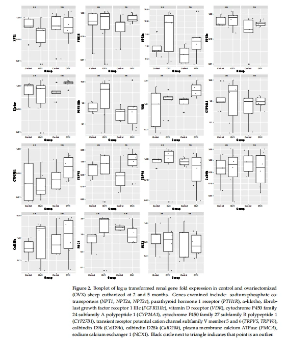
3.3. Effect of Ovariectomy and Glucocorticoid Treatment on Vitamin D and Phosphatonin-Related Gene Expression in the KIDNEY 5 Months Post-Operatively
The full results are presented in Figure 3. Expression of PTH1R was significantly greater in OVX sheep treated with glucocorticoids for 5 months compared with the other groups (p=0.04, adj R40.18), while TRPV5 (p=0.01, adj R20.31)and CYP24A1(p=0.005, adj R40.40) expression were significantly greater in OVX sheep treated with glucocorticoids for 2 months compared with the other groups.
Expression of klotho was significantly different between all groups (p =0.01, adj R20.48), with the lowest expression in control sheep. OVX sheep treated with glucocorticoids for 2 months had the highest expression (p =0.002), followed by OVX sheep (p=0.008)and then OVX sheep treated with glucocorticoids for 5 months (p=0.03). However, when adjusted for repeated measures, gene expression was not significantly different between these groups, with klotho showing the largest difference in OVX sheep treated with glucocorticoids for 5 months (p=0.1).
Similarly, the expression of the VDR was significantly different between all groups (p=0.03, adj R20.38). The greatest expression of the VDR was in OVX sheep treated with glucocorticoids for months (p=0.009), followed by OVX sheep (p=0.01), then OVX sheep treated with glucocorticoids for 5 months (p=0.05), and the lowest expression was present in control sheep.
3.4.PCA of Overall Gene Expression between Groups
PCA analysis of OVX, control, and euthanasia at 2 and 5 months failed to separate out the different groups (Figure S1).PCL and PC2 explained 69.6% of the variation. Similarly, PCA analysis of groups euthanized at 5 months also failed to separate out the different groups. PCL and PC2 explained 53.2% of the variation (Figure S2).PCA analysis showed that overall gene expression could not be explained by serum calcium and phosphorus of 25-hydroxyvitamin D concentrations(Figure S3).
3.5.Pairwise Correlation between all Analytes and Gene Expression
Regardless of ovariectomy status, the calcium content of the diet, time since ovariectomy (2m or 5m), or glucocorticoid treatment status, NPT1 gene expression was significantly and positively correlated (p<001) with PTH1R, NPT2c, Klotho, CYP24A1, TRPV5, CalD9k, and CalD28k gene expression. PTH1R was significantly positively correlated (p<001) with NPT2c, Klotho, CYP24A1, CYP27B1, TRPV5, and CalD28k. NPT2a was significantly positively correlated (p<0.001) with FGFR1WIc. Klotho was significantly positively correlated (p<0.001) with CYP24A1, CYP27B1, TRPV5, and CalD28k. CYP24A1 was significantly positively correlated (p<0.001) with TRPV5 and CalD28k. TRPV6 was significantly positively correlated (p<0.001) with Cal9Dk and PMCA, while Cal9DK was significantly positively correlated (p<0.001) with CalD28k and PMCA. None of the serum analytes measured (osteocalcin, CTX, 25OHD, Ca, and P) were correlated with the expression of any genes. The full pairwise correlations are shown in Figure 4.

4. Discussion
The results of this study suggest that treatment of ovariectomized sheep with glucocorticoids has had the greatest impact on gene expression, while ovariectomy itself and the time since ovariectomy (2 and 5 months) had little impact on gene expression. In a univariate analysis, significant differences in TRPV5, CYP24A1, and klotho gene expression were seen in ovariectomized sheep treated with glucocorticoids for 2 months followed by euthanasia at 5 months, and in PTH1R in ovariectomized sheep treated with glucocorticoids for5 months. Differences were most marked in klotho expression after adjustment for repeated measures.
Quantitative peripheral computed tomography of the proximal tibia of these sheep showed that ovariectomy decreased total volumetric bone mineral density (vBMD) by 8%, ovariectomy with glucocorticoid treatment for 5 months by 27%, and ovariectomy with glucocorticoids for 2 months and euthanasia at 5 months by 13% [6]. Interestingly, trabecular BMD, trabecular area and trabecular bone mineral content were similar to or greater than controls in ovariectomized sheep treated with glucocorticoids for 2 months and euthanized at 5 months, suggesting recovery of bone mass [6]. Glucocorticoids have several well-documented effects on bone that lead to the development of glucocorticoid-induced osteoporosis. These effects include inhibiting the formation of osteoblast progenitor cells and osteoblasts, increasing osteoblast apoptosis, and decreasing osteoid production, all of which decrease bone formation [25,26]. At the same time, glucocorticoids increase bone resorption by decreasing OPG expression, increasing RANKL expression, increasing osteoclast formation, decreasing osteoclast apoptosis, and increasing the secretion of cathepsins and matrix metalloproteinases by osteoclasts [25,26].
Klotho is an integral component of the phosphating system, whereby it binds to fibroblast growth factor 23(FGF23) in the kidney. The klotho-FGF23 complex then binds to the FGFR1IIC receptor, which initiates several cellular pathways and, ultimately, results in the upregulation of renal 24-hydroxylase(CYP24A1) expression and downregulation of renal la-hydroxylase (CYP27B1) and renal tubular Na/P II cotransporters (NPT2a and 2c) [10]. The increased expression (differences in which were most marked after adjusting for repeated measures) of klotho in sheep on glucocorticoids for 2 months and then euthanized at 5 months may explain the increased CYP241 expression also seen in this group of sheep, despite no significant differences in serum 25OHD concentrations.
Klotho is known as the anti-aging protein and klotho-deficient mice have low bone formation and bone resorption, leading to a low bone turnover osteopenia [27,28]. Additionally, specific klotho gene polymorphisms are associated with decreased bone mineral density in humans [29,30]. Soluble klotho upregulates early growth response protein 1 (EGR-1)and induces the expression of bone differentiation markers in osteoblast cell culture [31]Additionally glucocorticoids can suppress fibroblast growth factor 232]. Perhaps, the greater klotho expressions in sheep on glucocorticoids for 2 months and then euthanized at 5 months is reflective of the recovery process for this sheep post-glucocorticoid treatment and, given its role in bone formation and resorption, conceivably a key driver of bone recovery and, therefore, potentially a treatment target for osteoporosis in humans.
However, OVX sheep also had increased klotho expressions compared with control sheep. Similarly, one study in aromatase-deficient mice showed that estrogen deficiency increased klotho protein levels, and estradiol treatment decreased klotho expressions in these mice[17]. In mice, estrogen downregulates NPT2a in a process that is independent of klotho/FGF23 and PTH[18]. However, this effect was not seen in the ovariectomized sheep in this present study. Otherwise, there is little literature on the effect of estrogen deficiency on klotho. Further research should examine the effects of both ovariectomy and glucocorticoids on klotho expression, given its role in aging and in chronic kidney disease.
TRPV5 is an epithelial calcium channel chiefly expressed in the distal convoluted tubules and connecting tubules of the kidney [20,33]. The TRPV5 channel is constitutively open but undergoes calcium-dependent inactivation in association with calmodulin, a calcium-sensing protein [34]. TRPV5 expression can be increased by PTH and 1,25(OH)D3 [35]. One sheep that was on glucocorticoids for 2 months and then euthanized at 5 months was hypocalcemic, and serum calcium concentrations were significantly lower in sheep in this group compared with the other groups. It is likely, therefore, that the increased TRPV5 expression is a result of the decreased serum calcium concentration in these animals. Glucocorticoids have been shown to decrease intestinal calcium absorption by decreasing the expression of TRPV6 and CalD9k [36,37] and increasing urinary excretion of calcium [14], but clinically significant hypocalcemia in healthy individuals is uncommon. Similarly, the sheep with low serum calcium concentration was showing no clinical signs of hypocalcemic tetany. Alternatively, the increased expression of TRPV5 in sheep on glucocorticoids for2 months and then euthanized at 5 months could reflect the recovery process, whereby to replace the recovered trabecular BMD, area, and bone mineral content, increased resorption of calcium from the kidney is required.
In sheep treated with glucocorticoids for 5 months, PTH1R expression was increased compared with the other groups. Additionally, compared to other groups, there was little variation in expression between sheep in the group. While no differences were seen in serum calcium concentrations in sheep treated with glucocorticoids for 5 months, perhaps this could be associated with the increased action of PTH and its receptor. The effects of glucocorticoids on PTH secretion and action are unclear. Some studies in humans have found that glucocorticoid-treated patients have increased serum PTH concentrations, along with decreased calcium absorption, increased urinary calcium excretion and decreased bone mass [38,39]. However, most studies have found no changes in serum PTH concentrations in humans on glucocorticoid treatment [38,40,41]. One study has shown that perhaps the impact of glucocorticoids is on altering the secretion of PTH, with the increased pulsatile secretion of PTH in glucocorticoid-treated individuals[4]. Others have also shown increased PTH1R expression in mesenchymal stem cells and osteoblast-like cells in response to treatment with dexamethasone[43,44]. As such, the increase in renal PTH1R expression in the sheep treated with glucocorticoids for 5 months is likely due to the 5 months of glucocorticoid treatment.
While ovariectomy combined with a mildly low calcium diet resulted in minimal changes to gene expression, it did appear to alter the expression of the VDR, with ovariectomized sheep on a mildly low calcium diet having greater VDR expression compared to control sheep. This is in contrast to a study on ovariectomized rats whereby those fed a diet with normal calcium levels showed increased renal VDR expression, while those fed a low calcium diet had decreased VDR expression [45]. It is difficult in this present sheep study to separate out the effect of ovariectomy on renal expression of the VDR from the effect of the low calcium diet. However, there were no significant differences in serum calcium concentrations between groups (with the exception of the glucocorticoid group)suggesting the mildly low calcium diet had minimal effect and perhaps the effects on the VDR are due to the ovariectomy. However, further research will be required to investigate these relationships.
Two previous studies have examined the separate relationships between vitamin D-responsive genes, and phosphating-related genes in sheep kidneys [20,21]. However, relationships between the vitamin D and phosphating pathways have not been examined. Interestingly, the most significant correlations between genes reported in these two papers hold true for the gene relationships in this present sheep study as well, suggesting consistency in the results between studies and the relationships between these genes. cistanche salsa extract Of the interesting relationships not previously reported, as one might expect given its pivotal role in calcium metabolism, expression of PTH1R was highly correlated with CalD28k, CalD9k, TRPV5 CYP27B1, CYP24A1, VDR, Klotho, and NPT2c. Expression of klotho was strongly correlated with gene expression of TRPV5, which is consistent with other literature showing that klotho upregulates TRPV5 and promotes renal calcium reabsorption [46,47].
Unfortunately, the different groups could not be separated based on vitamin D responsive and phosphating gene expression using PCA analysis. This is likely due to the main limitation of this study—the small sample size. With only 3-6 animals in each of the groups, this has likely limited the ability to detect differences in gene expression between groups.
5. Conclusions
The results from this study show that the most marked differences in gene expression were in renal klotho expression in ovariectomized sheep treated with glucocorticoids for 2 months followed by a recovery period of 5 months. Given klotho is the"anti-aging" hormone and has a critical role in calcium and phosphate metabolism, it is possible that this hormone is critically involved in the recovery of trabecular bone mineral density and area in treated sheep and may, therefore, potentially be a treatment target for osteoporosis in humans. However, more research is required on the effects of glucocorticoids and ovariectomy on klotho.
This article is extracted from Animals 2022, 12, 67. https://doi.org/10.3390/ani12010067 https://www.mdpi.com/journal/animals





