

Optimized Processing Technology Of Cistanche Tubulosa Decoction Pieces And Its Anti‑Oxidative‑Stress Mechanism In RAW264.7 Macrophages
Sep 28, 2026
Abstract
Cistanche tubulosa is a precious tonic herb widely applied in Traditional Chinese Medicine. Phenylethanoid glycosides represented by echinacoside and acteoside are its core bioactive substances. This research aims to optimize modern processing parameters for Cistanche tubulosa decoction pieces to improve the yield of echinacoside and acteoside, and further explore its anti‑oxidative‑stress and anti‑inflammatory mechanism at cellular level.
Single‑factor experiments were conducted, followed by Box‑Behnken response‑surface optimization with slice thickness, steaming time and drying temperature as influencing factors. High‑performance liquid chromatography (HPLC) was adopted to detect the content of two marker compounds. The LPS‑induced RAW264.7 macrophage injury model was constructed to assess pharmacological activities. Reactive oxygen species (ROS), nitric oxide (NO), malondialdehyde (MDA), activities of antioxidant enzymes including SOD, CAT, LDH and glutathione (GSH) content were determined. ELISA was used to test the secretion level of pro‑inflammatory cytokines TNF‑α, IL‑1β and IL‑6. Western blot assay was performed to analyze the phosphorylation status of key proteins in p38/AKT/JNK/NF‑κB signaling pathway.
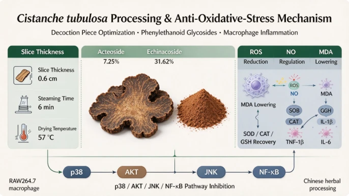
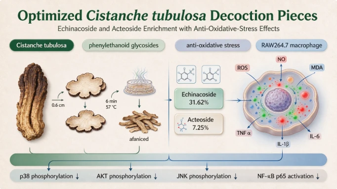
The optimal processing conditions were slice thickness of 0.6 cm, steaming time of 6 min and drying temperature of 57 °C. Under this condition, the content of acteoside reached 7.25 % and echinacoside reached 31.62 %. Extracts from optimized decoction pieces could significantly reduce the over‑production of intracellular ROS, NO and MDA in LPS‑stimulated RAW264.7 cells. It enhanced the activities of SOD, CAT, LDH and increased intracellular GSH level, meanwhile markedly inhibited the release of TNF‑α, IL‑1β and IL‑6. Western‑blot results indicated that the extract down‑regulated phosphorylation ratio of p‑p38/p38, p‑AKT/AKT, p‑JNK/JNK and p‑NF‑κB/NF‑κB.
The optimized steaming‑drying processing technology can effectively increase the recovery rate of main phenylethanoid glycosides of Cistanche tubulosa. Decoction pieces exert anti‑oxidative and anti‑inflammatory effects against LPS‑triggered cell damage through inhibiting p38/AKT/JNK/NF‑κB signaling pathway. This study provides practical technical parameters for standardized industrial processing of Cistanche tubulosa, and offers experimental evidence for the development of functional food and nutraceutical products derived from this herbal resource.
Keywords: Cistanche tubulosa; decoction‑piece processing technology; acteoside; echinacoside; anti‑oxidative stress; RAW264.7 macrophage; p38/AKT/JNK/NF‑κB pathway
1. Introduction
Cistanche tubulosa (Schenk) Wight belongs to Orobanchaceae family, and its medicinal part is the dried fleshy stem with scale leaves. In China, this plant is mainly planted in arid regions of northwest, with the largest planting area in southern Xinjiang. As a well‑known tonic herbal medicine, it functions to tonify kidney‑yang, replenish essence and blood, and moisten intestines to relieve constipation. Clinically, it is used for the treatment of kidney‑yang deficiency, insufficiency of essence‑blood, infertility, soreness and weakness of waist and knees as well as intestinal dryness constipation.
Phenylethanoid glycosides, mainly echinacoside and acteoside, are the primary active components of Cistanche tubulosa. According to Chinese Pharmacopoeia requirements, the total content of echinacoside and acteoside in qualified raw medicinal materials shall not be lower than 1.50 %, which serves as the critical quality evaluation indicator. Traditional processing approaches for Cistanche tubulosa mostly adopt soaking‑moistening, slicing‑drying or wine‑steaming methods. Plenty of studies focus on improving active‑component yield, while researches combining processing‑technology optimization with biological‑activity verification remain insufficient.
Oxidative stress acts as the common pathological basis for multiple metabolic and inflammatory diseases. Existing researches have proven that total glycosides extracted from Cistanche tubulosa can suppress inflammatory response and activate Nrf2/HO‑1 pathway to alleviate liver injury in sepsis animals. Lignan glycoside fractions from this herb can mitigate macrophage inflammation by regulating PI3K/AKT signaling. Nevertheless, the pharmacological property of modern processed decoction pieces and its underlying molecular mechanism still require further exploration.
In this study, Box‑Behnken response‑surface design was applied based on single‑factor tests, taking slice thickness, steaming time and drying temperature as variables to optimize processing workflow. After obtaining optimal technical parameters, LPS‑induced RAW264.7 oxidative‑damage cell model was established to evaluate anti‑oxidative‑stress and anti‑inflammatory capacity and dissect relevant signaling mechanism. This work intends to provide stable and operable processing parameters for pilot‑scale production, and lay experimental foundation for the comprehensive utilization of Cistanche tubulosa resources.
2. Materials and Methods
2.1 Plant Material, Reagents and Instruments
Fresh fleshy stems of Cistanche tubulosa were harvested in autumn from Hotan, Xinjiang. RAW264.7 mouse monocyte‑macrophage leukemia cells were purchased from Procell Life Science & Technology Co., Ltd (Wuhan, China).
HPLC‑grade acetonitrile and methanol were obtained from Merck company. Echinacoside (≥98 %) and acteoside (≥98 %) reference standards were supplied by Beijing North Weiye Metrology Institute. Dimethyl sulfoxide (DMSO) was purchased from Sigma‑Aldrich. DMEM high‑glucose medium, fetal bovine serum (FBS), penicillin‑streptomycin double‑antibody solution, BCA protein quantification kit, assay kits for NO, MDA, SOD, CAT, GSH and LDH, lipopolysaccharide (LPS), MTT reagent and ELISA kits for inflammatory cytokines were all commercial products.
Experimental instruments included Shimadzu high‑performance liquid chromatograph, rotary evaporator, Chinese‑herb pulverizer, thermostatic blast drying oven, steam cooker, full‑wavelength microplate reader, electronic analytical balance, refrigerated centrifuge, biosafety cabinet and inverted optical microscope.
2.2 Single‑factor Processing Experiment
Fresh Cistanche tubulosa fleshy stems were cut into slices with thickness of 0.2 cm, 0.4 cm, 0.6 cm, 0.8 cm, 1.0 cm and 1.2 cm. Slices were steamed for 3 min, 6 min, 9 min, 12 min, 15 min, 18 min and 21 min respectively, and dried under different temperature gradients including room temperature (25 °C), 40 °C, 50 °C, 60 °C, 70 °C and 80 °C. Dried decoction‑piece samples were crushed and sieved through 40‑mesh sieve for subsequent detection. Composite scoring formula was adopted for comprehensive assessment: Composite score = echinacoside content × 50 % + acteoside content × 50 %.
2.3 HPLC Determination of Acteoside and Echinacoside
Sample solution preparation: accurately weigh 1.0 g powder sample, add 100 mL 50 % methanol solution. After 30‑min soaking, ultrasonic extraction was performed for 40 min. The weight loss was supplemented with 50 % methanol after cooling. Supernatant was filtered by 0.22 μm microporous membrane before HPLC injection.
Mixed reference‑standard solution of acteoside and echinacoside was prepared with 50 % methanol and diluted into serial concentrations for standard‑curve plotting. Chromatographic conditions: COSMOSIL C₁₈ chromatographic column; mobile‑phase system consisted of 0.1 % phosphoric acid aqueous solution (A) and acetonitrile (B); detection wavelength was set as 330 nm; column temperature 35 °C; flow rate 1 mL/min. External‑standard method was used for quantitative calculation of target compounds.
2.4 Box‑Behnken Response‑surface Experimental Design
According to single‑factor experimental results, slice thickness (X₁), steaming time (X₂) and drying temperature (X₃) were selected as independent variables. Acteoside content (Y₁, %) and echinacoside content (Y₂, %) served as response values. Design‑Expert 13.0 software was used to establish three‑factor three‑level Box‑Behnken experimental scheme with total 17 groups of tests. Model fitting, variance analysis and parameter prediction were completed by software. Practical verification experiments were carried out in triplicate after adjusting theoretical optimal parameters to fit actual production operation.
2.5 Cell Culture and Sample Preparation
Decoction‑piece sample obtained under optimized processing condition was prepared into extract powder. The powder was dissolved in DMSO and further diluted with complete DMEM medium containing 10 % FBS and 1 % penicillin‑streptomycin for cell experiments. RAW264.7 cells were cultured in humidified incubator at 37 °C with 5 % CO₂.
2.6 MTT Assay for Cell Viability
Cell suspension was inoculated into 96‑well plates at cell density of 8 × 10⁴ cells/mL. Groups were set as solvent control group (0.1 % DMSO), LPS model group (1 μg/mL LPS), and multiple concentration groups of Cistanche tubulosa extract (3.125, 6.25, 12.5, 25, 50, 100, 200, 400 μg/mL). After 2‑hour pre‑incubation of samples, LPS with final concentration of 1 μg/mL was added except solvent‑control wells and incubated for another 24 h. MTT solution (5 mg/mL) was added for 4‑hour reaction. After removing supernatant, DMSO was added to dissolve formazan crystal. Absorbance value at 490 nm was detected by microplate reader to calculate cell viability. Safe concentration range was selected for follow‑up functional tests.
2.7 Intracellular ROS Measurement
RAW264.7 cells were seeded in 6‑well plates at density of 4 × 10⁵ cells per well. After adhering to the wall, cells were pretreated with gradient‑concentration Cistanche tubulosa extract (12.5, 25, 50, 100 μg/mL) for 2 h, then stimulated by 1 μg/mL LPS for 24 h. Intracellular ROS level was detected with fluorescent probe reagent kit. Fluorescence intensity was recorded under excitation wavelength 488 nm and emission wavelength 525 nm.
2.8 Detection of NO, MDA, SOD, CAT, LDH and GSH
Cell‑culture supernatant was collected to detect NO content via Griess method. Cells were washed with pre‑cooled PBS and lysed on ice. Cell lysate was centrifuged at 10 000 rpm under 4 °C for 5 min to acquire intracellular supernatant. MDA content, enzyme activities of SOD, CAT, LDH and GSH level were measured strictly in accordance with kit specifications. BCA method was applied for total‑protein normalization.
2.9 ELISA Detection of Pro‑inflammatory Cytokines
RAW264.7 cells were treated with sample and LPS for 16 h. Culture supernatant was collected and centrifuged at 10 000 rpm for 10 min. The secretion concentrations of TNF‑α, IL‑1β and IL‑6 were quantified by ELISA kits.
2.10 Western Blot Analysis
After 45‑minute intervention of samples and LPS, total cellular protein was extracted using RIPA lysis buffer. Protein concentration was quantified by BCA kit. Equal amount of protein sample (50 μg for each lane) was denatured, separated by SDS‑PAGE electrophoresis and transferred onto PVDF membrane. Membrane was blocked with 5 % skim‑milk TBST solution for 1 h, then incubated with primary antibodies overnight at 4 °C. After repeated washing with TBST, secondary antibody was incubated at room temperature for 1 h. Target protein bands were visualized by ECL chemiluminescence reagent. Band gray‑scale analysis was conducted to calculate phosphorylation‑protein / total‑protein ratio.
2.11 Statistical Analysis
GraphPad Prism 8.0.2 software was used for graph plotting and statistical analysis. One‑way ANOVA and t‑test were adopted for data comparison. P < 0.05 represented statistically significant difference; P < 0.01 and P < 0.001 stood for higher significance. All experimental data were expressed as mean ± standard deviation (\(\bar{x}\pm s\)).
3. Results
3.1 Single‑factor Experimental Results
Slice thickness exerted prominent influence on target‑glycoside content. Slices of 0.2 cm thickness were easy to be over‑steamed, which caused thermal decomposition of active ingredients. Over‑thick slices hindered the release of internal active substances. When steaming time was fixed at 10 min and drying temperature at 60 °C, 0.4 cm was the optimal slice‑thickness parameter in single‑factor screening.
Insufficient steaming time could not fully destroy plant‑cell structure and led to low glycoside yield. When steaming time exceeded 6 min, tissue disintegration occurred and echinacoside as well as acteoside degraded. Under fixed slice thickness of 0.4 cm and drying temperature of 60 °C, 6 min was the best steaming‑time condition.
Within a certain range, the content of phenylethanoid glycosides rose along with drying‑temperature increase, yet degradation took place once temperature exceeded 60 °C. With slice thickness of 0.4 cm and steaming time of 6 min, 60 °C showed the best comprehensive performance in single‑factor screening.
3.2 Response‑surface Optimization and Verification Test
Variance analysis showed that regression models for acteoside and echinacoside content were significant (P < 0.05, P < 0.01). The lack‑of‑fit term was non‑significant (P > 0.05), indicating good model fitting degree. The determination coefficient R² was close to 1, which proved high correlation between predicted value and actual experimental data.
The influencing order of factors for acteoside content was slice thickness > drying temperature > steaming time. For echinacoside content, the order was steaming time > drying temperature > slice thickness.
Theoretical optimal parameters calculated by software were slice thickness 0.600 cm, steaming time 5.685 min and drying temperature 57.009 °C. Considering practical production condition, parameters were adjusted to slice thickness 0.6 cm, steaming time 6 min and drying temperature 57 °C. Three groups of parallel verification experiments obtained acteoside content of 7.25 % and echinacoside content of 31.62 %. The deviation between practical value and theoretical predicted value was 3.89 % and 4.92 % respectively, which demonstrated the reliability of this response‑surface model.
3.3 Cell Viability Screening Result
Within 0‑100 μg/mL concentration range, Cistanche tubulosa decoction‑piece extract had no obvious cytotoxicity on RAW264.7 cells and exhibited slight proliferation‑promoting effect. When concentration reached 200 μg/mL and above, cell viability decreased significantly. Therefore, 12.5 μg/mL, 25 μg/mL, 50 μg/mL and 100 μg/mL were selected as working concentrations for subsequent cell experiments.
3.4 Effect on Intracellular ROS Level
Compared with solvent‑control group, LPS model group displayed remarkably elevated intracellular ROS fluorescence intensity (P < 0.001), proving the successful establishment of oxidative‑stress injury model. Pretreatment of Cistanche tubulosa extract reduced excessive ROS accumulation in a dose‑dependent way (P < 0.001 versus model group).
3.5 Regulation on NO, MDA and Antioxidant‑related Indicators
LPS stimulation induced sharp increase of NO and MDA content in RAW264.7 cells. Compared with LPS model group, extract intervention significantly lowered NO level with dose‑dependent tendency. 12.5 μg/mL extract did not produce obvious effect on MDA content, while 25‑100 μg/mL extract observably decreased MDA concentration (P < 0.001).
After LPS treatment, the activities of SOD, CAT, LDH and intracellular GSH content decreased markedly. 12.5 μg/mL extract could enhance SOD activity without improving CAT and LDH activities. Extract at concentration of 25‑100 μg/mL significantly elevated SOD, CAT, LDH enzyme activities and GSH content in concentration‑dependent manner.
3.6 Inhibitory Effect on Pro‑inflammatory Cytokine Secretion
LPS stimulation triggered massive release of TNF‑α, IL‑1β and IL‑6 in RAW264.7 macrophages. Cistanche tubulosa decoction‑piece extract pretreatment remarkably suppressed the secretion of above‑mentioned three pro‑inflammatory cytokines (P < 0.001), presenting prominent dose‑dependent anti‑inflammatory property.
3.7 Modulation on p38/AKT/JNK/NF‑κB Signaling Pathway
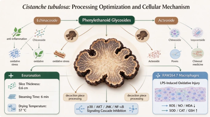
Compared with solvent‑control group, LPS intervention increased phosphorylation ratio of p‑p38/p38, p‑AKT/AKT, p‑JNK/JNK and p‑NF‑κB/NF‑κB. Medium and high‑dose extract groups significantly reduced p‑p38/p38 and p‑JNK/JNK ratio. All concentration groups down‑regulated p‑AKT/AKT and p‑NF‑κB/NF‑κB level (P < 0.05 or P < 0.001). Results illustrated that optimized Cistanche tubulosa decoction‑piece extract restrained the activation of p38/AKT/JNK/NF‑κB signaling pathway by inhibiting protein phosphorylation.
4. Discussion
Current processing requirements for Cistanche tubulosa recorded in Chinese Pharmacopoeia only describe qualitative operation steps without precise quantitative parameters. Previous researches indicated that different processing methods would cause great differences in chemical composition and pharmacological activity between crude and processed Cistanche tubulosa samples. Processed products show better antioxidant and immune‑regulatory performance compared with raw materials.
In this research, the modern steaming‑drying processing optimized via response‑surface methodology obtained much higher yield of echinacoside and acteoside than many traditional processing techniques. It suggests that precise control over slice thickness, steaming duration and drying temperature can effectively retain and improve phenylethanoid‑glycoside content of Cistanche tubulosa.
Oxidative stress and inflammation are closely connected pathological processes. Excessive ROS gives rise to lipid peroxidation and MDA accumulation; meanwhile it activates downstream inflammatory cascades and promotes the generation of NO and multiple pro‑inflammatory cytokines. Inflammatory mediators further accelerate ROS production and form a vicious pathological cycle. Our cellular experimental results demonstrated that optimized Cistanche tubulosa decoction‑piece extract could eliminate redundant ROS, reduce oxidative‑damage markers NO and MDA, restore the activity of antioxidant enzyme system represented by SOD, CAT, LDH and replenish intracellular GSH, as well as inhibit the release of pro‑inflammatory cytokines with obvious dose‑effect relationship.
p38, JNK belonging to MAPK family and AKT‑NF‑κB axis are core signal‑transduction hubs mediating macrophage oxidative‑inflammatory response. LPS stimulation promotes phosphorylation of p38, JNK and AKT, further activates NF‑κB and induces transcription of numerous pro‑inflammatory genes. Western‑blot data revealed that Cistanche tubulosa decoction‑piece extract blocked phosphorylation activation of p38, JNK, AKT and NF‑κB. It is inferred that high‑content echinacoside and acteoside from optimized processing jointly contribute to suppressing p38/AKT/JNK/NF‑κB signaling cascade, so as to relieve LPS‑caused oxidative‑stress and inflammatory injury in RAW264.7 macrophages.
There are still limitations existing in this study. All pharmacological evaluations were completed on in‑vitro cell model. Further animal‑level experiments are required to verify in‑vivo efficacy and metabolic characteristics of processed Cistanche tubulosa decoction pieces.
In conclusion, this study established quantifiable and scalable modern processing technology for Cistanche tubulosa decoction pieces. Under processing parameters of slice thickness 0.6 cm, steaming time 6 min and drying temperature 57 °C, the content of main phenylethanoid glycosides is greatly improved. Processed decoction pieces can alleviate oxidative‑stress and inflammatory damage in LPS‑stimulated RAW264.7 macrophages by inhibiting p38/AKT/JNK/NF‑κB signaling pathway. This research provides practical technical reference for industrialized production, and offers experimental basis for developing functional‑food and dietary‑supplement products of Xinjiang‑origin Cistanche tubulosa.
Acknowledgements
This work was supported by Youth Science and Technology Backbone Innovation Capacity Cultivation Project of Xinjiang Academy of Agricultural Sciences (xjnkq‑2022022) and Xinjiang "Tianchi Doctor Program" (2021).
References
[1] Lin J, Yu H, Zhao Y, et al. Extraction of echinacoside from Cistanche tubulosa and investigation of its protective effect on liver injury in sepsis rats. Food Science and Technology. 2023;43:e010523.
[2] Guo J, Tang S, Miao Y, et al. The anti‑inflammatory effects of lignan glycosides from Cistanche tubulosa stems on LPS/IFN‑γ‑induced RAW264.7 macrophage cells via PI3K/AKT pathway. Current Pharmaceutical Biotechnology. 2021;22(10):1380‑1391.
[3] Rani V, Deep G, Singh R K, et al. Oxidative stress and metabolic disorders: Pathogenesis and therapeutic strategies. Life Sciences. 2016;148:183‑193.
[4] Martínez‑de‑Toda I, Ceprián N, Díaz‑de‑Castro E. The role of oxi‑inflamm‑aging in human pathophysiology. Cells. 2021;10(11):2974.
[5] Chung Y C, Song S J, Lee A, et al. Isobavachin alleviates lipopolysaccharide‑induced inflammatory responses in macrophages and zebrafish by suppressing MAPK and NF‑κB signaling pathways. Journal of Ethnopharmacology. 2024;321:117501.





