

Part Ⅱ Association Between Indoxyl Sulfate And Dialysis Initiation And Cardiac Outcomes in Chronic Kidney Disease Patients
May 16, 2023
Results
1. Patient Characteristics
This study cohort consists of 89 pre-dialysis CKD stage 3 to 5 patients. The mean age was 63.5±15 years. The most common cause of CKD was diabetic nephropathy (44.9%) followed by hypertensive nephropathy (16.8%), and chronic glomerulonephritis (12.4%). The percentage of CKD stages 3, 4, and 5 was 29.2%, 32.6%, and 38.2%, respectively. Serum indoxyl sulfate levels were significantly higher in patients with worsening renal function. The mean serum indoxyl sulfate levels were 2.73 ± 1.97, 5.83 ± 3.35, and 18.44 ± 10.31 mg/L in stages 3, 4, and 5 CKD, respectively.
Patients with worse renal function had a higher prevalence of left atrial (LA) enlargement (19% in CKD stage 3, 46.1% in CKD stage 4, and 57.6% in CKD stage 5; p=0.02), left ventricular hypertrophy (LVH) (48% in CKD stage 3, 75.8% in CKD stage 4, and 82.3% in CKD stage 5; p=0.01), and impaired global longitudinal strain (GLS) (43.3%, 51.7%, and 70.6% in CKD stage 3,4, and 5, respectively; p=0.04). However, there were no significant differences in the prevalence of the remaining echocardiographic parameters, including left ventricular ejection fraction (LVEF), among pre-dialysis CKD stage 3 to 5 patients (Data not shown).

Click here to know the benefits of Cistanche
2. Comparison Between Low and High Indoxyl Sulfate Groups
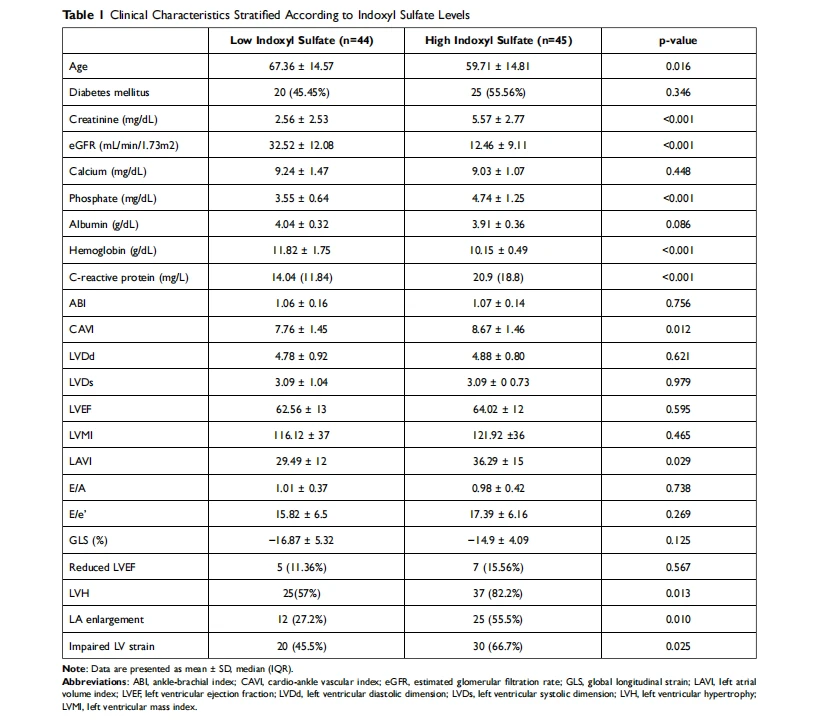
Patients were divided into 2 groups based on the median serum indoxyl sulfate levels (low indoxyl sulfate group, < 6.124 mg/L and high indoxyl sulfate group, ≥ 6.124 mg/L) (Table 1). The high indoxyl sulfate group showed significantly lower eGFR, higher phosphate levels, and greater CAVI than the low indoxyl sulfate group. Patients with high indoxyl sulfate had a higher prevalence of LA enlargement, LVH, and impaired GLS) than patients in the low indoxyl sulfate group.
3. Correlation Between Indoxyl Sulfate Levels and Clinical and Echocardiographic Parameters
The univariate correlations between serum indoxyl sulfate levels and clinical and echocardiographic parameters of the study population are shown in Table 2. A significant positive correlation was detected for creatinine, phosphate, and GLS, while a significant negative correlation was observed for hemoglobin. Figure 1 depicts a positive linear relationship between serum indoxyl sulfate levels and GLS (r=0.62, p = 0.01).

In multivariate linear regression analysis, creatinine (r = 1.49, p < 0.001), hemoglobin (r = −1.76, p = 0.002), and GLS (r= 0.42, p = 0.033) were independently correlated with indoxyl sulfate levels.

4. Predictor of Renal Outcomes
34 patients initiated renal replacement therapy during 24 months of follow-up. As detailed in Table 3, unadjusted predictors of progression to dialysis were age (p=0.005), creatinine (p<0.001), use of renin-angiotensin-aldosterone system (RAAS) inhibitors (p=0.001), serum indoxyl sulfate levels (p<0.001), LVEF (p=0.035), and GLS (p=0.010). Kaplan–Meier curve of the cumulative event-free survival for the dialysis dichotomized according to indoxyl sulfate levels (low and high) is reported in Figure 2. The high indoxyl sulfate group was associated with dialysis initiation when compared with the low indoxyl sulfate group (log-rank p < 0.001). After adjustment for age, using RAAS inhibitors, creatinine, and GLS; indoxyl sulfate and LVEF remained independent predictors for progression to dialysis (HR 1.04; 95% CI 1.01–1.08; p=0.022) (Table 3).


5. Predictors of Cardiovascular Events
There were 19 cardiovascular events during 24 months of follow-up. On univariate Cox proportional hazard analysis, LVEF and GLS were significantly associated with cardiovascular events together with age and CAVI. In contrast, there was no significant association between serum indoxyl sulfate levels and cardiovascular events (Table 4). Following adjustment with multivariate analysis, only GLS remained a significant predictor of cardiovascular events (HR 1.26; 95% CI 1.01–1.59; p=0.045) (Table 4). The cardiovascular events in the impaired GLS group were significantly higher than the preserved GLS group (p=0.015) (Figure 3). There was no significant difference in cardiovascular events between low and high indoxyl sulfate groups (p=0.082).


Discussion
In pre-dialysis CKD stage 3 to 5 patients, the results in the present prospective 2-year follow-up cohort study demonstrated that high serum indoxyl sulfate could predict progression to dialysis. In addition, indoxyl sulfate was significantly associated with GLS, an emerging echocardiography technique for measuring more subtle disturbances in LV systolic function that could forecast CVD events in pre-dialysis CKD patients.
Indoxyl sulfate has been reported as an atherosclerosis accelerator by increasing proinflammatory cytokines and oxidative stress, promoting endothelial dysfunction, inhibiting bone turnover, and inducing vascular calcification. Furthermore, a line of evidence illustrates that indoxyl sulfate could contribute to CKD progression. In in vitro and animal models of CKD, indoxyl sulfate was demonstrated to have direct toxic effects on tubular cells, leading to increased oxidative stress, inflammation, and fibrosis. Direct renal injury and vascular toxicity of indoxyl sulfate might accelerate renal progression. However, only a few prospective studies have evaluated the impact of serum indoxyl sulfate levels on CKD progression. The only prospective cohort in 269 adult CKD G1-5 patients, revealed a significant association between serum indoxyl sulfate levels and renal progression, defined as a reduction of eGFR by 50% or end-stage kidney disease requiring dialysis, after 21 month follow-up period. In the present study, we provided the result of a significant correlation between indoxyl sulfate and the commencement of dialysis which was independent of patients’ cardiovascular status, adjusted with CAVI, LVMI, and LVEF (Table 3). These findings suggest the temporal relationship between the nephrotoxicity of indoxyl sulfate and CKD progression.

Cistanche extract and Cistanche powder
Regarding cardiovascular changes, hemodynamic and metabolic alterations in CKD result in cardiac remodeling which occurs early and is significantly worse than non-CKD. Hypervolemia and renal anemia contribute to volume load while hypertension, vascular calcifications, and increased sympathetic activation in CKD result in pressure load. Both volume and pressure overloads can cause LV geometric changes. In our cohort, LV mass and the prevalence of LVH increased with deteriorating kidney function, whereas there was no significant difference in LVEF among CKD stages 3, 4, and 5. The LVH-enhancing effect observed in the present study was also detected in a previous study in patients with more advanced CKD while the prevalence of LV systolic dysfunction (reduced LVEF) in CKD patients varied according to different methodologies. Although the reduction in LVEF is a good predictor of CV events and mortality, there were some limitations in utilizing LVEF as a predictor. The methods of LVEF measurement involve inaccuracies associated with unclear delineation of endocardial borders and the assumption of geometric uniformity. Furthermore, LVEF may be insufficiently sensitive to identify mild or subclinical degrees of LV systolic dysfunction. These limitations have led to an interest in new echocardiographic techniques that provide more objective and sensitive measures of LV myocardial function. GLS is a relatively new echocardiographic application used increasingly in the general population. This technique is an offline application that follows the motion of myocardial tissue throughout the cardiac cycle by tracking acoustic reflections, known as speckles. As GLS quantifies longitudinal contraction, especially in the subendocardial fibers, the wall layer most susceptible to ischemia, reductions in longitudinal strain may be found before a reduction in LVEF. GLS is an effective tool for detecting early subtle disturbances of LV systolic function when LVEF is normal. In the present study, patients with more advanced CKD had a higher prevalence of impaired GLS but had no significant differences in reduced LVEF.
Indoxyl sulfate has strong proinflammatory and pro-hypertrophic effects on cardiac cells. In addition, this protein-bound uremic toxin contributed to cardiac fibrosis in vitro by stimulating the synthesis of transforming growth factor-beta and encouraging the NF-kB pathway activation. Therefore, accumulation of indoxyl sulfate in CKD patients might directly induce detrimental effects on the cardiac cells and cause adverse cardiac remodeling. Indoxyl sulfate also induces proinflammatory cytokines and oxidative stress, resulting in endothelial dysfunction and atherosclerosis. We demonstrated that patients with a high indoxyl sulfate group had a higher prevalence of LVH and impaired GLS than the low indoxyl sulfate group (Table 1). Of interest, there was a significant association between serum indoxyl sulfate levels and GLS in pre-dialysis CKD stage 3 to 5 patients (Table 2 and Figure 1). This finding was supported by a previous study in patients with CKD stages 3 and 4 by Krishnasamy et al who revealed that GLS was independently associated with free indoxyl sulfate level, body mass index, and arterial stiffness.

Cistanche pills
In terms of cardiac events, we revealed a significant association between impaired GLS and cardiovascular events (Table 4). Although certain previous studies reported a correlation between GLS and cardiovascular events in non-CKD patients with heart failure, there were sparse data assessing the prognostic value of GLS in pre-dialysis CKD. In a cross-sectional study of 106 CKD patients stage 1–5 with preserved LVEF, Panoulas et al found that patients with impaired GLS (more than −16%) had an increased risk of major adverse cardiac events. However, this significant association was lost after adjustment for age, gender, diabetes, hypertension, and eGFR. In another prospective cohort of 106 pre-dialysis CKD patients with preserved LVEF, Sulemane et al demonstrated that GLS was a significant independent predictor of the composite endpoint of all-cause mortality, acute coronary syndrome, stable angina requiring revascularization, Hospitalization for heart failure, and stroke.
As indoxyl sulfate is associated with GLS, serum indoxyl sulfate is supposed to be a good predictor of cardiac events in CKD patients. However, we did not find a significant association between serum indoxyl sulfate and cardiovascular events during the follow-up period (Table 4). On the contrary, Shimazu et al revealed that serum indoxyl sulfate was a significant predictor of cardiac events in patients with dilated cardiomyopathy. Moreover, a previous 3-year prospective cohort of CKD patients showed an association between indoxyl sulfate and major adverse cardiac events. The different outcomes might be explained by the disparities in populations, low incidence of cardiac events, and shorter follow-up times in our study.
There are some limitations in this study. First, the numbers of participants and the occurrence of cardiovascular events were relatively small. Therefore, the power to demonstrate the relationship between indoxyl sulfate and cardiovascular events might be limited. Second, the follow-up time is relatively short for detecting cardiovascular outcomes. Further studies with larger sample sizes and longer follow-up times are still needed.

Standardized Cistanche
Conclusions
The persistently heightened serum indoxyl sulfate in CKD is associated with early impaired left ventricular systolic function detected by GLS. In addition, high serum indoxyl sulfate and reduced LVEF are significant predictors for dialysis initiation while impaired GLS significantly forecasts cardiovascular events in CKD patients.
References
20. Watanabe K, Tominari T, Hirata M, et al. Indoxyl sulfate, a uremic toxin in chronic kidney disease, suppresses both bone formation and bone resorption. FEBS Open Bio. 2017;7(8):1178–1185.
21. Milanesi S, Garibaldi S, Saio M, et al. Indoxyl sulfate induces renal fibroblast activation through a targetable heat shock protein 90-dependent pathway. Oxid Med Cell Longev. 2019;2019:2050183.
22. Miyazaki T, Ise M, Seo H, Niwa T. Indoxyl sulfate increases the gene expressions of TGF-beta 1, TIMP-1, and pro-alpha 1(I) collagen in uremic rat kidneys. Kidney Int Suppl. 1997;62:S15–22.
23. Chen CN, Chou CC, Tsai PSJ, Lee YJ. Plasma indoxyl sulfate concentration predicts the progression of chronic kidney disease in dogs and cats. Vet J. 2018;232:33–39. doi:10.1016/j.tvjl.2017.12.011
24. Wu IW, Hsu KH, Lee CC, et al. P-cresyl sulfate and indoxyl sulfate predict progression of chronic kidney disease. Nephrol Dial Transplant. 2011;26(3):938–947.
25. Kaesler N, Babler A, Floege J, Karmann R. Cardiac remodeling in chronic kidney disease. Toxins. 2020;12(3):161.
26. Nitta K, Iimuro S, Imai E, et al. Risk factors for increased left ventricular hypertrophy in patients with chronic kidney disease. Clin Exp Nephrol. 2013;17(5):730–742.
27. London GM, Pannier B, Guerin AP, et al. Alterations of left ventricular hypertrophy in and survival of patients receiving hemodialysis: follow-up of an interventional study. J Am Soc Nephrol. 2001;12(12):2759–2767.
28. Sulemane S, Panoulas VF, Nihoyannopoulos P. Echocardiographic assessment in patients with chronic kidney disease: a current update. Echocardiography. 2017;34(4):594–602.
29. Pecoits-Filho R, Barberato SH. Echocardiography in chronic kidney disease: diagnostic and prognostic implications. Nephron Clin Pract. 2010;114 (4):c242–7.
30. Amzulescu MS, De Craene M, Langet H, et al. Myocardial strain imaging: a review of general principles, validation, and sources of discrepancies. Eur Heart J Cardiovasc Imaging. 2019;20(6):605–619.
31. Hung SC, Kuo KL, Wu CC, Tarng DC. Indoxyl sulfate: a novel cardiovascular risk factor in chronic kidney disease. J Am Heart Assoc. 2017;6:2.
32. Krishnasamy R, Isbel NM, Hawley CM, et al. Left ventricular global longitudinal strain (GLS) is a superior predictor of all-cause and cardiovascular mortality when compared to ejection fraction in advanced chronic kidney disease. PLoS One. 2015;10(5):e0127044.
33. Otani K, Higa Y, Kitano T, Nabeshima Y, Takeuchi M. Prediction of cardiac events using fully automated GLS and BNP titers in patients with known or suspected heart failure. PLoS One. 2020;15(6):e0234294.
34. Park JJ, Park JB, Park JH, Cho GY. Global longitudinal strain to predict mortality in patients with acute heart failure. J Am Coll Cardiol. 2018;71 (18):1947–1957.
35. Panoulas VF, Sulemane S, Konstantinou K, et al. Early detection of subclinical left ventricular myocardial dysfunction in patients with chronic kidney disease. Eur Heart J Cardiovasc Imaging. 2015;16(5):539–548.
36. Sulemane S, Panoulas VF, Bratsas A, Grapsa J, Brown EA, Nihoyannopoulos P. Subclinical markers of cardiovascular disease predict adverse outcomes in chronic kidney disease patients with normal left ventricular ejection fraction. Int J Cardiovasc Imaging. 2017;33(5):687–698.
37. Shimazu S, Hirashiki A, Okumura T, et al. Association between indoxyl sulfate and cardiac dysfunction and prognosis in patients with dilated cardiomyopathy. Circ J. 2013;77(2):390–396.
38. Fan PC, Chang JC, Lin CN, et al. Serum indoxyl sulfate predicts adverse cardiovascular events in patients with chronic kidney disease. J Formos Med Assoc. 2019;118(7):1099–1106.
Kullaya Takkavatakarn 1, Jeerath Phannajit 1, Suwasin Udomkarnjananun 1, Suri Tangchitthavorngul 1, Pajaree Chariyavilaskul 2,3, Patita Sitticharoenchai 4, Kearkiat Praditpornsilpa 1, Somchai Eiam-Ong 1, Paweena Susantitaphong 1,5
1 Division of Nephrology, Department of Medicine, Faculty of Medicine, King Chulalongkorn Memorial Hospital, Chulalongkorn University, Bangkok, Thailand;
2 Clinical Pharmacokinetics and Pharmacogenomics Research Unit, Faculty of Medicine, Chulalongkorn University, Bangkok, Thailand;
3 Department of Pharmacology, Faculty of Medicine, Chulalongkorn University, Bangkok, Thailand;
4 Division of Cardiology, Department of Medicine, Faculty of Medicine, King Chulalongkorn Memorial Hospital, Chulalongkorn University, Bangkok, Thailand;
5 Research Unit for Metabolic Bone Disease in CKD Patients, Faculty of Medicine, Chulalongkorn University, Bangkok, Thailand





