

Part Two Diffusion-Weighted MRI in The Genitourinary System
Jul 05, 2023
Diffusion-Weighted Prostate Imaging
DWI represented a breakthrough in oncological imaging, including prostate imaging [43,44]. It now constitutes the cornerstone of prostate cancer imaging which is realized by multi-parametric MRI (mpMRI) [45]. The “multi-parametric” perspective corresponds to three sequences acquired in a single MRI session. It first includes the anatomical T2W sequence depicting the prostatic zonal architecture in at least two different planes, primarily in the axial and secondly in the sagittal and/or the coronal planes by using high-resolution images. Then, two functional sequences are acquired in the axial plane. The considered DW sequence is performed before contrast administration and followed by DCE imaging, which is sometimes referred to as perfusion imaging. It has to be noted that the latter requires intravenous gadolinium contrast injection at a high flow rate. When no dynamic contrast-enhanced imaging is performed, the pair of T2W and DW sequences is referred to as bi-parametric MRI (bpMRI). BpMRI corresponds to the minimal standard of a contributive examination without loss in diagnostic performance, as illustrated in a study conducted on 431 patients [46].
Most of the prostate mpMRI examinations are based on ADC maps provided by only two different b-values and a monoexponential model where tissue diffusion is presumably free with Gaussian distributed distances [47]. However, more complex diffusion models have been developed to explore microarchitectural tissue properties [48]. Increasing the number of coefficient numbers in mathematical models improves the description of the water diffusion in the prostate as demonstrated by statistical methods, such as the Akaike Information Criterion [47,49,50]. For example, the addition of a second parameter in the bi-exponential model better illustrates the difference in diffusion properties due to multiple tissue compartments. The contribution of multiexponential models was investigated in the study of Bourne et al. using MR microimaging over prostatic formalin-fixed samples [49]. The voxel signal behavior first follows a rapid exponential decay and then, from b-values higher than 200 s/mm2, a slow exponential decay. Such observations led to the biexponential “three-coefficients” model, called the intravoxel incoherent motion (IVIM) model which takes into account the blood flow in the capillary network [51]. The IVIM model requires many b-values to produce three parametric maps of the perfusion fraction, the pure diffusion, and the pseudo diffusion coefficient, respectively [47]. Pseudodiffusion originates from microscopic circulation in tissue predominant at low b-values (<200 s/mm2 ) and can be differentiated from the pure diffusion prevailing at higher b-values secondary to Brownian motion within the extravascular space [51]. Furthermore, the diffusion in prostatic tissue at b-values higher than 1000 s/mm2 is influenced by the spatial partitioning by cellular membranes which separate extracellular and intracellular spaces. This leads to another representation of a bi-exponential model with specific coefficients named ADCslow and ADChigh introduced for prostate DW imaging by Mulkern [52]. While multiexponential models add new terms for the DW signal decay, other models of higher complexity were studied in prostate MR imaging, such as the stretched exponential model or diffusion kurtosis imaging (DKI), which are not currently used in the clinical routine [53].

Click here to know what are the Cistanche benefits
To this day, the monoexponential model keeps the high ground in routinely performed prostate mpMRI, due to its rapid processing and interpretation based on the ADC map. Such a nonparametric model is well integrated as a routine clinical standard [47]. Furthermore, its diagnostic performance was considered superior to the IVIM model [48,54]. In a recent study, the DKI model was not found superior to the monoexponential model for the detection of clinically significant cancers [55]. However, as suggested in the prostate imaging–reporting and data system (PI-RADS) guidelines, it is essential to interpret the ADC map in conjunction with the native DW images [45]. Figure 6 illustrates a neoplastic nodule arising in the peripheral zone of the prostate, appearing with lower ADC values in combination with a hyperintense focus on high b-values images. Such findings are typical for reduced diffusion inside a tumor area as a consequence of the high cellularity. Cellular density quantitatively measured by histological techniques was shown to be negatively correlated to ADC values [56,57]. Studies based on the comparison between axial DW images and histological slides have reported a negative correlation between ADC values and the differentiation grade of the tumor tissue—the Gleason score [58], which is more conspicuous in the peripheral zone than in the transition zone [59].

Figure 6. MR images of a prostate MRI at 3T in a 78-year-old man with a 7.7 ng/mL PSA. (A) The axial T2W morphologic image shows a hypointense area (arrows) expanding in the peripheral zone of the prostatic parenchyma on both sides with a predominant tissular infiltration on the left side(B) The corresponding ADC map obtained by fitting a monoexponential model using the b-50 (C)and b-1500 (D) images. As a typical example of high-grade prostate adenocarcinoma, the b-50 (C) tob-1500 (D) images show an increasing signal in the neoplastic tissue due to the diffusion restriction properties in histology-proven Gleason 9 prostate carcinoma. These MR findings were reported as aspirants 5.
The essential role of the diffusion technique in prostate imaging is well illustrated by the PI-RADS score. This scoring system was established by a conjoined representation of the American College of Radiology (ACR), the European Society of Urogenital Radiology (ESUR), and the AdMeTech Foundation. The PI-RADS score ranges from 1 to 5, with normal MR findings (1) to MR findings highly suspicious of malignant prostate neoplasia (5). The 1.0 version was published in 2012 then followed by the 2.0 version in 2014 and the 2.1 version in 2019 [60–62]. Across versions 1.0 to 2.0, major changes revealed the role of diffusion imaging in prostate cancer detection. In version 1.0, all sequences performed in prostate MRI (T2W, DWI, and DCE) were considered equal for cancer detection. Since PI-RADS version 2.0, DWI constitutes a key factor of appreciation. In the peripheral zone of the prostate, the PI-RADS score of tumor suspicion is primarily based on the diffusion findings as illustrated in Figure 6. In the transition zone, the parenchyma may be modified by stromal and glandular hyperplasia resulting in a complex and chaotic tissue that requires the anatomical information provided by T2W sequences. However, DWI is essential to complete the analysis, allowing to upgrade of “equivocal areas” to higher PI-RADS score lesions. In a recent study, the performance of the PI-RADS score in prostate cancer detection was characterized by a high negative predictive value (NPV) of 94.1% for a PI-RADS cut-off at 3 and an NPV of 85.5% with a cutoff at 4 [63]. Prostate MRI examination provides a valuable tool for cancer risk stratification. However, a meta-analysis found the NPV of mpMRI to range from 63% to 98% illustrating the remaining limitations to detecting significant prostate cancer lesions in some patients [64,65]. The new version 2.1 of the PI-RADS score holds DWI as a reference for the detection of significant prostate cancer in the peripheral zone and as a highly valuable support tool for the transition zone next to the dominant T2W sequence.

Cistanche extract
In the PI-PRADS scoring system, the score's assessment is commonly based on qualitative and visual interpretation of the mpMRl. In such a frame, the radiologist'sexperience and score reproducibility must be clarified. In a study of 2017 by Kwon et al.authors report an excellent interobserver agreement (k > 0.870) in the DWI score and a good to the excellent agreement (k > 0.771) in the final PI-RADS score by using PI-RADS v2.0between two radiologists of 11 years and 1-year experience, respectively (66]. However previous studies reported lower interobserver agreement scores (66-68). Therefore, to limit variability in mpMRI interpretation, optimization of MR protocol is the first and necessary step. The Prostate imaging Quality (PI-QUAL) score [69] was specifically implemented for assessing the image quality of MR examinations, also including the assessment of DWI vulnerable to artifacts from rectal air. In addition, the learning curve and the length of the radiologist's training have an improving effect on the inter-reader agreement (70,71]. The European Society of Urogenital Radiology (ESUR) and EuropeanAssociation of Urology (EAU) Section of Urologic Imaging (ESUl) consensus offered some recommendations suggesting radiologists be trained with a threshold of supervised cases before independent reporting, as well as a minimal yearly threshold of readings [70]. The expert panel and studies suggest a minimum of 100 training cases before obtaining an AUC on par with more experimented readers [70,71]. However, training requirements may be drastically modified by the introduction of new machine learning algorithms to assist prostate MRI analysis [72,73].
By incorporating DWI sequences into mpMRI of the prostate, the technique has evolved to a dominant role in the clinical setting. Up to this point, clinical MR imaging of the prostate was limited to local staging before random bioptic sampling using trans-rectal ultrasound. MRI now holds more duties in the management of prostate cancer, including prostate cancer detection before biopsies, pre-operative staging, active surveillance of biopsy-proven low-grade cancer, and detection of local recurrence after radical prostatectomy or other local therapy schemes. Siddiqui et al. demonstrated the importance of a prostate MRI examination before prostate biopsies to improve the detection of high-grade tumor lesions. Indeed, targeted biopsy based on MR images coregistered with ultrasound images during the procedure led to 30% more high-risk cancer detection in comparison with the standard procedure [74]. This finding translates into 20–30% of significant prostate cancer lesions being missed by standard systematic transrectal biopsies [75,76]. Furthermore, the role of prostate MRI in local staging was recently assessed by Caglic et al. [77]. According to them, mpMRI yields sensitivity and specificity of 66.2% and 84.6% in extracapsular extension and 83.3% and 97.8% in seminal vesicle invasion, with comparable results using the parametric MR examination. From this aspect, mpMRI provides vital local information by allowing the urologist to appreciate the surgical tactic for preserving the neurovascular bundles in Da-Vinci robot-assisted radical prostatectomy. After radical surgery, mpMRI can be used to analyze the operation situation in case of biochemical recurrence. However, the performance of the examination depends on Prostate Specific Antigen (PSA) levels and the Gleason grade of the initial neoplasia, as per the study of Venkatesan et al. which reported 87.2% negative examinations in cases of PSA less than 0.5 ng/mL in lower grade tumor and 88.9% positive examinations in cases of PSA higher than 1.5 ng/mL in higher grade tumor [78]. From this observation, the diagnostic contribution of mpMRI is most relevant in intermediate combinations of PSA levels and Gleason-grade tumors.
In conclusion, DWI represents a major advancement in prostate cancer detection by MRI. MRI is recognized as a key factor in the actual scoring system PI-RADS 2.1, updated last in 2019. The quantitative ADC map derived from the acquisition of b-values images by fitting the monoexponential model is routinely used in the clinical setting and allows to obtain of representative tissue characterization. Low ADC values are correlated with high Gleason score tumor tissues composed of strong cellular areas. In the future, the role of DWI in bi-parametric MRI combining T2W and DW images without contrast administration must be more clearly defined as a potential method for better cancer risk stratification and for guiding prostate-targeted biopsies.

Cistanche capsules
Diffusion-Weighted Bladder Imaging
Bladder MRI in neoplastic disease is emerging while the local staging of urothelial carcinoma is essentially based on the cystoscopic findings and histological analysis of the tumor’s transurethral resection. The main criterion for the therapeutic decision is the integrity or invasion of the detrusor muscular layer, directing treatment towards radical surgery or more conservative endoscopic local resection. This duality leads to the major distinction between superficial, non-invasive muscle carcinoma and a musculo-invasive tumor. A score, the Vesical Imaging Reporting And Data System (VI-RADS) was introduced in 2018 [79] in addition to grades on a scale from 1 to 5. While the PI-RADS score in the prostate cancer mpMRI is dedicated to the probability of significant cancer, the VI-RADS score in bladder cancer grades the local staging of the tumoral lesion in the depth of the bladder wall. Each sequence of the mpMRI is assessed for the presence of a discontinuity in the bladder’s normal muscular layer, helping to assign a probability of muscular invasion between 1 (muscle invasion unlikely) and 5 (muscle invasion very likely). T2W images provide a first assessment because of their superior anatomical depiction of the bladder layers before DWI and DCE imaging contributes to the final category VI-RADS [79,80]. The scoring system’s performance was reviewed by Jury et al. [80] based on six previous reports. Setting a cut-off at VI-RADS 4, a sensitivity and specificity range between 76–91% and 76–93%, respectively, with an interobserver agreement score over 0.7 [80]. It is clear that DWI is an essential part of the mpMRI in bladder cancer staging and further clinical validations of the VI-RADS score could call for an increasing contribution of the technique in routine practice.
Diffusion-Weighted Penile Imaging
In current practice, penile MRI is most frequently required by urologists for the local staging of tumors that mostly grow in the glans or prepuce, for penile fracture, and Peyronie’s disease. High-resolution T2W images allow precise depiction of tumoral extension inside the corpora cavernosal or urethral wall, as well as traumatic tears or inflammatory thickening in the tunica albuginea of the corpora cavernosa. Under such well-defined clinical conditions, DWI has a less preponderant role. However, primary malignancies, most commonly squamous cell carcinomas, can be well identified using DWI and ADC maps. Penile neoplastic infiltration shows lower ADC values, in inverse proportion to the tumor’s histologic grade, according to the study of Barua et al. in 26 patients [81]. In cases of severe phimosis, tumors could be obscured [82] by the covering skin, making MRI examination particularly appropriate for a local exploration in search of neoplastic tissue before surgery.
Diffusion-Weighted Testicular Imaging
As for other organs, DWI is integrated into standard MRI protocols to characterize intratesticular masses, contributing to a scrotal multiparametric protocol similar to prostate imaging. Studies have investigated the role of DWI in the differentiation between non-germ cell tumors arising in the interstitial testicular tissue from germ cell neoplasms, or between seminomatous or non-seminomatous tumors [83,84]. Indeed, DWI provides functional information about the microstructural histological architecture which can combine with the microvascular tissue properties brought by DCE imaging to define the diagnosis. However, MRI only contributes in some specific cases because of the ultrasound’s high ability to demonstrate malignancies as vascularized and solid masses [85]. MRI may be performed when ultrasound findings are inconclusive in mass-like lesions, such as hematoma, segmental infarction, or granulomatous inflammation or infection [85,86]. With the support of the European Society of Urogenital Radiology (ESUR), recommendations are edited by the Scrotal and Penile Imaging Working Group on the appropriate indications of scrotal mpMRI [87]. From a promising perspective, studies have demonstrated a link between ADC and the testis spermatogenic function [88], although no clinical validation yet followed. Interestingly, a study by Ntorkou et al. on 49 men discussed ADC’s potential capability to predict a successful Microdissection Testicular Sperm Extraction for sperm retrieval in patients presenting nonobstructive azoospermia [89].

Cistanche supplement
Diffusion-Weighted Renal Imaging
DWI has currently no established indication in the clinical diagnosis and management of kidney disease. However, multiple studies have reported promising observations supporting the possible role of DWI in various renal diseases. The technical description of the different acquisition or analysis methods specific to renal DWI is beyond the scope of this review and can be obtained elsewhere [90,91].
In oncology, ADC is not able to robustly differentiate between malignant and benign renal tumors but may be helpful to characterize tumor subtypes [92]. In a meta-analysis of 1126 renal lesions from 13 studies, clear-cell renal cell carcinomas (RCC) demonstrated higher ADC values than non-clear cell RCCs, low-fat angiomyolipomas, papillary RCCs, and chromophobe RCCs, but lower ADC values than oncocytomas [93]. Although these results were recently confirmed using either ADC [92] or kurtosis tensor MR imaging—a more advanced type of DWI analysis [94]—the moderate sensitivity and specificity reported by these works may be insufficient for DWI to be a reliable single test to differentiate RCC subtypes [93]. In a study of 46 patients with Von Hippel–Lindau (VHL) disease, ADC at baseline was negatively correlated with the volume doubling time of 100 clear cell RCCs [95]. As active surveillance and kidney-sparing surgery are important components of the management of VHL patients, assessment of tumor growth by ADC may be an important development of DWI in the future [96].
Acute pyelonephritis (APN) can be diagnosed by DWI with a reduction of the ADC value in the area of inflammatory cell infiltration [97–99]. Pyelonephritic foci appear dark on the ADC map. The DWI performance to detect APN is similar to both contrast-enhanced CT (CECT) [100] and renal scintigraphy [101–103] and probably better than nuclear imaging for the detection of multiple inflammatory foci [103]. This could be advantageous in ambiguous situations where the diagnosis of APN is uncertain, especially in transplant kidneys and children. Therefore, DWI could replace traditional diagnostic tools without the need for iodine contrast media and sparing radiation doses. As emphasized by a recent report of an imaging task force of the European Society of Pediatric Radiology [104], DWI is currently not part of the current clinical guidelines, and further studies are needed to better define the clinical role of DWI in the case of APNs.
In acute graft dysfunction, ADC values are reduced in case of either acute rejection (AR), acute tubular necrosis (ATN), or immunosuppressive toxicity, but DWI is not able to differentiate between these pathologies [98,105–110]. An interesting application of DWI may be the selection of patients with acute transplant dysfunction who can benefit from a renal biopsy. In a retrospective study of 40 transplant kidneys, a combination of qualitative and quantitative DW-MRI parameters predicted the severity of histopathologic findings by comparison to normal or mild changes [111]. These results were confirmed in a prospective study of 33 transplant patients who needed an intervention [112]. DWI helped differentiate patients with or without the need for clinical management changes following a renal biopsy and may be an important step in a paradigm shift toward virtual biopsy [113].
Both cortical and medullary ADC values of diabetic kidneys are reduced by comparison to ADC values of well-functioning kidneys [114–116] and are correlated to the clinical stages of diabetic nephropathy [117]. However, the clinical utility of DWI for diabetic patients is not yet demonstrated and the results of large ongoing clinical trials, such as the Prognostic Imaging Biomarkers for Diabetic Kidney Disease (iBEAt) (ClinicalTrials.gov Identifier: NCT03716401) will help in this matter.
One of the most promising applications of DWI is the estimation of renal fibrosis in chronic kidney disease (CKD) patients, which is a key prognostic marker for renal function decrease and the progression of CKD. This assertion has two main arguments. First, ADC values correlate with renal function [118–123] and DWI is an accurate non-invasive imaging technique for the early diagnosis and staging of CKD, as shown by a meta-analysis [124]. Second, numerous clinical studies have directly demonstrated that cortical ADC values as well as other diffusion-related parameters are linearly correlated to renal fibrosis in CKD patients, as assessed by renal biopsy [105,125–133]. As fibrosis increases, the cortical ADC values decrease much more than the medullary ADC values, resulting in an inversion of the cortico-medullary difference of ADC or so-called ∆ADC which can be observed in the images [129] (see Figure 7). DWI can also differentiate between the different levels of fibrosis [127,128].
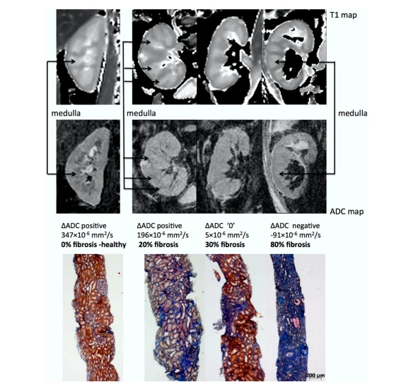
Figure 7. Representative biopsy and MR images in chronic kidney disease patients. MorphologicalMOLLI Tl maps were used for the positioning of the regions of interest (top row) and ADC maps (lower row) for three patients showing different AADC cases: positive, zero, and negative; along with the corresponding fibrosis levels from histology (Masson trichrome staining). The inversion of the corticomedullary ADC difference meets the increasing degree of renal fibrosis. Adapted from Figure 7 of Ref. [129] with permission. Copyright 2016 Springer Nature.
The exact physiological and mechanistic explanation for the link between DWI values and renal fibrosis is not fully understood but may be the result of two main synergetic events occurring in the development of renal fibrosis in CKD: (1) an increase in cellular density and the extra-cellular matrix which reduces free water motion and (2) a reduction of microvascular perfusion and filtration-induced water mobility expected with impaired
renal function(134]. Interestingly, the correlation of DWI with renal fibrosis is independent of the renal function as measured by eGFR, further supporting the role of DWI as a surrogate of renal fibrosis (127]. Whether DWI may allow reducing the number of renal biopsies by predicting the amount of interstitial fibrosis in CKD is currently not known and needs further evaluation.
Another potential application of DWI in CKD is related to the prediction of its evolution. Recently, three independent research groups observed that baseline DWI was correlated to the decline of renal function (126,135,136]. In a study with 197 CKD patients prediction of the worst renal outcome as shown in Figure 8 was independent of baseline age, sex, eGFR, and proteinuria, strongly suggesting that DWl could be an independent prognostic marker of CKD.

Figure 8. Kaplan-Meier survival curves, stratified according to the corticomedullary difference of the apparent diffusion coefficient AADC in chronic kidney disease patients. The primary outcome was a decline of eGFR > 30% or renal replacement therapy. A negative AADC was associated with a rapid decline in renal function. Cl, confidence interval; HR, hazard ratio. Adapted from Figure 2 ofRef.[126] with permission. Copyright 2016 Springer Nature.
In conclusion, DWI shows strong promise to characterize renal tumors, detect APN, and quantify renal fibrosis in diabetic and CKD patients. With the ongoing clinical trials as well as international efforts for methodological uniformization (137], its diagnostic and predictive power is likely to be significantly improved. The use of DWI may be further amplified by the development of multiparametric MRI allowing simultaneous ad ditionalassessment of renal blood flow and oxygenation as well as other morphometric parameters such as the relaxation time Tl on top of DWI (128].
Study Limitations
This review is based on a literature analysis performed using the PubMed database. For each genitourinary organ, the most relevant studies are in terms of recency and representation. native impact in the field of DWI was selected. Queries were performed by searching the database for keywords regarding the involved organ and involving diffusion imaging and/or MRI. However, this work does not report studies in the way a structured meta-analysis does by including scientific articles meeting systematic criteria. The selected works’ pertinence was established from the viewpoint of our actual practice and from the MR examinations routinely performed at our institution. In such a setting, the study selection contains an acknowledged part of subjectivity mostly relying on the authors’ combined experience.

Cistanche tubulosa
Conclusions
DWI is already a well-established imaging technique for the assessment of the genitourinary system with the potential for significant improvement. In female pelvis cancer and prostate cancer, DWI is part of the international guideline for the diagnosis, staging, and assessment of recurrence. DWI is even a determinant in the standardized risk scores O-RADS and PI-RADS. In the kidney, the most promising applications of DWI may be the quantification of renal fibrosis and the prediction of disease evolution in CKD. To further integrate DWI into the diagnosis strategies and patient management, standardizing the acquisition and analysis protocols will be the next challenge.
References
43. Blackledge, M.D.; Leach, M.; Collins, D.; Koh, D.-M. Computed Diffusion-weighted MR Imaging May Improve Tumor Detection. Radiology 2011, 261, 573–581. [CrossRef] [PubMed]
44. Lim, H.K.; Kim, J.K.; Kim, K.A.; Cho, K.-S. Prostate Cancer: Apparent Diffusion Coefficient Map with T2-weighted Images for Detection—A Multireader Study. Radiology 2009, 250, 145–151. [CrossRef] [PubMed]
45. American College of Radiology. PIRADS Prostate Imaging—Reporting and Data System v2.1. 2019. Available online: https: //www.acr.org/-/media/ACR/Files/RADS/Pi-RADS/PIRADS-V2-1.pdf (accessed on 1 February 2021).
46. Pesapane, F.; Acquasanta, M.; Di Meo, R.; Agazzi, G.M.; Tantrige, P.; Codari, M.; Schiaffino, S.; Patella, F.; Esseridou, A.; Sardanelli, F. Comparison of Sensitivity and Specificity of Biparametric versus Multiparametric Prostate MRI in the Detection of Prostate Cancer in 431 Men with Elevated Prostate-Specific Antigen Levels. Diagnostics 2021, 11, 1223. [CrossRef] [PubMed]
47. Wichtmann, B.D.; Zöllner, F.G.; Attenberger, U.I.; Schönberg, S.O. Multiparametric MRI in the Diagnosis of Prostate Cancer: Physical Foundations, Limitations, and Prospective Advances of Diffusion-Weighted MRI. RöFo-Fortschr. Geb. Röntgenstrahlen Bildgeb. Verfahr. 2021, 193, 399–409. [CrossRef]
48. Quentin, M.; Blondin, D.; Klasen, J.; Lanzman, R.S.; Miese, F.-R.; Arsov, C.; Albers, P.; Antoch, G.; Wittsack, H.-J. Comparison of different mathematical models of diffusion-weighted prostate MR imaging. Magn. Reson. Imaging 2012, 30, 1468–1474. [CrossRef]
49. Bourne, R.M.; Panagiotaki, E.; Bongers, A.; Sved, P.; Watson, G.; Alexander, D.C. Information theoretic ranking of four models of diffusion attenuation in fresh and fixed prostate tissue ex vivo. Magn. Reson. Med. 2013, 72, 1418–1426. [CrossRef]
50. Liang, S.; Panagiotaki, E.; Bongers, A.; Shi, P.; Sved, P.; Watson, G.; Bourne, R. Information-based ranking of 10 compartment models of diffusion-weighted signal attenuation in fixed prostate tissue. NMR Biomed. 2016, 29, 660–671. [CrossRef]
51. Le Bihan, D.; Breton, E.; Lallemand, D.; Aubin, M.L.; Vignaud, J.; Laval-Jeantet, M. Separation of diffusion and perfusion in intravoxel incoherent motion MR imaging. Radiology 1988, 168, 497–505. [CrossRef]
52. Mulkern, R.V.; Barnes, A.S.; Haker, S.J.; Hung, Y.P.; Rybicki, F.J.; Maier, S.E.; Tempany, C.M. Biexponential characterization of prostate tissue water diffusion decay curves over an extended b-factor range. Magn. Reson. Imaging 2006, 24, 563–568. [CrossRef]
53. Brancato, V.; Cavaliere, C.; Salvatore, M.; Monti, S. Non-Gaussian models of diffusion-weighted imaging for detection and characterization of prostate cancer: A systematic review and meta-analysis. Sci. Rep. 2019, 9, 16837. [CrossRef] [PubMed]
54. Döpfert, J.; Lemke, A.; Weidner, A.; Schad, L.R. Investigation of prostate cancer using diffusion-weighted intravoxel incoherent motion imaging. Magn. Reson. Imaging 2011, 29, 1053–1058. [CrossRef] [PubMed]
55. Park, H.; Kim, S.H.; Lee, Y.; Son, J.H. Comparison of diagnostic performance between diffusion kurtosis imaging parameters and mono-exponential ADC for determination of clinically significant cancer in patients with prostate cancer. Abdom. Radiol. 2020, 45, 4235–4243. [CrossRef]
56. Gibbs, P.; Liney, G.P.; Pickles, M.D.; Zelhof, B.; Rodrigues, G.; Turnbull, L.W. Correlation of ADC and T2 Measurements with Cell Density in Prostate Cancer at 3.0 Tesla. Investig. Radiol. 2009, 44, 572–576. [CrossRef] [PubMed]
57. Kwak, J.T.; Sankineni, S.; Xu, S.; Turkbey, B.; Choyke, P.L.; Pinto, P.A.; Merino, M.J.; Wood, B.J. Correlation of magnetic resonance imaging with digital histopathology in the prostate. Int. J. Comput. Assist. Radiol. Surg. 2016, 11, 657–666. [CrossRef] [PubMed]
58. Dhatt, R.; Choy, S.; Co, S.J.; Ischia, J.; Kozlowski, P.; Harris, A.C.; Jones, E.C.; Black, P.C.; Goldenberg, S.L.; Chang, S.D. MRI of the Prostate with and Without Endorectal Coil at 3 T: Correlation with Whole-Mount Histopathologic Gleason Score. Am. J. Roentgenol. 2020, 215, 133–141. [CrossRef]
59. Surov, A.; Meyer, H.J.; Wienke, A. Correlations between Apparent Diffusion Coefficient and Gleason Score in Prostate Cancer: A Systematic Review. Eur. Urol. Oncol. 2020, 3, 489–497. [CrossRef]
60. Barentsz, J.O.; Richenberg, J.; Clements, R.; Choyke, P.; Verma, S.; Villeirs, G.; Rouviere, O.; Logager, V.; Fütterer, J.J. ESUR prostate MR guidelines 2012. Eur. Radiol. 2012, 22, 746–757. [CrossRef] [PubMed]
61. Turkbey, B.; Rosenkrantz, A.B.; Haider, M.A.; Padhani, A.R.; Villeirs, G.; Macura, K.J.; Tempany, C.M.; Choyke, P.L.; Cornud, F.; Margolis, D.J.; et al. Prostate Imaging Reporting and Data System Version 2.1: 2019 Update of Prostate Imaging Reporting and Data System Version 2. Eur. Urol. 2019, 76, 340–351. [CrossRef]
62. Weinreb, J.C.; Barentsz, J.O.; Choyke, P.L.; Cornud, F.; Haider, M.A.; Macura, K.J.; Margolis, D.; Schnall, M.D.; Shtern, F.; Tempany, C.M.; et al. PI-RADS Prostate Imaging—Reporting and Data System: 2015, Version 2. Eur. Urol. 2016, 69, 16–40. [CrossRef]
63. Tsai, W.C.; Field, L.; Stewart, S.; Schultz, M. Review of the accuracy of multi-parametric MRI prostate in detecting prostate cancer within a local reporting service. J. Med Imaging Radiat. Oncol. 2020, 64, 379–384. [CrossRef] [PubMed]
64. Felker, E.R.; Margolis, D.J.; Nassiri, N.; Marks, L.S. Prostate cancer risk stratification with magnetic resonance imaging. Urol. Oncol. Semin. Orig. Investig. 2016, 34, 311–319. [CrossRef]
65. Fütterer, J.J.; Briganti, A.; De Visschere, P.; Emberton, M.; Giannarini, G.; Kirkham, A.; Taneja, S.S.; Thoeny, H.; Villeirs, G.; Villers, A. Can Clinically Significant Prostate Cancer Be Detected with Multiparametric Magnetic Resonance Imaging? A Systematic Review of the Literature. Eur. Urol. 2015, 68, 1045–1053. [CrossRef] [PubMed]
66. Kwon, M.-R.; Kim, C.K.; Kim, J.-H. PI-RADS version 2: Evaluation of diffusion-weighted imaging interpretation between b = 1000 and b = 1500 s mm–2. Br. J. Radiol. 2017, 90, 20170438. [CrossRef]
67. Rosenkrantz, A.B.; Ginocchio, L.; Cornfeld, D.; Froemming, A.T.; Gupta, R.T.; Turkbey, B.; Westphalen, A.C.; Babb, J.; Margolis, D.J. Interobserver Reproducibility of the PI-RADS Version 2 Lexicon: A Multicenter Study of Six Experienced Prostate Radiologists. Radiology 2016, 280, 793–804. [CrossRef]
68. Muller, B.G.; Shih, J.H.; Sankineni, S.; Marko, J.; Rais-Bahrami, S.; George, A.K.; De La Rosette, J.J.M.C.H.; Merino, M.J.; Wood, B.; Pinto, P.; et al. Prostate Cancer: Interobserver Agreement and Accuracy with the Revised Prostate Imaging Reporting and Data System at Multiparametric MR Imaging. Radiology 2015, 277, 741–750. [CrossRef]
69. Giganti, F.; Kirkham, A.; Kasivisvanathan, V.; Papoutsaki, M.-V.; Punwani, S.; Emberton, M.; Moore, C.M.; Allen, C. Understanding PI-QUAL for prostate MRI quality: A practical primer for radiologists. Insights Into Imaging 2021, 12, 59. [CrossRef] [PubMed]
70. de Rooij, M.; Israël, B.; Tummers, M.; Ahmed, H.U.; Barrett, T.; Giganti, F.; Hamm, B.; Løgager, V.; Padhani, A.; Panebianco, V.; et al. ESUR/ESUI consensus statements on multi-parametric MRI for the detection of clinically significant prostate cancer: Quality requirements for image acquisition, interpretation, and radiologists’ training. Eur. Radiol. 2020, 30, 5404–5416. [CrossRef]
71. Gatti, M.; Faletti, R.; Calleris, G.; Giglio, J.; Berzovini, C.; Gentile, F.; Marra, G.; Misischi, F.; Molinaro, L.; Bergamasco, L.; et al. Prostate cancer detection with parametric magnetic resonance imaging (bpMRI) by readers with different experience: Performance and comparison with multiparametric (mpMRI). Abdom. Radiol. 2019, 44, 1883–1893. [CrossRef]
72. Bertelli, E.; Mercatelli, L.; Marzi, C.; Pachetti, E.; Baccini, M.; Barucci, A.; Colantonio, S.; Gherardini, L.; Lattavo, L.; Pascali, M.A.; et al. Machine and Deep Learning Prediction of Prostate Cancer Aggressiveness Using Multiparametric MRI. Front. Oncol. 2022, 11, 802964. [CrossRef]
73. Hosseinzadeh, M.; Saha, A.; Brand, P.; Slootweg, I.; de Rooij, M.; Huisman, H. Deep learning–assisted prostate cancer detection on bi-parametric MRI: Minimum training data size requirements and effect of prior knowledge. Eur. Radiol. 2021, 32, 2224–2234. [CrossRef] [PubMed]
74. Siddiqui, M.; Rais-Bahrami, S.; Turkbey, B.; George, A.K.; Rothwax, J.; Shakir, N.; Okoro, C.; Raskolnikov, D.; Parnes, H.L.; Linehan, W.M.; et al. Comparison of MR/Ultrasound Fusion–Guided Biopsy with Ultrasound-Guided Biopsy for the Diagnosis of Prostate Cancer. JAMA J. Am. Med Assoc. 2015, 313, 390–397. [CrossRef] [PubMed]
75. Boesen, L. Multiparametric MRI in detection and staging of prostate cancer. Dan. Med. J. 2017, 64, 5327.
76. Serefoglu, E.C.; Altinova, S.; Ugras, N.S.; Akincioglu, E.; Asil, E.; Balbay, D. How reliable is a 12-core prostate biopsy procedure in the detection of prostate cancer? Can. Urol. Assoc. J. 2013, 7, 293. [CrossRef]
77. Caglic, I.; Sushentsev, N.; Shah, N.; Warren, A.Y.; Lamb, B.W.; Barrett, T. Comparison of parametric versus multiparametric prostate MRI for the detection of extracapsular extension and seminal vesicle invasion in biopsy naïve patients. Eur. J. Radiol. 2021, 141, 109804. [CrossRef]
78. Venkatesan, A.M.; Mudairu-Dawodu, E.; Duran, C.; Stafford, R.J.; Yan, Y.; Wei, W.; Kundra, V. Detecting recurrent prostate Cancer using multiparametric MRI, the influence of PSA and Gleason grade. Cancer Imaging 2021, 21, 3. [CrossRef] [PubMed]
79. Panebianco, V.; Narumi, Y.; Altun, E.; Bochner, B.H.; Efstathiou, J.A.; Hafeez, S.; Huddart, R.; Kennish, S.; Lerner, S.; Montironi, R.; et al. Multiparametric Magnetic Resonance Imaging for Bladder Cancer: Development of VI-RADS (Vesical Imaging-Reporting and Data System). Eur. Urol. 2018, 74, 294–306. [CrossRef]
80. Juri, H.; Narumi, Y.; Panebianco, V.; Osuga, K. Staging of bladder cancer with multiparametric MRI. Br. J. Radiol. 2020, 93, 20200116. [CrossRef]
81. Barua, S.K.; Kaman, P.K.; Baruah, S.J.; TP, R.; Bagchi, P.K.; Sarma, D.; Singh, Y. Role of Diffusion-Weighted Magnetic Resonance Imaging (DWMRI) in Assessment of Primary Penile Tumor Characteristics and Its Correlations with Inguinal Lymph Node Metastasis: A Prospective Study. World J. Oncol. 2018, 9, 145–150. [CrossRef]
82. Lindquist, C.M.; Nikolaidis, P.; Mittal, P.K.; Miller, F.H. MRI of the penis. Abdom. Radiol. 2020, 45, 2001–2017. [CrossRef]
83. Tsili, A.C.; Sofikitis, N.; Stiliara, E.; Argyropoulou, M.I. MRI of testicular malignancies. Abdom. Radiol. 2019, 44, 1070–1082. [CrossRef] [PubMed]
84. Tsili, A. Apparent diffusion coefficient values and dynamic contrast enhancement patterns in differentiating seminomas from nonseminomatous testicular neoplasms. Eur. J. Radiol. 2015, 84, 1219–1226. [CrossRef]
85. Moreno, C.C.; Small, W.C.; Camacho, J.C.; Master, V.; Kokabi, N.; Lewis, M.; Hartman, M.; Mittal, P. Testicular Tumors: What Radiologists Need to Know—Differential Diagnosis, Staging, and Management. Radiographics 2015, 35, 400–415. [CrossRef] [PubMed]
86. Parenti, G.C.; Feletti, F.; Carnevale, A.; Uccelli, L.; Giganti, M. Imaging of the scrotum: Beyond sonography. Insights Imaging 2018, 9, 137–148. [CrossRef] [PubMed]
87. Tsili, A.C.; Bertolotto, M.; Turgut, A.T.; Dogra, V.; Freeman, S.; Rocher, L.; Belfield, J.; Studniarek, M.; Ntorkou, A.; Derchi, L.E.; et al. MRI of the scrotum: Recommendations of the ESUR Scrotal and Penile Imaging Working Group. Eur. Radiol. 2018, 28, 31–43. [CrossRef] [PubMed]
88. Emad-Eldin, S.; Salim, A.M.A.; Wahba, M.H.; ElAhwany, A.T.; Abdelaziz, O. The use of diffusion-weighted MR imaging in the functional assessment of the testes of patients with clinical varicocele. Andrologia 2019, 51, e13197. [CrossRef]
89. Ntorkou, A.; Tsili, A.C.; Goussia, A.; Astrakas, L.G.; Maliakas, V.; Sofikitis, N.; Argyropoulou, M.I. Testicular Apparent Diffusion Coefficient and Magnetization Transfer Ratio: Can These MRI Parameters Be Used to Predict Successful Sperm Retrieval in Nonobstructive Azoospermia? Am. J. Roentgenol. 2019, 213, 610–618. [CrossRef]
90. Caroli, A.; Schneider, M.; Friedli, I.; Ljimani, A.; De Seigneux, S.; Boor, P.; Gullapudi, L.; Kazmi, I.; Mendichovszky, I.A.; Notohamiprodjo, M.; et al. Diffusion-weighted magnetic resonance imaging to assess diffuse renal pathology: A systematic review and statement paper. Nephrol. Dial. Transplant. 2018, 33 (Suppl. S2), ii29–ii40. [CrossRef]
91. Jiang, K.; Ferguson, C.M.; Lerman, L.O. Noninvasive assessment of renal fibrosis by magnetic resonance imaging and ultrasound techniques. Transl. Res. 2019, 209, 105–120. [CrossRef]
92. Serter, A.; Onur, M.R.; Coban, G.; Yildiz, P.; Armagan, A.; Kocakoc, E. The role of diffusion-weighted MRI and contrast-enhanced MRI for differentiation between solid renal masses and renal cell carcinoma subtypes. Abdom. Radiol. 2021, 46, 1041–1052. [CrossRef] [PubMed]
93. Tordjman, M.; Mali, R.; Madelin, G.; Prabhu, V.; Kang, S. Diagnostic test accuracy of ADC values for identification of clear cell renal cell carcinoma: Systematic review and meta-analysis. Eur. Radiol. 2020, 30, 4023–4038. [CrossRef] [PubMed]
94. Zhu, J.; Luo, X.; Gao, J.; Li, S.; Li, C.; Chen, M. Application of diffusion kurtosis tensor MR imaging in the characterization of renal cell carcinomas with different pathological types and grades. Cancer Imaging 2021, 21, 30. [CrossRef] [PubMed]
95. Farhadi, F.; Nikpanah, M.; Paschall, A.K.; Shafiei, A.; Tadayoni, A.; Ball, M.W.; Linehan, W.M.; Jones, E.C.; Malayeri, A.A. Clear Cell Renal Cell Carcinoma Growth Correlates with Baseline Diffusion-weighted MRI in Von Hippel–Lindau Disease. Radiology 2020, 295, 583–590. [CrossRef] [PubMed]
96. Goh, V.; Prezzi, D. Predicting Growth Kinetics in Hereditary Renal Cancer with Diffusion-weighted MRI. Radiology 2020, 295, 591–592. [CrossRef] [PubMed]
97. Vivier, P.-H.; Salem, A.; Beurdeley, M.; Lim, R.P.; Leroux, J.; Caudron, J.; Coudray, C.; Liard, A.; Michelet, I.; Dacher, J.-N. MRI and suspected acute pyelonephritis in children: Comparison of diffusion-weighted imaging with gadolinium-enhanced T1-weighted imaging. Eur. Radiol. 2013, 24, 19–25. [CrossRef]
98. Thoeny, H.C.; De Keyzer, F. Diffusion-weighted MR Imaging of Native and Transplanted Kidneys. Radiology 2011, 259, 25–38. [CrossRef]
99. Faletti, R.; Cassinis, M.C.; Fonio, P.; Grasso, A.; Battisti, G.; Bergamasco, L.; Gandini, G. Diffusion–Weighted Imaging and Apparent Diffusion Coefficient Values Versus Contrast–Enhanced MR Imaging in the Identification and Characterisation of Acute Pyelonephritis. Eur. Radiol. 2013, 23, 3501–3508. [CrossRef]
100. Sriman, R.; Venkatesh, K.; Mathew, C.; Pankaj, M.; Shankar, R. Validity of diffusion-weighted magnetic resonance imaging in the evaluation of acute pyelonephritis in comparison with contrast-enhanced computed tomography. Pol. J. Radiol. 2020, 85, e137–e143.
101. Aoyagi, J.; Kanai, T.; Odaka, J.; Ito, T.; Saito, T.; Betsui, H.; Furukawa, R.; Nakata, W.; Yamagata, T. Non-enhanced magnetic resonance imaging versus renal scintigraphy in acute pyelonephritis. Pediatr. Int. 2018, 60, 200–203. [CrossRef]
102. Bosáková, A.; Šalounová, D.; Havelka, J.; Kraft, O.; Sirucek, P.; Kocvara, R.; Hladik, M. Diffusion-weighted magnetic resonance imaging is more sensitive than dimercaptosuccinic acid scintigraphy in detecting parenchymal lesions in children with acute pyelonephritis: A prospective study. J. Pediatr. Urol. 2018, 14, 269.e1–269.e7. [CrossRef]
103. Simrén, Y.; Stokland, E.; Hansson, S.; Sixt, R.; Svensson, P.-A.; Lagerstrand, K. Diffusion-weighted imaging is a promising method to detect acute pyelonephritis in non-sedated free breathing infants. J. Pediatr. Urol. 2020, 16, 320–325. [CrossRef] [PubMed]
104. Damasio, M.B.; Müller, L.-S.O.; Augdal, T.A.; Avni, F.E.; Basso, L.; Bruno, C.; Kljuˇcevšek, D.; Littooij, A.S.; Franchi-Abella, S.; Lobo, L.M.; et al. European Society of Paediatric Radiology abdominal imaging task force: Recommendations for contrast-enhanced ultrasound and diffusion-weighted imaging in focal renal lesions in children. Pediatr. Radiol. 2019, 50, 297–304. [CrossRef] [PubMed]
105. Li, Y.; Lee, M.M.; Worters, P.W.; MacKenzie, J.D.; Laszik, Z.; Courtier, J.L. Pilot Study of Renal Diffusion Tensor Imaging as a Correlate to Histopathology in Pediatric Renal Allografts. Am. J. Roentgenol. 2017, 208, 1358–1364. [CrossRef] [PubMed]
106. Lanzman, R.S.; Ljimani, A.; Pentang, G.; Zgoura, P.; Zenginli, H.; Kröpil, P.; Heusch, P.; Schek, J.; Miese, F.R.; Blondin, D.; et al. Kidney Transplant: Functional Assessment with Diffusion-Tensor MR Imaging at 3T. Radiology 2013, 266, 218–225. [CrossRef] [PubMed]
107. Hueper, K.; Gutberlet, M.; Rodt, T.; Gwinner, W.; Lehner, F.; Wacker, F.; Galanski, M.; Hartung, D. Diffusion tensor imaging and tractography for assessment of renal allograft dysfunction—initial results. Eur. Radiol. 2011, 21, 2427–2433. [CrossRef] [PubMed]
108. Fan, W.-J.; Ren, T.; Li, Q.; Zuo, P.-L.; Long, M.-M.; Mo, C.-B.; Chen, L.-H.; Huang, L.-X.; Shen, W. Assessment of renal allograft function early after transplantation with isotropic resolution diffusion tensor imaging. Eur. Radiol. 2015, 26, 567–575. [CrossRef]
109. Eisenberger, U.; Thoeny, H.C.; Binser, T.; Gugger, M.; Frey, F.J.; Boesch, C.; Vermathen, P. Evaluation of renal allograft function early after transplantation with diffusion-weighted MR imaging. Eur. Radiol. 2010, 20, 1374–1383. [CrossRef]
110. Abou-El-Ghar, M.E.; El-Diasty, T.A.; El-Assmy, A.M.; Refaie, H.F.; Refaie, A.F.; Ghoneim, M.A. Role of diffusion-weighted MRI in the diagnosis of acute renal allograft dysfunction: A prospective preliminary study. Br. J. Radiol. 2012, 85, e206–e211. [CrossRef]
111. Steiger, P.; Barbieri, S.; Kruse, A.; Ith, M.; Thoeny, H.C. Selection for biopsy of kidney transplant patients by diffusion-weighted MRI. Eur. Radiol. 2017, 27, 4336–4344. [CrossRef]
112. Ni, X.; Wang, W.; Li, X.; Li, Y.; Chen, J.; Shi, D.; Wen, J. Utility of Diffusion-Weighted Imaging for Guiding Clinical Management of Patients with Kidney Transplant: A Prospective Study. J. Magn. Reson. Imaging 2020, 52, 565–574. [CrossRef]
113. Hussain, S.M. Editorial for “Utility of Diffusion-Weighted Imaging for Guiding Clinical Management of Patients with Kidney Transplant: A Prospective Study”. J. Magn. Reson. Imaging 2020, 52, 575–576. [CrossRef] [PubMed]
114. Chen, X.; Xiao, W.; Li, X.; He, J.; Huang, X.; Tan, Y. In vivo, evaluation of renal function using diffusion-weighted imaging and diffusion tensor imaging in type 2 diabetics with normoalbuminuria versus microalbuminuria. Front. Med. 2014, 8, 471–476. [CrossRef] [PubMed]
115. Lü, L.; Sedor, J.R.; Gulani, V.; Schelling, J.R.; O’Brien, A.; Flask, C.A.; Dell, K.M. Use of Diffusion Tensor MRI to Identify Early Changes in Diabetic Nephropathy. Am. J. Nephrol. 2011, 34, 476–482. [CrossRef] [PubMed]
116. Razek, A.A.K.A.; Al-Adlany, M.A.A.A.; Alhadidy, A.M.; Atwa, M.A.; Abdou, N.E.A. Diffusion tensor imaging of the renal cortex in diabetic patients: Correlation with urinary and serum biomarkers. Abdom. Radiol. 2017, 42, 1493–1500. [CrossRef]
117. Cakmak, P.; Yagci, A.B.; Dursun, B.; Herek, D.; Fenkci, S.M. Renal diffusion-weighted imaging in diabetic nephropathy: Correlation with clinical stages of the disease. Diagn. Interv. Radiol. 2014, 20, 374–378. [CrossRef]
118. Carbone, S.F.; Gaggioli, E.; Ricci, V.; Mazzei, F.; Mazzei, M.A.; Volterrani, L. Diffusion-weighted magnetic resonance imaging in the evaluation of renal function: A preliminary study. Radiol. Med. 2007, 112, 1201–1210. [CrossRef]
119. Ding, J.; Chen, J.; Jiang, Z.; Zhou, H.; Di, J.; Xing, W. Assessment of renal dysfunction with diffusion-weighted imaging: Comparing intra-voxel incoherent motion (IVIM) with a mono-exponential model. Acta Radiol. 2015, 57, 507–512. [CrossRef]
120. Özçelik, Ü.; Çevik, H.; Bircan, H.Y.; Karakayalı, F.Y.; I¸sıklar, I.; Haberal, M. Evaluation of Transplanted Kidneys and Comparison with Healthy Volunteers and Kidney Donors with Diffusion-Weighted Magnetic Resonance Imaging: Initial Experience. Exp. Clin. Transpl. 2017. [CrossRef]
121. Emre, T.; Kiliçkesmez, Ö.; Büker, A.; Inal, B.B.; Do ˘gan, H.; Ecder, T. Renal function, and diffusion-weighted imaging: A new method to diagnose kidney failure before losing half function. Radiol. Med. 2016, 121, 163–172. [CrossRef]
122. Xu, X.; Fang, W.; Ling, H.; Chai, W.; Chen, K. Diffusion-weighted MR imaging of kidneys in patients with chronic kidney disease: Initial study. Eur. Radiol. 2010, 20, 978–983. [CrossRef]
123. Yalçin- ¸Safak, K.; Ayyildiz, M.; Ünel, S.Y.; Umarusman-Tanju, N.; Akça, A.; Baysal, T. The relationship of ADC values of renal parenchyma with CKD stage and serum creatinine levels. Eur. J. Radiol. Open 2016, 3, 8–11. [CrossRef] [PubMed]
124. Liu, H.; Zhou, Z.; Li, X.; Li, C.; Wang, R.; Zhang, Y.; Niu, G. Diffusion-weighted imaging for staging chronic kidney disease: A meta-analysis. Br. J. Radiol. 2018, 91, 20170952. [CrossRef] [PubMed]
125. Beck-Tölly, A.; Eder, M.; Beitzke, D.; Eskandary, F.; Agibetov, A.; Lampichler, K.; Hamböck, M.; Regele, H.; Kläger, J.; Nackenhorst, M.; et al. Magnetic Resonance Imaging for Evaluation of Interstitial Fibrosis in Kidney Allografts. Transpl. Direct 2020, 6, e577. [CrossRef] [PubMed]
126. Berchtold, L.; Crowe, L.A.; Combescure, C.; Kassaï, M.; Aslam, I.; Legouis, D.; Moll, S.; Martin, P.-Y.; de Seigneur, S.; Vallée, J.-P. Diffusion-magnetic resonance imaging predicts the decline of kidney function in chronic kidney disease and in patients with a kidney allograft. Kidney Int. 2022, 101, 804–813. [CrossRef] [PubMed]
127. Berchtold, L.; Friedli, I.; Crowe, L.A.; Martinez, C.; Moll, S.; Hadaya, K.; De Perrot, T.; Combescure, C.; Martin, P.-Y.; Vallée, J.-P.; et al. Validation of the corticomedullary difference in magnetic resonance imaging-derived apparent diffusion coefficient for kidney fibrosis detection: A cross-sectional study. Nephrol. Dial. Transpl. 2020, 35, 937–945. [CrossRef]
128. Buchanan, C.E.; Mahmoud, H.; Cox, E.F.; McCulloch, T.; Prestwich, B.L.; Taal, M.W.; Selby, N.; Francis, S.T. Quantitative assessment of renal structural and functional changes in chronic kidney disease using multi-parametric magnetic resonance imaging. Nephrol. Dial. Transpl. 2019, 35, 955–964. [CrossRef]
129. Friedli, I.; Crowe, L.A.; Berchtold, L.; Moll, S.; Hadaya, K.; De Perrot, T.; Vesin, C.; Martin, P.-Y.; De Seigneux, S.; Vallée, J.-P. New Magnetic Resonance Imaging Index for Renal Fibrosis Assessment: A Comparison between Diffusion-Weighted Imaging and T1 Mapping with Histological Validation. Sci. Rep. 2016, 6, 30088. [CrossRef]
130. Inoue, T.; Kozawa, E.; Okada, H.; Inukai, K.; Watanabe, S.; Kikuta, T.; Watanabe, Y.; Takenaka, T.; Katayama, S.; Tanaka, J.; et al. Noninvasive Evaluation of Kidney Hypoxia and Fibrosis Using Magnetic Resonance Imaging. J. Am. Soc. Nephrol. 2011, 22, 1429–1434. [CrossRef]
131. Mao, W.; Ding, Y.; Ding, X.; Fu, C.; Zeng, M.; Zhou, J. Diffusion kurtosis imaging for the assessment of renal fibrosis of chronic kidney disease: A preliminary study. Magn. Reson. Imaging 2021, 80, 113–120. [CrossRef]
132. Zhang, J.; Yu, Y.; Liu, X.; Tang, X.; Xu, F.; Zhang, M.; Xie, G.; Zhang, L.; Li, X.; Liu, Z.-H. Evaluation of Renal Fibrosis by Mapping Histology and Magnetic Resonance Imaging. Kidney Dis. 2021, 7, 131–142. [CrossRef]
133. Zhao, J.; Wang, Z.; Liu, M.; Zhu, J.; Zhang, X.; Zhang, T.; Li, S.; Li, Y. Assessment of renal fibrosis in chronic kidney disease using diffusion-weighted MRI. Clin. Radiol. 2014, 69, 1117–1122. [CrossRef] [PubMed]
134. Akashi, T.; Terayama, N.; Okada, E. Solitary Renal Pelvic Lesion as the Primary Manifestation of Hodgkin Lymphoma: A Case Report. Urol. Case Rep. 2017, 13, 87–88. [CrossRef] [PubMed]
135. Liu, Y.; Zhang, G.-M.-Y.; Peng, X.; Li, X.; Sun, H.; Chen, L. Diffusion kurtosis imaging as an imaging biomarker for predicting prognosis in chronic kidney disease patients. Nephrol. Dial. Transpl. 2021, gfab229. [CrossRef] [PubMed]
136. Srivastava, A.; Cai, X.; Lee, J.; Li, W.; Larive, B.; Kendrick, C.; Gassman, J.J.; Middleton, J.P.; Carr, J.; Raphael, K.L.; et al. Kidney Functional Magnetic Resonance Imaging and Change in eGFR in Individuals with CKD. Clin. J. Am. Soc. Nephrol. 2020, 15, 776–783. [CrossRef]
137. Ljimani, A.; Caroli, A.; Laustsen, C.; Francis, S.; Mendichovszky, I.A.; Bane, O.; Nery, F.; Sharma, K.; Pohlmann, A.; Dekkers, I.A.; et al. Consensus-based technical recommendations for clinical translation of renal diffusion-weighted MRI. Magn. Reson. Mater. Phys. Biol. Med. 2020, 33, 177–195. [CrossRef]
Thomas De Perrot 1, Christine Sadjo Zoua 1 , Carl G. Glessgen 1 , Diomidis Botsikas 1 , Lena Berchtold 2 , Rares Salomir 1 , Sophie De Seigneux 2 , Harriet C. Thoeny 3 and Jean-Paul Vallée 1
1 Division of Radiology, Geneva University Hospitals and University of Geneva, 1205 Geneva, Switzerland; christine.sadjo@hcuge.ch (C.S.Z.); carl.glessgen@hcuge.ch (C.G.G.); diomidis.botsikas@hcuge.ch (D.B.); raresvincent.salomir@hcuge.ch (R.S.); jean-paul.vallee@hcuge.ch (J.-P.V.)
2 Division of Nephrology, Geneva University Hospitals, 1205 Geneva, Switzerland; lena.berchtold@hcuge.ch (L.B.); sophie.deseigneux@hcuge.ch (S.D.S.)
3 Division of Radiology, Hôpital Cantonal Fribourgois, 1752 Villars-sur-Glâne, Switzerland; harriet.thoeny@h-fr.ch





