

Quantitative Proteomics Reveals Significant Differences Between Mouse Brain Formations 2
Aug 23, 2022
Please contact oscar.xiao@wecistanche.com for more information
12. Cholinergic Transmission
Acetylcholine transmission is mediated by two classes of receptors: the nicotinic receptors, which are acetylcholine-gated ion channels for sodium cations, and muscarinic receptors, which are metabotropic receptors coupled to the activity of trimeric G proteins.
Unexpectedly, in our study, we were not able to detect peptides that could be unequivocally attributed to nicotinic receptors. On the other hand, we determined the titer of several members of muscarinic cholinergic receptors. 12.1.Muscarinic Receptors——Charm
In the hippocampus and cortex, but not in the cerebellum, we measured the titer of Chrml, Chrm3, and Chrm4 (Figure 5A-C).In both brain formations, Chrml was the most abundant muscarinic receptor subunit. Aging reduced the level of Chrml and Chrm3 in the hippocampus, whereas in the cortex, the concentrations of Chrm3 and Chrm4 were decreased (Figure 5A, B).

12.2.Acetylcholine Metabolism
Choline acetyltransferase(Chat) is an enzyme involved in acetylcholine synthesis. We found that its expression was 2-fold increased in the hippocampus of aged animals (Figure 5A). In contrast to Chat, the titer of acetylcholine esterase(Ache),an enzyme de-grading the neurotransmitter, was not affected by aging in this brain structure(Figure 5A).

Please click here to know more
In the cortex, the concentrations of Chat and Ache were significantly higher than in the hippocampus and cerebellum; however, they were not influenced by aging (Figure 5A-C).
We also did not observe age-related changes in the concentrations of Chat and Ache in the cerebellum, where the amount of the proteins was the lowest from all the analyzed brain regions.
12.3.Vesicular Acetylcholine Transporter—Slc18a3
Slc18a3 is a protein responsible for acetylcholine loading into synaptic vesicles. cistanche life extension Our study reveals that the concentration of Slc18a3 was the highest in the cortex of young mice (Supplementary Table S1),and that aging reduced the level of Slc18a3 by about 80%(Figure 5B).In the hippocampus, the protein amount was not affected by aging (Figure 5A), and in the cerebellum, the protein was detected only in aged animals(Supplementary Table S1).
13. Monoamines Receptors,Signal Transmission and Metabolism
13.1.Receptors
Although we could measure the titer of several membrane proteins involved in monoamine signaling (e.g, in monoamine reuptake), we detected only a few members of monoamine receptors. We did not find peptides specifically attributed to dopamine receptors, and among numerous groups of serotonin receptors, we were able to unequivocally measure the titer of only one serotonin receptor (Htrla) in the hippocampus of old animals (Supplementary Table S1).
In the hippocampus, we measured the titer of two members of alpha-adrenergic receptors: a2a(Adra2a)and a2c(Adra2c)(Figure 6A).Their concentration was lower in old animals; however, the changes were notstatistically significant. In the cortex, Adra2c was present both in young and in old animals, but AdraZa was expressed only in young animals (Figure 6B).In the cerebellum, the only detected alpha-adrenergic receptor was AdraZa in young animals (Figure 6C).In this study, we were not able to unequivocally assign any peptides to beta-adrenergic receptor proteins, Adrb.
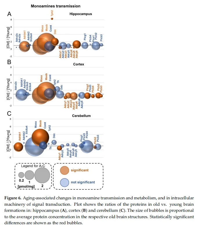
In contrast to beta-adrenergic receptors, we found a significant decrease in the expression of kinases involved in the desensitization of these receptors, Adrbkl, in the hippocampus and cerebellum (Figure 6A-C). cistanche nz This suggests that beta-adrenergic receptors may be ubiquitously expressed in the brain, but because of the methodology used in our experiment, the peptides related to Adrb receptors could not have been assigned to the proteins.

cistanche can anti-aging
13.2.Monoamine Reuptake
The concentration of the serotonin transporter (Slc6a4) responsible for the neurotransmitter reuptake was present in all the studied brain structures and was practically unaffected by aging (Figure 6A-C). We obselrved a statistically significant increase in Slc6a4 in the hippocampus of aged mice; however, this increase was very low (Figure 6A).In contrast to the hippocampus, we found about a 3.5 times higher titer of Slc6a4 in aged cerebella, but the increase was not statistically significant (Figure 6C).
We determined the titer of a dopamine transporter (Slc6a3) in the cortex (Figure 6B). Its concentration was not affected by aging.
14.Monoamine Deactivation
14.1.Monoamine Oxidase—Mao
Monoamine oxidase catalyzes the deamination of amines and is involved in the degradation of monoamines released by neurons and glia cells. There are two isoforms of Mao, Maoa and Maob, whose expression is attributed, respectively, to neurons and glial cells [36].
The results of our study reveal that both Mao isoenzymes were ubiquitously expressed in all the brain formations (Figure 6A-C).In the hippocampus, the concentration of Maoa was reduced by aging, while the level of Maob was significantly elevated in the aged animals (Figure 6A).cistanche penis size The same trend could be observed for the Mao isoforms in the cortex (Figure6B).
In the cerebellum, the titer of Maoa was unaffected by aging, but the concentration of Maob was more than four times higher in old animals than in young ones (Figure 6C). 14.2.Catechol O-Methyltransferase——Comt
Catechol O-methyltransferase catalyzes O-methylation and, thus, the inactivation of monoamines such as adrenaline, dopamine and serotonin. In our analysis, we found that the enzyme was relatively abundant in all the studied brain formations, and that its level was not affected by aging(Figure 6A-C).
15.Monoamine Synthesis
15.1.Tryptophan Hydroxylase 2—Tph2
Tryptophan hydroxylase is the enzyme catalyzing the first step of serotonin synthesis. Our study shows that the level of Tph2 in all the brain structures of old animals was similar (Figure 6A-C).In the hippocampus of young animals, the titer of Tph2 was much lower than in other brain formations, but aging reslulted in a significant, more than8-fold, increase in the titer of the enzyme in this structure (Figure 6A). In the cortex and cerebellum, aging had no effect on the expression of the enzyme (Figure 6B,C).
15.2.Tyrosine Hydroxylase—Th
Th is involved in the first step of monoamine (such as dopamine, adrenaline and nora-drenaline) synthesis from tyrosine. The titer of Th was the highest in the cortex (Figure 6C), and the presence of the enzyme was not detected in the hippocampus (Figure 6A). cistanche powder Aging had no effect on the expression of Th in both brain formations (Figure 6B,C).

15.3. Aromatic-L-amino-acid decarboxylase—Ddc
Ddc (also known as DOPA decarboxylase and AADC, and 5-hydroxytryptophan decarboxylase) participates in neurotransmitter synthesis, catalyzing the decarboxylation of various substrates such as DOPA, phenylalanine, histidine and 5-hydroxytryptamine. In our studies, we found that Ddc was more than two times higher in the hippocampi of aged animals (Figure 6A), whereas in the cortex and cerebellum, the protein amount was not significantly modified by aging (Figure 6B,C).
16. Signal Transduction
Stimulation of several metabotropic receptors is associated with modification of the activity of adenylate cyclase and/or phospholipases and changes in the concentration of secondary messengers such as cAMP and inositol trisphosphates. 16.1.Adenylyl Cyclase——Adcy
Adcy is the enzyme that catalyzes the formation of cAMP from ATP,and its activity is regulated after stimulation of metabotropic cholinergic and catecholaminergic receptors. Our study reveals that in the hippocampus, Adcy2 and Adcy9 were the most abundant isoforms of the cyclase (Figure 6A). We found that the concentration of the main form of the enzyme in the hippocampus, Adcy9, was significantly increased in old mice, while the titer of Adcy1, Adcy3, Adcy6 and Adcy8 decreased in old animals (Figure 6A).Aging had no effect on the abundance of the main forms of Adcy, Adcy5 and Adcy9, in the cortex, but it reduced the titer of Adcy1, Adcy2, Adcy3 and Adcy6 (Figure 6B). We also observed a reduction in the main Adcy isoform in the cerebellum, Adcy (Figure 6C).
16.2.Phospholipase C—Plc
Phospholipase C (Ple), an enzyme that hydrolyzes phospholipids, is involved in intracellular signal transmission after stimulation of the muscarinic cholinergic receptor (Chrm) and alpha-adrenergic receptor(Adra1)[37].
In our study, we found several members of the Ple class in all the studied brain structures (Figure 6A-C). The most abundant Plc isoforms in the hippocampus were Plcgl and Plch2, whose titer was not affected by aging (Figure 6A). In contrast to the hippocampus, almost all Ple isoforms, except for Plcd3, were downregulated in the cortex of aged mice (Figure 6B).In the cerebellum, Plcb4 was the predominant isoform of Plc, but its titer was unaffected by aging (Figure 3C).The only Ple whose concentration was different in the cerebellum of old mice was Plch2(Figure 6C).
17.Cytomatrix Active Zone——CAZ
The CAZ is a presynaptic region involved in neurotransmitter release [38]. Pro-teins within this region mediate, directly and indirectly, synaptic vesicle fusion with the presynaptic membrane. Among this group of proteins,several functional classes may be distinguished: SNAREs (soluble N-ethylmaleimide-sensitive factor attachment protein re-ceptors), which can be divided into v-SNAREs (vesicle-associated SNAREs) and t-SNAREs (target membrane-associated SNAREs),and proteins involved in calcium-mediated dock-ing of synaptic vesicles [39].

In our analysis, we did not observe numerous significant age-related changes in the concentration of CAZ proteins in the hippocampus and cerebellum (Figure7A,C).The only age-affected proteins in the hippocampus were Snap25 and Vap, whose titers were lower in old mice (Figure7A). cistanche salsa extract On the other hand, the concentration of several CAZ proteins was significantly modified, usually elevated, by aging in the cortex (Figure 7B). The most prominent changes were related to v- and t-SNARE proteins such as syntaxins (Stx), synap-totagmine (Syt), synaptobrevins (Vamp), synaptogyrins (Syngr) and synaptophysins (Syp)(Figure7B).This was accompanied by an increase in the concentrations of proteins involved in the calcium-dependent machinery of synaptic vesicle docking and anchoring, such as
18.Postsynaptic Density——PSD
The postsynaptic density is the protein-rich region attached to the postsynaptic mem-brane where proteins involved in signal reception and transmission, as well as modulation of synaptic plasticity, are located [40]. In our study, we found several members of PSD proteins in all the studied brain structures (Supplementary Table S1), and changes in the titers of some of them (receptors, proteins involved in the synthesis of secondary messengers, etc.) are described in previous sections of the paper. The concentration of PSD proteins in the cerebellum was not significantly altered by aging. Psd and Shank3 were the only exceptions: their titer was, respectively, reduced and elevated in old animals (Figure 7C).In contrast to the cerebellum, aging significantly reduced the concentration of several members of PSD proteins in the cortex and hippocampus (Figure7A,B).Among them were such proteins important for synaptic plasticity as Dlg4/Psd95,Syngap1, Shankl and Shank2 in the cortex(Figure7B),and Psd,Dlg, Dlgap and Shank in the hippocampus (Figure7A).A detailed list of these changes is provided in Supplementary Table S1.
19. Trans-Synaptic Cell Adhesion Molecules—CAMs
Trans-synaptic cell adhesion molecules regulate synaptic plasticity via the organization of the synaptic connection, control synapse morphology and regulate receptor functions[41]
We quantitatively measured the titers of almost 140 proteins annotated to the trans-synaptic cell adhesion molecules (Supplementary Table S1). Overall, the level of most of the CAMs was affected by aging in the hippocampus and cortex (Figure 8A,B), but relatively small changes were observed in the cerebellum (Figure 8C). We observed a significant decrease in the concentration of several cell adhesion molecules such as cadherins (Cdh), catenins (Ctnn), ephrins (Eph), receptor-type tyrosine-protein phosphatase (Ptpr), neurexins (Nrxn) and the Lin7 protein, both in the hippocampus and in the cortex of old animals (Figure 8A,B).The full set of proteins presented in Figure 8 is described in Table 1, and the titer of various isoforms of Cdh, Ctnn, Eph and Nrxn is presented in Supplementary Table S1.
The most significant changes in the cerebellum were associated with the expression of Lgi, whose concentration was more than two times higher in old mice (Figure 8C).Lgis are secreted proteins regulating receptor distribution and cellular interactions in the nervous system [73]. Although in the cerebellum, the titer of most of the cell adhesion proteins was unaffected by aging, the titer of some of them such as ephrins (Eph), liprin-alpha (Ppfia)and neurexins (Nrxn) was reduced (Figure 8C). We also found a significant elevation in the Lgi protein in the hippocampus and cortex (Figure 8A-C).The roles of individual CAMs are summarized in Table 1.
20. Extracellular Matrix (ECM) Perineuronal Net (Proteins
PNNs are extracellular matrix structures that cover the surface of the neuronal cell body and protrusions in the central nervous system and stabilize synapses in adult brains. PNNs are composed mainly of chondroitin sulfate proteoglycans [74].
We found more than 70 members of the PNNs in young and old murine brain structures (Supplementary Table S1). Generally, we observed elevated levels of PNN proteins in aged brains, and these increases were attributed mainly to an enormous increase in Hapln (hyaluronan and proteoglycan link protein), the main component of PNNs (Figure9A-C). Except for Japan, the titer of collagens(Col) was increased in the hippocampus and cerebellum, and laminins (Lam) were elevated in all the studied brain structures (Figure 9A-C). Unexpectedly, we found that some of the PNN proteins were downregulated in aged mice, e.g, the concentrations of talin-2(TIn2) and semaphorins (Sema)in the hippocampus and cortex (Figure 9A,B).
On analyzing the ECM protein, we found an interesting dependence: the titers of most large proteoglycans (such as Agrn, Ncan, Vcan and Bcan) were significantly increased in the hippocampus and cerebellum of old animals, but not in the cortex (Figure 9A-C). The cortex also appears to be the most stable brain formation in the context of the expression of ECM remodeling enzymes. We found several members of ADAM proteases in all the studied brain structures. Only the ADAM23 concentration was elevated by aging in the cortex (Figure 9B), ADAM10, ADAM11 ADAM22, and ADAM23 were increased in the hippocampus of old mice, while the titers of ADAM22 and ADAM23 were elevated in the cortex (Figure 9A,C).
Among metalloproteinases, we could measure the concentration of only Mmp17, and it was slightly reduced by aging in the hippocampus and cerebellum (Figure 9A-C).The roles of CAM protein groups are summarized in Table2.
21. Discussion
Aging-associated changes in the protein composition of brain formations are still far from being well understood. Several valuable pieces of data have been delivered by studies using immunocytochemical and immunohistochemical techniques. They demonstrated a diverse expression of several proteins in various brain structures, and even in different populations of neurons [97-99]. However, because of the methodological limitations, such studies have been restricted to a small number of proteins, and they could deliver only semi-quantitative data on protein expression. They also could not deliver the real concentration of proteins, given in absolute values (e.g, in mol/g protein), in the studied samples.
In this paper, we used a mass spectrometry-based technique, the label-free total protein approach method, to quantitatively describe proteins involved in signal transmission in the hippocampus, cerebral cortex, and cerebellum of young and old mice. All brain formations are heterogeneous structures composed of various cells, among which neurons and astrocytes are the most abundant. Due to that, the results presented in this paper cannot be unequivocally annotated to neurons or glial cells. However, in several cases, the expression of the analyzed proteins is known to be related almost exclusively to one type of cell, e.g, GABA-synthetizing enzymes in the hippocampus, which are associated mainly with interneurons (for review, see [100]).
Our analysis demonstrated that the molecular machinery involved in the excitatory and inhibitory transmission (respectively, glutamatergic and GABAergic) in the hippocampus and cortex was significantly altered (reduced) by aging. In turn, the expression of glutamate and GABA receptors in the cerebellum was practically unchanged; however, the titer of GADs (the enzymes involved in GABA synthesis) was strongly elevated in aged mice.
The decreased concentration of proteins involved in glutamatergic and GABAergic transmission might indicate a decreased neuronal plasticity of the hippocampus and cortex in aged animals.
The decreased titer of Camk4 might be the next marker of the lower synaptic plasticity of old animals as Camk4 is a protein that is indispensable for the formation of long-term memory [101]. We observed not only a significant reduction in Cam4 in all the studied brain formations but also a very high level of this kinase in the cerebellum, which is in line with studies showing the substantial role of active Camk4 for cerebellar long-term depression, regarded to be the main form of synaptic plasticity in this brain structure[102-104]. We did not find any age-related differences in thetiter of Camk2, which is known to be directly involved in synaptic enhancement. However, Camk2 is a protein expressed at a very high level and involved in a variety of cellular events, and hence the lack of statistically significant changes in the whole structures is not unexpected.
We did not observe numerous changes in the concentration of other kinases involved in synaptic transmission and plasticity, such as PKA and Mark. However, we found that the concentration of Prkac, a catalytic subunit of PKA, is significantly reduced by aging in the hippocampus. This might suggest lower excitatory transmission and plasticity of aged hippocampi because PKA-dependent phosphorylation of the AMPA subunits directly controls the synaptic incorporation of AMPA receptors [105].
Unexpectedly, we were not able to measure the titers of nicotinic acetylcholine receptors (as well as dopamine and serotonin receptors, except for Htrla). Since we identified several other membrane proteins, the lack ofthese receptors in our analysis is not an effect of the sample preparation method but results from an actual absence of unique peptides that could be unequivocally annotated to those receptors.
In contrast to nicotinic receptors, we identified muscarinic acetylcholine receptors (Chrm) in the hippocampus and cortex, and we found that their titer was significantly reduced in old animals. Interestingly, the concentration of the enzyme Chat involved in acetylcholine synthesis was significantly increased in the hippocampus and cerebellum, and the cerebellum was the only brain structure in which we were not able to measure the Chrm concentration.
The most pronounced difference between young and old animals regarding adrenergic transmission was the very high increase in the titer of monoamine oxidase b, an enzyme responsible for the deactivation of amines. This may suggest that adrenergic transmission and, overall, the catecholaminergic signaling are reduced in old animals. However, we also found a significant aging-associated reduction in the Adrbkl concentration, a kinase that is involved in the desensitization of adrenergic receptors. The downregulation of Adrbkl should lead to an increase in the sensitivity of adrenergic receptors—an adaptation to reduced amounts of neurotransmitters caused by a strongly increased activity of Moab.
The capacity to release neurotransmitters also depends on the presence of proteins involved in synaptic vesicle trafficking. Our study demonstrates that the concentration of proteins engaged in neurotransmitter release, such as v-SNAREs, t-SNAREs and exocytosis-associated proteins, was relatively constant during aging in the hippocampus and cerebel-lum. In contrast, the titer of proteins involved in neurotransmitter release was significantly elevated in old cortices, which may suggest that the amount of active synapses in old cortices is higher than that in young cortices.However, the increase in the presynaptic part of the neurotransmitter release apparatus inthe cortex was not correlated with an elevation in the postsynaptic proteins forming the machinery of signal reception and transmission. We observed a significant reduction in the titer of these proteins both in the cortex and in the hippocampus.
As synaptic plasticity also depends on the expression of proteins organizing synapse morphology, we checked the concentration of the trans-synaptic cell adhesion molecules and extracellular matrix proteins that form perineuronal nets.
We found that the titers of most CAMs were decreased in aged hippocampi and cortices, while in the cerebellum, the concentrations of these proteins were relatively stable. Although the expression of several members of CAMs has been studied in the context of Alzheimer's disease [106-108], our analysis provides the first global picture of age-dependent changes in CAM expression. In contrast to almost all other CAMs, we found a significant increase in the Lgi proteiln level. Lgi proteins are known to participate in synapse formation and maturation but also in the myelination process (for review, see[42]). Crucial Lgi binding partners in synapse development are ADAM proteins such as ADAM11,ADAM22 and ADAM23 [42]. In our studies, we detected significant increases in the concentrations of all these ADAM isoforms in the hippocampus. This finding suggests a higher number of mature, stable synapses in aged hippocampi than in young hippocampi.
In contrast, we found that the majority of the perineuronal net proteins were more abundant in the aged brain formations. The differences were most pronounced for the Hapln, Acan and Bgn proteins, whose titers were elevated in all the brain structures.
Moreover, we also observed a significant elevation in proteoglycans in the hippocampus and cerebellum. This is in apparent opposition to previous immunohistochemical studies which suggested that in the mouse brain, chondroitin sulfate proteoglycans that mainly comprise the Hapln protein do not change with aging [109]. This contradiction may result from the different age of the young animals used in this (1-month-old) and the previous study (4 months old)[109]. Such an interpretation is roughly in line with observations that the expression of some proteoglycans steadily increases in the rat brain up to 5 months of age, but then the titer of some proteins decreases(for review, see [110]).
Although we found that some proteins or protein group concentrations were affected by aging in a similar manner in all the studied brain formations," simultaneous aging does not seem to be a rule. We observed that the cerebellar proteome displayed the smallest number of changes during aging, while the hippocampal and cortical proteomes were unstable.
Concluding, our analysis is the first in-depth and comprehensive quantitative proteomic study describing changes in the concentration of proteins critical for signal transmission and synaptic plasticity in the hippocampus, cortex, and cerebellum of young and old mice. The data presented here provide a general picture of the effect of physiological aging on synaptic plasticity and might suggest potential drug targets for anti-aging therapies.
22. Conclusions
Aging is known to change brain functions, and our studies demonstrate that these changes are related to an altered expression of several receptors, signal transduction proteins, and structural proteins involved in synapse formation. Aging-related changes in the proteome were observed in all the examined brain structures, e.g, in the hippocampus, cerebral cortex, and cerebellum. With aging, the most stable brain structure is the cerebellum, while the hippocampus and cortex exhibit a similar amount of differentially expressed proteins involved in neurotransmission and neuroplasticity. Our study reveals that there is no single universal pattern of aging-related changes in proteomes; instead, each of the analyzed brain formations represents its own mode of change.
This article is extracted from Cells 2021, 10, 2021. https://doi.org/10.3390/cells10082021 https://www.mdpi.com/journal/cells




