

SIRT5-Related Desuccinylation Modification Contributes To Quercetin-Induced Protection Against Heart Failure And High-Glucose-Prompted Cardiomyocytes Injured Through Regulation Of Mitochondrial Quality Surveillance Part 1
Mar 18, 2022
Please contact oscar.xiao@wecistanche.com for more information
Myocardial fibrosis represents the primary pathological change associated with diabetic cardiomyopathy and heart failure, and it leads to decreased myocardial compliance with impaired cardiac diastolic and systolic function. Quercetin, an active ingredient in various medicinal plants, exerts therapeutic effects against cardiovascular diseases. Here, we investigate whether SIRT5- and IDH2-related desuccinvlation is involved in the underlying mechanism of myocardial fibrosis in heart failure while exploring related therapeutic drugs for mitochondrial quality surveillance. Mouse models of myocardial fibrosis and heart failure, established by transverse aortic constriction (TAC), were administered with quercetin (50 mg/kg) daily for 4 weeks. HL-1 cells were pretreated with quercetin and treated with high glucose(30 mM)in vitro. Cardiac function, western blotting, quantitative PCR, enzyme-linked immunosorbent assay, and immunofluorescence analysis were employed to analyze mitochondrial quality surveillance, oxidative stress, and inflammatory response in myocardial cells, whereas IDH2 succinylation levels were detected using immunoprecipitation. Myocardial fibrosis and heart failure incidence increased after TAC, with abnormal cardiac ejection function. Following high-glucose treatment, HL-1 cell activity was inhibited, causing excess production of reactive oxygen species and inhibition of mitochondrial respiratory complex I/II activity and mitochondrial antioxidant enzyme activity, as well as increased oxidative stress and inflammatory response, imbalanced mitochondrial quality surveillance and homeostasis, and increased apoptosis. Quercetin inhibited myocardial fibrosis and improved cardiac function by increasing mitochondrial energy metabolism and regulating mitochondrial fusion/fission and mitochondrial biosynthesis while inhibiting the inflammatory response and oxidative stress injury. Additionally, TAC inhibited SIRT5 expression at the mitochondrial level and increased IDH2 succinylation. However, quercetin promoted the desuccinylation of IDH2 by increasing SIRT5 expression. Moreover, treatment with si-SIRT5 abolished the protective effect of quercetin on cell viability. Hence, quercetin may promote the desuccinylation of IDH2 through SIRT5, maintain mitochondrial homeostasis, protect mouse cardiomyocytes under inflammatory conditions, and improve myocardial fibrosis, thereby reducing the incidence of heart failure.

Please click here to know more
1. Introduction
Heart failure is a condition that leads to ventricular filling or impaired ejection function due to various organic or functional heart diseases, and it represents the end stage of various cardiovascular diseases, particularly those associated with diabetic cardiomyopathy. Thus, heart failure is referred to as"the last battleground of diabetic cardiovascular dis-ease" in clinical practice [1]. In the past few decades, most research has focused on the maintenance of diabetic myocardial injury, cardiac hemodynamics, and regulation of the islet system. Diabetes in patients with heart failure has been shown to occur significantly earlier than in patients without coronary heart disease, hypertension, or diabetes. Myocardial fibrosis is vulnerable to factors such as oxidative stress, inflammatory response, mitochondrial energy metabolism disorder, and cellular aging [2]. As the main cause of ventricular remodeling [3], myocardial fibrosis can promote decreased myocardial compliance and impaired cardiac systolic and diastolic function. Ultimately, long-term volume or pressure overload can lead to heart failure. Thus, progression from diabetic cardiomyopathy to heart failure is accompanied by severe myocardial fibrosis or cardiac hypertrophy [4,5]. As such, improving myocardial fibrosis has been shown to delay heart failure and improve heart function and has become a new approach for treating diabetic cardio-myopathy and heart failure [6].
Mitochondria, the energy metabolism centers of cells, produce energy through oxidative phosphorylation to meet the high energy needs of the heart [7,8]. Many important physiological activities occurring in the heart, such as myocardial contraction and maintenance of intracellular homeostasis, require ATP [9-11]. Mitochondria have their own quality control system to maintain and restore structure and energy metabolism by regulating mitochondrial fission, fusion, biogenesis, and mitophagy [12,13]. This surveillance system can protect mitochondria and cardiomyocytes from stress stimulation [8,13]. Moreover, myocardial damage because of diabetic cardiomyopathy may be related to mitochondrial dysfunction caused by an imbalance in mitochondrial quality surveillance.

Cistanche can improve immunity
Therefore, normal cardiac physiological functioning requires intact and fully functioning mitochondria. Indeed, excessive production of mitochondrial reactive oxygen species(ROS) or damage to the myocardial antioxidant system is related to ventricular hypertrophy and wall remodeling, which can lead to myocardial fibrosis and heart failure. Studies have revealed abnormalities in the morphology and structure of myocardial mitochondria in patients with diabetes mellitus complicated with heart failure and diabetic cardiomyopathy. Moreover, mitochondrial membrane damage and structural abnormalities have been reported in canine models of heart failure. Decreased mitochondrial membrane potential, abnormal mitochondrial permeability transition pore (mPTP)opening, decreased ATP synthesis, and excessive ROS production has also been detected [14,15]. Hence, conducting mitochondrial quality surveillance by targeting mitochondrial mass and homeostasis may be useful for inhibiting oxidative stress and inflammatory responses, thereby improving fibrosis [16]. Oxidative stress and inflammatory responses are important factors that induce myocardial fibrosis and often coexist at the same lesion site in various cardiovascular diseases [16,17]. When a variety of cardiovascular complications or metabolic cardiomyopathy occur accompanied by acute or chronic myocardial injury, the immune system is activated, releasing numerous inflammatory factors that induce oxidative stress injury, activate myocardial fibroblasts, and cause abnormal collagen metabolism, myocardial cell necrosis, and tissue degeneration, thus ultimately leading to myocardial fibrosis and heart failure [18, 19]. Sirtuin 5(SIRT5)is widely distributed in the nucleus, cytoplasm, and mitochondria. Although SIRT5 exhibits deacetylase activity, it can regulate lysine succinylation [20]. Through its N-terminal peroxisome localization signal PTS2, SIRT5 can enter peroxisomes to reduce intracellular H, O, production, thus playing a major role in cell oxidation. Moreover, SIRT5 reportedly promotes desuccinylation as a protective mechanism in acute myocardial infarction [21-23]. Specifically, acute cardiac ischemia and hypoxia may upregulate the expression of SIRT5 through the PGC-lα/PPAR-y pathway, leading to subsequent desuccinylation of key proteins involved in cardiomyocyte energy metabolism, thus exerting a protective effect on these cells [24]. Posttranslational modification of SIRT5 occurs primarily in the mitochondria. Also occurring within the mitochondria, NADP+-dependent isocitrate dehydrogenase 2(IDH2)can affect the normal operation of the glutathione-(GSH-)related mitochondrial antioxidant system, including the activities of glutathione peroxidase(GPX)[25]. Moreover, IDH2 provides NADPH to glutathione reductase and thioredoxin reductase, thereby eliciting a regulatory effect to protect mitochondria from oxidative stress [26]. However, few studies have evaluated the regulation of SIRT5 and IDH2 succinylation in heart failure.
Quercetin is a flavonoid that is widely present in nature. Pharmacological studies have reported that quercetin can delay vascular endothelial functional damage and cardiac terminal damage [27]. Quercetin also has a regulatory role in the prevention of myocardial fibrosis [28] and can further regulate islet function. Moreover, we previously found that quercetin can regulate mitophagy and endoplasmic reticulum stress through SIRT1/TMBIM6, improve mitochondrial energy metabolism, and protect human cardiac myocytes [27]. However, the regulatory mechanisms underlying the effect of quercetin on SIRT5 succinylation and its protective effect on myocardial cells remain unclear. Therefore, we hypothesized that succinylation regulated by SIRT5 affects the metabolic growth of cells via mitochondrial quality surveillance and mitochondrial homeostasis. We found that SIRT5 deletion may lead to increased succinylation, which in turn affects cardiomyocyte activity and myocardial fibrosis.
2. Materials and Methods
2.1.Animals and Drug Treatment.
All experimental procedures were performed in accordance with the NIH Guide for the Care and Use of Laboratory Animals and were approved by the Shandong University of Traditional Chinese Medicine Institutional Animal Care and Use Committee. Briefly, 30 male wild-type C57BL/6] mice(8-week-old)were obtained from the Experimental Animal Center of Shandong University of Traditional Chinese Medicine and randomly divided into three groups:(1)sham operation, (2)transverse aortic constriction(TAC), and (3)TAC+quer-cetin. The TAC+quercetin group was intraperitoneally administered with 50mg/kg quercetin daily (Shanghai Yuanye Biotechnology Company, Shanghai, China)for 15 days. Sham and TAC mice were administered with a corresponding intraperitoneal injection of normal saline.
2.2. Establishment of the Animal Model.
A congestive heart failure model was established using TAC [29]. Briefly, the mice were anesthetized by intravenous injection of pentobarbital(50mg/kg; Sigma-Aldrich, St. Louis, MO, USA), and a ventilator was connected after which thoracotomy was performed. The aorta was ligated with an 8-0 silk thread between the right artery and left common carotid artery and reduced to 25-30% of the original cross-sectional area with a 27 G needle.
2.3. Cell Culture.
HL-1 myocytes were provided by the Experimental Center of Shandong University of Traditional Chinese Medicine. The cells were cultured in a Claycomb medium containing 10% fetal bovine serum,100U/mL penicillin/streptomycin,0.1 mM noradrenaline, and 2mM L-glutamine. The cells were cultured at 37℃ and 5% CO, [30]. Glucose was purchased from Sigma-Aldrich. Quercetin was obtained from the Shanghai Yuanye Biotechnology Company. The cells were randomized into the four experimental groups:(1)control,(2)high glucose(HG),(3)HG+querce-tin, and(4)HG+quercetin+si-SIRT5. HL-1 cells were treated with high glucose (30mmol/L) and quercetin(150mg/L). For small interfering RNA (siRNA) transfection, Lipofectamine RNAiMAX(Invitrogen, Carlsbad, CA, USA) was used to transfect 50nM siRNA into the HL-1 cells 24h before treatment. All siRNAs were obtained from Jikai Biology (Shanghai, China).
2.4. Flow Cytometry.
To analyze the apoptosis level, HL-1 cells were resuspended in phosphate-buffered saline(Gibco, Grand Island, NY, USA), fixed with 70% ethanol for 24h, washed, and then placed in 50μg/mL propidium iodide solution. After 30min, the cells were resuspended, and flow cytometry was performed to detect apoptosis as described previously.
2.5.Cell Viability Assay.
The viability of HL-1 cells was evaluated using an MTT assay. Cells were seeded into 12-well plates at a density of 50,000 cells/well. After 22h, the cells were supplemented with fresh growth medium and incubated for 24h, after which cell viability was determined by MTT assay [31].
2.6.Enzyme-Linked Immunosorbent Assay.
An enzyme-linked immunosorbent assay (ELISA) kit was used to quantitatively analyze the activity of antioxidant enzymes and inflammatory factors in mouse myocardial tissue homogenates and HL-1 cells. Briefly, myocardial tissue homogenates and HL-1 cell suspensions were collected.
2.7.Oxygen Consumption Rate.
The oxygen consumption rate of HL-1 cells in different groups was measured using a Seahorse XF Cell Mitochondrial Pressure Test kit(Agilent Technologies, Santa Clara, CA, USA), whereas the extracellular acidification rate was measured with a Seahorse XF glycolysis rate assay kit(Agilent Technologies). Both assays were performed according to the manufacturer's instructions. 2.8. Real-Time Quantitative PCR. Total RNA was extracted using TRIzol reagent, and RNA integrity was analyzed by agarose gel electrophoresis. First-strand cDNA was synthesized using an iScript" cDNA synthesis kit(1 μL of total RNA; Bio-Rad, Hercules, CA, USA)in a total volume of 20μL. A CFX96 RT-PCR system(Bio-Rad) was used to analyze the level of cDNA twice using 500×10-nM specific primers. In each experiment, continuously diluted mixed cDNA was used to evaluate the efficiency of the PCR analysis. Gene expression was quantified relative to the geometric mean of housekeeping gene expression amplified in the same sample as the studied gene, and gene expression was method [32]. determined using the 2-AACT

2.9.Statistical Analysis.
Data are expressed as the mean±standard deviation of the mean. One-way analysis of variance was used to verify the differences between multiple groups, and the Student-Newman-Keuls post hoc test was performed. Two-tailed t-tests were used to compare two groups. Statistical analysis was performed using SPSS 22.0 software (SPSS, Inc., Chicago, IL, USA). Statistical significance was set at P<0.05.
3. Results
3.1. Quercetin Alleviates Myocardial Hypertrophy and Cardiac Dysfunction after TAC. C57BL/6J male mice were randomly divided into three groups (control, TAC operation, and TACoperation+quercetin). Quercetin was admin-istered to the TAC+quercetin group (50mg/kg, every 12h). Seven weeks after TAC, approximately 20% of the mice in the TAC model group and 10% of those in the quercetin treatment group died. Compared with that in the sham operation group, cardiac function in the TAC group deteriorated remarkably after 3 weeks(Figures 1(a)-1(i)). However, cardiac function in mice treated with quercetin was improved compared to mice not treated with quercetin (Figures 1(a)-1()).
We then assessed myocardial hypertrophy in TAC mice after TAC+quercetin treatment using hematoxylin and eosin, WGA, and TUNEL staining 8 weeks after TAC (Figures 1(j)-1(m)). Compared with that in the sham operation group, the degree of cardiac hypertrophy and cardiomyocyte hypertrophy or death in TAC mice was significantly increased (Figures 1(j)-1(m)); however, this effect was significantly decreased following quercetin treatment (Figures 1(j)-1(m)).
3.2. Quercetin Attenuates Myocardial Fibrosis and Inflammatory Levels after TAC.
We detected collagenase I level in the myocardium of different groups by immunohistochemistry. The collagenase I/II level in the myocardium of the model group was significantly increased after TAC (Figures 2(h)-2(i)); this effect was reversed by quercetin (Figures 2(h)-2(I). We also assessed the degree of myocardial fibrosis in different groups by Masson staining. The degree of myocardial fibrosis in the model group was


significantly increased after TAC; treatment with quercetin reversed this phenomenon and protected the myocardial tissue (Figures 2(a) and 2(b)).
Myocardial fibrosis is a complex pathological process primarily caused by long-term pressure overload and inflammatory reactions. Many proinflammatory factors, such as TGF-β, TNF-α, IL-13, IL-18, and MMPs, can participate in myocardial fibrosis. Hence, we detected the abundance of select pro-inflammatory factors(TNF-α, IL-13, and IL-18)by ELISA, while transcription levels of TGF-βand MMP-9 were detected by RT-PCR. The expression levels of TNF-α, IL-13, and IL-18 and mRNA levels of TGF-β and MMP-9 in the model group were higher than those in the control group(Figures 2(c)-2(g)). Quercetin reversed this phenomenon and inhibited inflammation (Figures 2(c)-2(g)).To verify whether the therapeutic mechanism of quercetin is related to SIRT5 and IDH2, we detected the mRNA and protein expression of SIRT5 and IDH2. The mRNA and protein expression levels were significantly inhibited after TAC(Figures 2(m)-2(p). Quercetin reversed these effects, indicating that quercetin targets SIRT5 and IDH2 to protect against myocardial fibrosis and heart failure(Figures 2(m)-2(p)).

To further verify the protective mechanism of quercetin on myocardial fibrosis or a myocardial injury after TAC, we investigated ROS production in the myocardial tissue and found that it was significantly increased after TAC (Figures 2(k)and 2(I), whereas quercetin inhibited ROs overproduction(Figures 2(k) and 2()). These findings suggest that the protective effect of quercetin on myocardial fibrosis or myocardial injury is related to the regulation of redox homeostasis.
3.3. Quercetin Reduces High-Glucose-Induced HL-1 Inflammation Injury by Regulating Oxidative Stress. The aforementioned experimental results preliminarily showed that quercetin can improve cardiac function and inhibit the level of myocardial fibrosis in mice after TAC. Its protective effect might be related to the regulation of SIRT5 and anti-inflammatory factors; however, whether these events occur through a direct regulatory mechanism remained unclear. To study the regulatory mechanism of quercetin under high-glucose conditions, we induced HL-1 cell injury by high glucose, treated these cells with quercetin, and knocked down SIRT5 with siRNA. Cell activity was detected using the MTT and CCK-8 assays, apoptosis levels were detected using flow cytometry, and superoxide dismutase (SOD), GSH, TrxR, and malondialdehyde (MDA)activities were detected using ELISA. We used CCK-8 to analyze the viability of HL-1 cells under different glucose concentrations. As shown in Figure 3(j), cell viability decreased significantly at a high-glucose concentration of 30mM. MTT and flow cytometry revealed that high-glucose conditions inhibited cell activity, increased apoptosis and ROS production, and accelerated cell death (Figures 3(a)-3(e)). Compared with that in the control group, high-glucose stimulation increased MDA activity and inhibited the activity of antioxidant enzymes, such as SOD, GSH, and TrxR (Figures 3(f)-3(i)).
Quercetin inhibited the activity of MDA, increased the activity of SOD, GSH, and TrxR(Figures 3(f)-3(I), inhibited apoptosis and ROS production (Figures 3(a)-3(e)), and improved cell activity(Figure 3(c)). However, si-SIRT5 +quercetin treatment further enhanced the activity of MDA, increased the level of apoptosis and ROS production, and inhibited cellular activity (Figures 3(a)-3(i)). Collectively, these results demonstrate that quercetin regulates the imbalance in the redox state stimulated by high glucose and protects HL-1 cardiomyocytes. This regulatory effect may be directly related to SIRT5.
3.4. Quercetin Reduces High-Glucose-Induced HL-1 Inflammation Injury by Promoting SIRT5-Related Desucinylation Modification.
Oxidative stress directly affects the structure and function of myocardial cells and can directly activate the signaling molecules associated with myocardial fibrosis, such as MMPs, leading to hypertrophy and apoptosis of myocardial cells, which is related to excessive production of ROS, accompanied by an inflammatory reaction. Excess ROS production can damage mitochondrial macromolecules at or near their formation sites. Mitochondrial structural damage and functional collapse in heart failure are related to increased levels of ROS, primarily manifested as increased mitochondrial lipid peroxide and decreased enzyme activities of mitochondrial respiratory complexes I, III, and IV. However, energy metabolism in
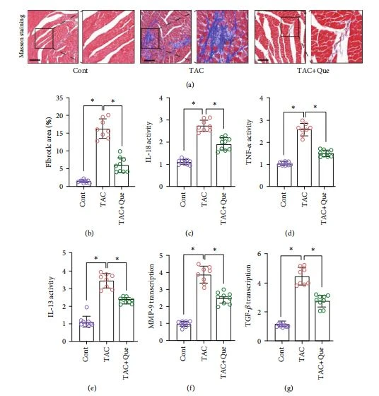


FiGURE 2: Quercetin(Que) attenuates myocardial fibrosis and inflammatory levels after transverse aortic constriction (TAC). (a) Masson staining. (b)Fibrotic area (%). (c-e)Expression levels of IL-18, TNF-α, and IL-13 were detected using ELISA.(f, g)mRNA expression of MMP_9 and TGF-B was detected using RT-PCR.(h-j)Expression of collagenase(collage) I/Ill was detected using immunohistochemistry. (k, I) Changes of ROS. (m-p) Protein/RNA expression of SIRT5 and IDH2 was detected.Mean± SD;*P<0.05.
mitochondria is regulated by succinylation. Previous experimental results established that the therapeutic effect of quercetin on myocardial fibrosis and heart failure is related to SIRT1.
In the current study, analysis of the mRNA levels of SIRT5 and IDH2 in high-glucose-stimulated HL-1 cells and immunoprecipitation showed that IDH2 expression in HL-1 cells was significantly inhibited following high-glucose stimulation, whereas the succinylation level of IDH2 was significantly increased(Figures 4(a)-4(d)). Moreover, following quercetin intervention, the mRNA expression of SIRT5 and IDH2 and level of IDH2 desuccinylation increased(Figures 4(a)-4(d)). To further verify the effect of SIRT5-mediated succinylation on the mitochondrial respiratory chain, we assessed mitochondrial respiratory complexes I, III, and IV. After high-glucose stimulation, the levels of these complexes were decreased; however, quercetin reversed this phenomenon (Figures 4(e)-4(g)).
The regulatory effect of quercetin on the mitochondrial respiratory complex and IDH2 succinylation was also inhibited following si-SIRT5 treatment(Figures 4(a)-4(g)). Therefore, SIRT5-mediated desuccinylation of IDH2 may be an important regulatory mechanism of myocardial fibro-sis and heart failure. Moreover, quercetin may improve the inflammatory response and oxidative stress injury through SIRT5-mediated desuccinylation of IDH2; however, the regulatory mechanism of mitochondrial homeostasis requires further analysis.
3.5. Quercetin Reduces High-Glucose-Induced HL-1 Inflammation Injury by Regulating Mitochondrial Energy Metabolism and NLRP3.
Mitochondrial energy metabolism is regulated by succinylation. However, heart failure or myocardial fibrosis caused by various factors involves an important pathological mechanism of oxidative stress and mitochondrial energy metabolism disorder in the inflammatory state. Therefore, we verified the regulatory effects of quercetin on mitochondrial energy metabolism and homeostasis. Immunofluorescence analysis showed that NLRP3 was highly expressed after high-glucose stimulation, which was reversed by quercetin (Figures 5(f) and 5(g)). Moreover, si-SIRT5 treatment eliminated the regulatory effect of quercetin on NLRP3 (Figures 5(f) and 5(g)).
Regulation of mitochondrial homeostasis is closely related to NLRP3 activation. In fact, NF-xB can limit the activation of NLRP3 by eliminating damaged mitochondria [33, 34]. We found that quercetin affected mitochondrial homeostasis by regulating NLRP3. High-glucose-stimulated HL-1


cells exhibited serious disorders of mitochondrial energy metabolism. The ATP level, basal respiration, maximal respiration, and respiratory reserve of mitochondria were significantly reduced. Moreover, the mPTP opening was abnormally increased (Figures 5(a)-5(e)). Quercetin reversed these effects and restored mitochondrial energy metabolism and mPTP closure (Figures 5(a)-5(e)). However,si-SIRT5 treatment reversed the regulation induced by quercetin on mitochondrial energy metabolism and mPTP(Figures 5(a)-5(e)). Therefore, excess production of ROS accompanied by inflammation and oxidative stress can lead to mitochondrial oxidative stress injury, and NLRP3 can interact with mitochondrial energy metabolism. Mitochondria may be the"Trojan horse" of inflammation, which is consistent with the results of a previous study [35].
This article is extracted from Hindawi Oxidative Medicine and Cellular Longevity Volume 2021, Article ID 5876841, 17 pages https://doi.org/10.1155/2021/5876841





