

The Pathological Features Of Lupus Nephritis Ushered in A Major Update, And Two New Injury Subtypes Deserve Attention!
Jun 28, 2023
The relationship between pathology and clinical medicine can be described as a "bridge", which can predict disease prognosis and guide clinical management. Pathological diagnosis is an essential tool for clinicians in the face of immune-mediated nephropathy. Taking lupus nephritis (LN) as an example, when dealing with LN patients with different pathological types, clinicians need to combine the patient's clinical manifestations and pathological types to adjust treatment and management strategies. Therefore, the pathological classification of LN is crucial for the management of LN.

Click to cistanche deserticola vs tubulosa for kidney disease
At the 60th Congress of the European Renal Association (ERA), Professor Maddalena Maria Bolognesi from the University of Milan summarized the new progress of LN pathological classification. She made it clear that recent studies have shown that LN pathology is associated with vasculitis and membranoproliferative glomerulonephritis (MPGN) injury patterns. These two pathological injury patterns are not only associated with glomerular lesions but also with the expression of immune cells. This finding may influence changes in pathological classification and ultimately improve clinical management.

Need for updated pathology criteria for LN
First, the criteria for LN are not set in stone. In 1974, the World Health Organization established the initial standard for LN, but its pathological classification is relatively simple, with only 5 types. In 2004, the pathological classification of LN was increased to 6 types, and in 2018, the new standard suggested that the pathological classification of LN should be increased to type III+IV. Changes in these standards are closely related to clinical evidence and basic research progress. So, what causes the current LN pathological classification to be updated?

A study in 2022 showed that the existing scoring system for pathological classification of LN cannot effectively guide clinical treatment. This study aimed to evaluate the predictive value of pathological chronicity score and activity score in patients with LN at baseline renal biopsy for their long-term renal impairment. A total of 203 LN patients were included in the study and followed up for 14 years. The results of the study showed that the chronic score was significantly correlated with long-term renal impairment in LN patients (P<0.001), while the activity score was not associated with long-term renal impairment. Cox model analysis showed that baseline serum creatinine, arterial hypertension/arterial hypertension, chronic glomerulopathy, and delayed renal biopsy were predictors of long-term renal impairment in LN patients. This study suggests that the value of chronicity and activity scores for LN management should be re-examined.

In addition, a poll in 2015 showed that nearly half (15/34) of nephrology experts believed that in actual clinical work, LN pathological classification is of limited help to clinicians. A meta-analysis found that LN pathological manifestations, such as LN classification, activity score, chronicity score, glomerulosclerosis, immune cell infiltration, etc., had limited correlation with clinical symptoms and prognosis of patients (intraclass correlation [ICC] Or k value <0.6; Figure 1).

Figure 1 Correlation of LN renal biopsy features with clinical symptoms and prognosis
Remarks: The closer the ICC or K value is to 1, the higher the correlation between the pathological manifestations of LN and the clinical manifestations and prognosis.
The series of the evidence above shows that the update of LN pathological criteria is imminent and imperative.
Immunohistochemical staining and pathological classification of LN
So, how to improve the pathological criteria of LN? Immunohistochemical staining may be one answer. First, with the improvement of immunohistochemical staining techniques, basic research workers discovered a new mechanism of kidney injury in patients with type III/IV LN.
In 2022, a study on LN showed that patients with type III/IV LN have unique mechanisms of renal injury and that patients with type IV LN could be further divided into subtypes IVS and IVG based on these injury mechanisms and immunohistochemical staining ( figure 2). These two types correspond to two injury mechanisms, MPGN and vasculitis, respectively.

Fig. 2 Classification decision diagram of patients with III/IV LN
Remarks: Endocapillary_over means intracapillary cell hyperplasia; Endocapillary_absent means no intracapillary cell hyperplasia; wire loop means ring lesion; Karyorrhexis means nuclear fragmentation.
After reviewing the clinic, it is found that many patients with type III, type III+V and type IV LN can be further subdivided. Taking patients with “pure” type III/IV LN as an example, they can also be subdivided into 214 categories, but the subgroups are optimally classified into 2 groups (ie, types IVS and IVG; Figure 3). Overall, this new classification method can more accurately describe the type of damage to the patient's kidney and help doctors manage LN patients.

Figure 3 Subgroup classification of patients with "pure" type III LN
Characteristics of two injury subtypes and their clinical implications
01 feature
Now that the injury subtypes of IVS and IVG have been clarified, what are their characteristics under the microscope? See Figures 4, 5, and 6.

Figure 4 Characteristics of MPGN subtypes
Remarks: ① MPGN subtype is a pattern of injury, not a diagnosis of patients with MPGN; ② Immunofluorescence is characterized by intraepithelial immune complex deposition and subendothelial mesangial insertion; ③ Glomerular lobules can be seen under a low-power microscope Abnormal morphology and capillaries; ④ Under a high-power microscope, "platinum ear lesions", ring lesions and/or transparent thrombi can be seen.

Figure 5 Characteristics of vasculitis subtypes
Remarks: Under low magnification, segmental hypercellularity, fibrinous necrosis, and crescents in the capillaries can be seen in glomeruli.

Figure 6 Potential pathological mechanisms of vasculitis subtypes
Remarks: Or similar to ANCA vasculitis injury
02 clinical impact
Both subtypes have clinically significant implications. First, LN patients with different pathological types have different chronic and/or active scores. In contrast, among patients with MPGN and vasculitis subtypes, their activity scores were significantly different (P < 0.001; Fig. 7), suggesting that these two subtypes have different clinical characteristics and outcomes, or may require different management models.

Figure 7 Differences in active and chronic scores of the 2 subtypes
Immune cells and LN typing
At the conference, Professor Maddalena Maria Bolognesi also reported on her unpublished research. The purpose of this study was to evaluate the relationship between different immune cells, inflammatory factors, and ISN/RPS classification, and combine immunophenotype with routine renal biopsy results to improve the reliability of LN diagnosis.
A total of 119 LN patients were enrolled in the study, with a median age of 37 years (range: 24-67), women accounting for 89.9%, median baseline creatinine of 71umol/L, and proteinuria (UPCR) of 3mg/g.
There are 4 items in the research results, namely, ① the distribution of B lymphocytes (CD20) among different types is not different, and they are generally located in the gap; Renal biopsy samples of LN patients all increased, but only CD68 reached statistical significance; ③T lymphocytes (CD3) were more common in the interstitium, and macrophages (CD68 and CD163) were more common in glomeruli (P<0.001 and P = 0.01); ④ The protein related to the interferon signaling pathway, MX1 and the mesangial marker CD 248, were all expressed in the glomeruli of patients with class IV LN.
According to the above differences, LN patients can be classified pathologically based on the results of CD68, CD20, CD34, CD163, MX1, and other immunofluorescence examinations (Figure 8). Meanwhile, immune cells were also associated with the MPGN subtype and vasculitis subtype classification (Fig. 9, 10).
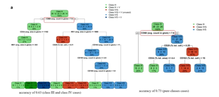
Figure 8 LN pathological classification based on immune cells
Remarks: Whether the CD68 count is <7.6 is an important index for LN classification (marked in red box).

Figure 9 CD68 and CD34 are important indicators for the classification of MPGN and vasculitis subtypes
Remarks: CD68 count ≥ 20 is a key index for the classification of MPGN and vasculitis subtypes (marked in red box)

Figure 10 Immunohistochemical staining and microscopy of MPGN and vasculitis subtype glomerular cells
Note: It can be seen from the figure that under immunofluorescence, the glomeruli of the two subtypes have completely different characteristics
In conclusion, recent studies have found that MPGN and vasculitic subtypes are present in LN patients and are significantly associated with glomerular lesions and immune cell infiltration. Therefore, stage III/IV patients can be further classified and facilitate their clinical management.
In addition to the above research report, Professor Maddalena Maria Bolognesi believes that with the development of artificial intelligence, researchers can quickly identify the pathological types of LN through artificial intelligence, machine learning, and other methods, and find the correlation between pathological types and clinical manifestations, which will bring great benefits Contribute to the management and prognosis of LN patients.
References:
1. Maddalena Maria Bolognesi. Pathology in lupus nephritis – 2023 revised. Jun 15, 2023.





