Apocynin Ameliorates Monosodium Glutamate Induced Testis Damage By Impaired Blood-Testis Barrier And Oxidative Stress ParametersⅢ
May 30, 2023
4. Discussion
In this study, the healing effect of APO on MSG-induced testicular damage has been evaluated by biochemical, microscopical, and immunohistochemical parameters. Testosterone and LH levels decreased and FSH levels increased, oxidative stress parameters such as MDA level increased, while GSH and SOD levels decreased in the MSG group. In MSG-induced testicular injury, the histopathological score and the number of damaged seminiferous tubules and spermatozoa were increased. The proliferative index was low and the apoptotic index was higher in the MSG group. The germinal epithelium of the seminiferous tubules was decreased in thickness.

Click to jade cistanche for testosterone
Those degenerative changes have been reversed by the administration of APO. The MSG group depicted a decrease in the distribution of ZO-1 and occluding as tight junction proteins of the blood-testis barrier. MSG affected the oxidative stress markers. In this group, there was an increase in the distribution of the protein NOX-2, which was involved in the synthesis of ROS. Ultrastructure examinations of the MSG group indicated prominent damage in the cytoplasm of seminiferous tubule germinal epithelial cells of the seminiferous tubules with separated tight junctions. Biochemical, histological, immunohistochemical, and ultrastructural analyses showed that these parameters improved with APO treatment in the MSG + APO group.
Experimental studies have shown that oral ingestion of MSG can be toxic to reproduction; however, due to the high doses used in the studies, the toxic effects on the reproductive system due to consumption of MSG were unlikely to translate to human consumption [33,34] The human ingested daily dose of MSG is thought to be between 1200 and 3000 mg/kg [35]. In this study, the dose of 120 mg/kg body weight of MSG was used for experimental design, based on an allometric extrapolation of the average daily intake of this substance in humans [5].
The hypothalamic–pituitary–gonadal (HPG) axis plays a key role in processes related to the development and maturation of the male reproductive system. FSH is a factor in the maintenance of intragonadal testosterone synthesis and spermatogenesis. LH plays a role in the development of the male genital system and the process of sex determination by stimulating Leydig cells [36]. The stimulation causes Leydig cells to secrete the necessary levels of testosterone. The proper testosterone level is controlled by GnRH activity through a negative feedback mechanism [36].
With this mechanism, the HPG axis functions in a controlled manner [37,38]. In some population-based studies, sex hormones have been shown to affect sperm morphology, concentration, and motility. In a study of subfertile couples, an inverse relationship was found between LH and sperm motility and morphology [39]. In clinical studies, FSH levels were found to correlate negatively with sperm concentration [40]. In other experimental studies, following the administration of MSG, a decrease in sperm motility, concentration, and LH hormone levels was revealed. Studies have also shown that MSG causes a decrease in testosterone hormone levels and sperm concentrations [34,41].
In our present study, FSH, LH, and testosterone levels in the APO group were similar to the control group. A statistically significant decrease in testosterone hormone was observed in the MSG group. A significant increase in FSH hormone levels was observed in the MSG group. In the MSG + APO group, the FSH level was lower than in the group MSG. Although a decrease in LH hormone was observed in the MSG group, there was no statistical difference in LH hormone between the experimental groups. MSG could do damage to Leydig cells for testosterone secretion. Decreased testosterone and LH levels are also associated with sperm count. Thus, low testosterone and LH levels could be related to decreased sperm count and abnormal sperm morphology [42].
In contrast, an elevated FSH level indicates that there is a disturbance in spermatogenesis causing altered FSH signaling. MSG has been shown that it may cause a disturbance in hormonal pathways in the hypothalamus [4]. As a result of our study, it was observed that the administration of MSG interfered with the HPG axis by affecting FSH, LH, and testosterone levels, as well as testicular and sperm morphology, sperm concentration, and motility. Administration of apocynin reduced the impaired testosterone, FSH, and LH levels to near normal levels and this improvement in hormone levels also affected the morphological parameters. SOD is an important enzyme that protects cells from damage caused by internal and external superoxide ions [43].
MDA is an aldehyde formed by free radicals during lipid peroxidation and is an indicator of both cell membrane damage and the severity of the effect of oxygen radicals on reactive cells. A decrease in SOD and an increase in MDA can trigger oxidative stress, leading to cell damage and cell death [24]. GSH is involved in the reduction of ROS such as hydrogen peroxide in the cell [44]. NOX is a source of ROS and apocynin is known as a NOX inhibitor [45].

In studies of MSG-induced testicular injury, MDA levels in testicular tissue increased and SOD levels decreased when MSG was administered [4,46]. In another study, ingestion of MSG was also shown to decrease GSH levels in testicular tissue [4]. In a study in which apocynin was used as a healing agent, it was shown that apocynin reduced MDA levels, which were increased due to tissue damage and caused an increase in GSH and SOD levels in the testicular damage group [27]. In another experimental study of the hypoxia-induced testicular degeneration model, NOX-2 activation and MDA levels were increased and GSH levels were decreased due to the effect of hypoxia. However, testicular tissue damage was shown to be reduced by the application of APO [47].
Our present study concluded an increase in MDA levels with a decrease in GSH and SOD levels in the MSG group. We could suggest that APO improved these parameters in the MSG + APO group through NOX-2 inhibition. Some preclinical studies have revealed the toxic effects of MSG on many organ systems, such as the liver, kidneys, and reproductive systems [3]. An experimental study states that MSG can cause infertility due to its harmful effect on the ovaries and oocytes [48]. The toxic effect of MSG on the testes with an imbalance of sex hormones has been noted [34]. In a similar study, the levels of oxidative stress markers were increased in the testicular tissues of rats administered MSG, and testicular morphology and sperm morphology were also negatively affected by MSG [41].
It has been observed that MSG leads to a degeneration in testicular morphology with a negative effect on testosterone [4,49]. In testicular injury models, a decrease in the thickness of the seminiferous tubule germinal epithelium was demonstrated [50,51]. In an MSG-induced testicular injury model, the thickness of the seminiferous tubule epithelium was decreased [52]. In our study, the thickness of the seminiferous tubular germinal epithelium of the MSG group was decreased compared to the other experimental groups. There was an increase in the germinal epithelial thickness of the MSG + APO group compared to the MSG group. MSG is stated to lead to obesity, as an appetizer [53]. There are question marks about whether MSG consumption can be a risk factor in the rapid increase in epidemic obesity.
In an open-cohort study among Chinese adults, it was shown that those who used MSG had a higher body mass index compared with those who did not use MSG [54]. In other experimental studies, it has been observed that the weight of MSG-given rats increased and made them prone to obesity [55,56]. However, in our study, there was no statistically significant difference in body weight between the experimental groups, weighted every week during the experiment. In a related study where MSG has been administrated to rats, a decrease was observed in testicular weights in the MSG group compared to the control group [4,52]. However, testicular weight/body-weight ratios of our current study, indicated a decrease in the MSG group compared to other experimental groups, as statistically insignificant. In studies examining the effects of MSG on sperm concentrations, MSG was found to cause a decrease in sperm viability [4,49,52].
In another testicular damage model, the curative effect of apocynin on sperm parameters was examined, and an increase in sperm concentration was observed in the apocynin healing group compared to the damage group [27]. In our study, when comparing sperm counts between experimental groups, it was found that the sperm count was lowest in the MSG group, while the sperm count in the MSG + APO group was statistically higher than in the MSG group. It is stated that the use of apocynin is safe in animal studies. In addition, APO has potent antioxidant and anti-inflammatory effects in many experimental models [57]. Experimental testicular injury models reported the effect of apocynin on the reduction of oxidative stress [58,59].
In chemotherapy-based experimental studies, the healing effect of apocynin was also highlighted on testes tissue. In these studies, it was observed that apocynin leads to a decrease in the number of apoptotic cells and an increase in testosterone levels [27,60]. As one of the factors of male infertility, oxidative stress leads to sperm dysfunction. Oxidative stress-induced spermatozoon damage affects 30–80% of infertile men [61]. Enzymes in the NOX family are responsible for cellular ROS synthesis, and apocynin is a NOX inhibitor [45]. In an experimental model of testicular injury in which apocynin was used as a healing agent, the highest NOX-2 immunoreactivity was observed in the testicular injury group. APO treatment concluded in a decrease in the number of NOX-2 immunopositive cells [27].

Literature data revealed a limited number of experimental testicular damage studies evaluating the amount and distribution of NOX-2. In our study, NOX-2 immunoreactivity was highest in the MSG group, with a decreased immunoreactivity in the MSG + APO group compared to the MSG group. There was no significant difference between the APO group and the control group, presenting the lowest NOX-2 immunoreactivity. Apoptosis is programmed cell death, which is essential for tissue homeostasis [62]. Regulation of apoptosis is a critical factor in the development, differentiation, and function of germ cells [61]. Exposure to factors that lead to oxidative stress negatively affects spermatogenesis by negatively affecting the structure of chromatin in spermatozoa [63].
Infertile men have been found to have higher oxidative stress and apoptotic cells in their seminal fluid compared to fertile men. In experimental models of testicular damage, the increased number of apoptotic cells in damaged testis tissue was reversed by the administration of APO as a curative substance [23,51]. In an experimental study of testicular injury, apocynin was shown to have a healing effect on DNA damage caused by NOX -induced oxidative stress [64]. Similarly, in our study, the proliferation index was decreased in the MSG group, with an increase in the apoptotic index.
In the MSG + APO group, a decrease in the apoptotic index and an increase in the proliferative index were evaluated. The blood-testis barrier protects the developing germ cells against the external environment and provides support for the germ cells to divide and develop, which is very important for fertility [65]. Sertoli cells, which are specialized epithelial cells found in the seminiferous tubules, form the basis of the blood-testis barrier with tight junctions. Occludin, ZO-1, ZO-2, and ZO-3 are molecules found in the tight junctions of the blood-testis barrier. ZO proteins are involved in binding the occludin proteins to the actin in the cytoskeleton [66,67]. It was observed that spermatogenesis was negatively affected, and atrophic seminiferous tubules formed in the occludin knockout mouse model.
Our study revealed the highest immunoreactivities of ZO-1 and occludin in the control group and APO group, whereas the MSG group had significantly lower immunoreactivity. ZO-1 and occludin immunoreactivities were higher in the MSG + APO group, compared to the MSG group. Transmission electron microscopic examinations of experimental studies depicted degenerated testicular germinal epithelial cells and lipid droplets [41,54] with deteriorated tight junctions in the blood-testis barrier [68]. In our study, transmission electron microscopic examinations revealed normal testicular seminiferous tubule ultrastructure in the control and APO groups. Vacuolization, lipid droplets, and degenerations in the tight junctions of the blood-testis barrier were evident in the MSG group. In the MSG + APO group, apocynin was found to have a healing effect at the level of ultrastructural damage.

5. Conclusions
Our study concluded with a regulating role of APO, for the formation of reactive oxygen derivatives and the HPG axis in rat testicular injury induced by MSG. APO maintained the balance between the proliferative and apoptotic indexes, preserved the integrity of the blood-testis barrier, and contributed to the healing of testicular tissue damage caused by oxidative damage. It is also thought that apocynin administration may have a healing effect on infertility by reversing the harmful effects of MSG both on sperm motility and number.
How does Cistanche boost testosterone?
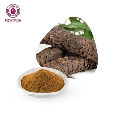
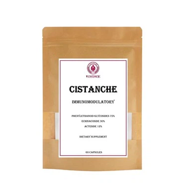
Cistanche is an herb traditionally used in Chinese medicine to boost energy, libido, and overall vitality. It is believed that Cistanche may increase testosterone levels by inhibiting the activity of an enzyme called aromatase, which converts testosterone into estrogen. Cistanche has also been found to increase the production of luteinizing hormone (LH) in the body. LH plays a crucial role in regulating testosterone production in the testes. By increasing LH levels, Cistanche may indirectly boost testosterone production. Furthermore, Cistanche also contains several phytochemicals such as echinacoside and acteoside, which possess antioxidant properties. These compounds may help to protect the testes and other reproductive organs from oxidative damage, thereby improving their function and increasing testosterone production. Overall, the exact mechanisms by which Cistanche increases testosterone levels are not fully understood, but it is likely an interplay of inhibiting aromatase, increasing LH production, and providing antioxidant support to the reproductive system.
Merve Acikel-Elmas 1,*, Salva Asma Algilani 1 , Begum Sahin 1 , Ozlem Bingol Ozakpinar 2 , Mert Gecim 2 , Kutay Koroglu 3 and Serap Arbak 1
1 Department of Histology and Embryology, School of Medicine, Acibadem Mehmet Ali Aydinlar University, Icerenkoy Mah., Kayisdagi Cad. No. 32, Atasehir, Istanbul 34752, Turkey
2 Department of Biochemistry, Faculty of Pharmacy, Marmara University, Basibuyuk Yolu, 4/A, Basibuyuk, Istanbul 34854, Turkey
3 Department of Histology and Embryology, School of Medicine, Marmara University, Basibuyuk Yolu No. 9 D:2, Maltepe, Istanbul 34854, Turkey