Cistanche Phenylethanoid Glycosides: Pre‑Clinical Findings On T‑Cell Lymphoma And Anti‑Tumour Mechanisms
Nov 11, 2026
Introduction
T‑cell lymphoma represents a diverse family of non‑Hodgkin lymphomas originating from abnormal T‑lymphocyte clones. These haematological malignancies display high biological heterogeneity, and clinical outcomes for many patient subgroups remain unsatisfactory. Even with modern immunotherapy and chemotherapy protocols, overall survival rates stay relatively low, creating an urgent global demand for innovative therapeutic candidates and complementary research directions. Natural bioactive compounds sourced from traditional medicinal plants have drawn growing scientific attention as potential starting points for anti‑tumour drug discovery.
Cistanche deserticola, commonly known as desert ginseng, is a well‑documented tonic herb in Traditional Chinese Medicine (TCM). Historically, practitioners used it to support vitality, kidney function and intestinal health. Contemporary pharmacological investigations have revealed a broad spectrum of biological activities for its extracts: antioxidant, anti‑inflammatory, hepatoprotective, neuroprotective, immunomodulatory and anti‑proliferative effects against multiple cancer cell linesPMC. Among its many constituents, phenylethanoid glycosides (PhGs) stand out as the primary bioactive class. A four‑compound blend termed CPhGs consists of echinacoside, acteoside (verbascoside), cistanoside A, and 2‑acetylacteoside. Recent pre‑clinical laboratory and animal‑model work has explored how this combined mixture acts against malignant T‑cells, opening new research avenues for T‑cell lymphoma intervention.
Important preliminary note: All effects described in this article derive from in‑vitro cell experiments and mouse xenograft studies. No large‑scale human clinical trials have validated CPhGs as lymphoma treatment. This article serves purely as science‑focused educational content, not medical guidance. Cistanche‑based supplements cannot replace standard oncological care. Readers must consult qualified physicians for all cancer‑related decisions.
What Are Cistanche Phenylethanoid Glycosides (CPhGs)
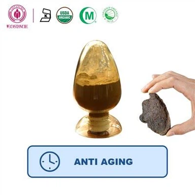
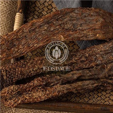
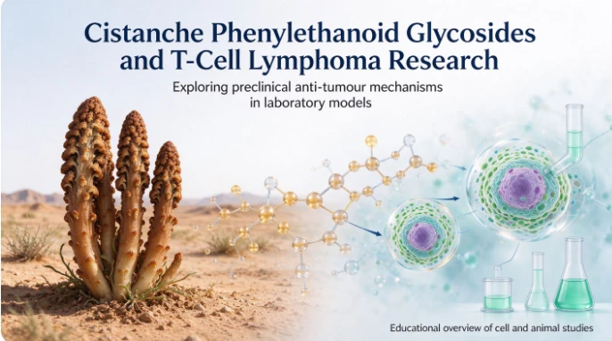
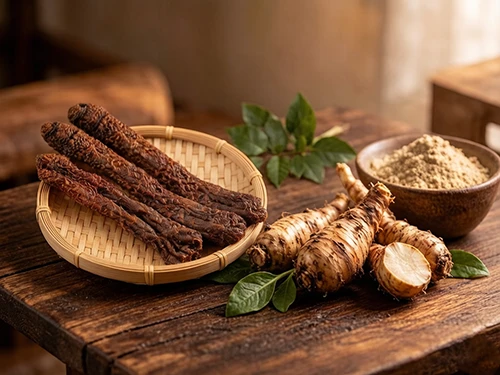
Cistanche grows as a parasitic plant across the arid desert zones of Northwest China and Central Asia. Its fleshy scaly stems are harvested and processed for medicinal purposes. Phenylethanoid glycosides are signature water‑soluble molecules formed by phenylethanol moieties linked with sugar groups. More than 20 different PhG variants have been isolated from Cistanche deserticola.
The four components of CPhGs selected for lymphoma research each carry distinct biological traits:
Echinacoside (ECH): Powerful antioxidant, modulates inflammatory signalling pathways, shows neuro‑ and hepatoprotective properties.
Acteoside (verbascoside, VB): Well‑studied for anti‑inflammatory activity, can trigger programmed cell death in multiple tumour cell lines.
Cistanoside A: Less‑investigated member of the PhG family, contributes to the overall synergistic profile of total PhG extracts.
2‑acetylacteoside: Structural derivative of acteoside with overlapping bioactivity.
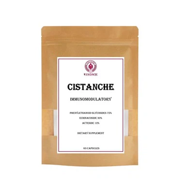
Most herbal studies historically tested total crude extracts. However, this particular body of research used a well‑defined equal‑proportion mixture of these four purified standards, permitting more reproducible laboratory testing. It should be emphasised that commercial cistanche supplements vary widely in PhG concentration and component ratios, so lab‑observed results do not directly translate to off‑the‑shelf consumer products.
Background: T‑Cell Lymphoma and Programmed Cell‑Death Biology
T‑cell lymphoma arises when mature T‑lymphocytes acquire oncogenic mutations, leading to uncontrolled replication, resistance to natural cell‑death signals, and potential tissue invasion or metastasis. In healthy bodies, defective or aged cells self‑eliminate via regulated programmed cell death; cancer cells frequently disable these protective biological brakes. Two major forms of programmed cell death feature prominently in this cistanche research: apoptosis and pyroptosis.
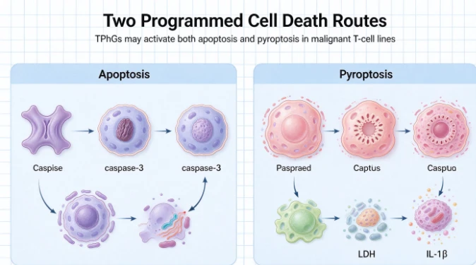
Apoptosis, also called "silent cell suicide", is non‑inflammatory programmed cell death. It operates through two primary routes:
Intrinsic (mitochondrial) pathway: Internal cell stress activates mitochondrial changes, regulated by protein families including BAX (pro‑apoptotic) and Bcl‑2 (anti‑apoptotic).
Extrinsic (death‑receptor) pathway: External signal molecules bind surface death receptors, starting caspase‑8‑driven cascades. Both pathways converge on effector caspase‑3, dismantling the cell neatly without spilling cellular contents into surrounding tissue.
Pyroptosis is an inflammatory type of programmed cell death. Key execution proteins belong to the gasdermin family (GSDMD, GSDME). Upon cleavage by activated caspases, gasdermin‑N fragments insert pores into cell membranes. The cell swells, ruptures, and releases pro‑inflammatory intracellular contents such as LDH and interleukin‑1β (IL‑1β)PMC. Three known pyroptosis signalling cascades exist:
Canonical: caspase‑1 / GSDMD, downstream of NLRP3 inflammasome assembly.
Non‑canonical: caspase‑4/5 / GSDMD.
Alternative pyroptosis bypass: caspase‑3‑mediated cleavage of GSDME.
The NLRP3 inflammasome creates scientific complexity: while its activation can trigger pyroptotic cancer‑cell killing, NLRP3‑driven IL‑1β maturation can simultaneously promote tumour‑cell proliferation, migration and invasion. This dual‑edged characteristic makes targeting NLRP3 a tricky objective for anti‑tumour pharmacology.
Pre‑Clinical Observations: CPhGs Inhibit Malignant T‑Cell Proliferation
In laboratory work, researchers exposed four well‑established human malignant T‑cell lines (Jurkat, HH, MyLa, Hut78) to gradient concentrations of CPhGs over multiple time points. Microscopic visual inspection, CCK‑8 cell‑viability assays and flow‑cytometry measurements consistently demonstrated that CPhGs suppressed cell survival in both dose‑dependent and time‑dependent manners.
With rising CPhGs concentration, malignant T‑cells exhibited obvious morphological changes: cellular shrinkage, rounding, and reduced cell clumping. Longer incubation amplified these cytotoxic effects. Calculated IC50 values (concentration reducing cell viability by half) fell in the approximate range of 48 μM to 67 μM across these four cell lines.
These in‑vitro outcomes provided initial proof‑of‑concept: the four‑component PhG blend could slow replication and drive death of malignant T‑cells in controlled culture conditions. Scientists then moved on to dissect which cell‑death pathways were responsible for this cytotoxicity.
Mechanism One: CPhGs Induce Intrinsic and Extrinsic Apoptosis
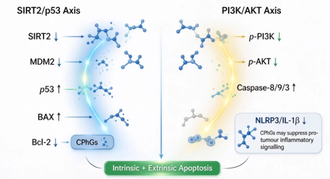
Multiple molecular assays including qRT‑PCR gene‑expression quantification and Western‑Blot protein‑detection revealed that CPhGs trigger apoptosis by modulating two central signalling axes: SIRT2/p53 and PI3K/AKT.
SIRT2 / p53 signalling axis SIRT2 is a histone deacetylase with context‑dependent oncogenic or tumour‑suppressing roles. In these malignant T‑cell models, CPhGs lowered SIRT2 and its downstream partners p300 and MDM2 expression. MDM2 normally marks tumour‑suppressor p53 for degradation. By suppressing SIRT2‑MDM2 signalling, CPhGs stabilised and elevated p53 protein abundance. Elevated p53 further shifted the balance of Bcl‑2‑family proteins: pro‑apoptotic BAX levels rose, anti‑apoptotic Bcl‑2 declined, favouring mitochondrial‑triggered intrinsic apoptosis. PTEN tumour‑suppressor protein expression also increased under CPhGs treatment.
PI3K / AKT signalling axis The PI3K‑AKT pathway is frequently hyper‑activated in haematological malignancies, delivering strong pro‑survival and pro‑proliferation signals to cancer cells. CPhGs did not alter total PI3K and AKT protein amounts, yet significantly reduced their phosphorylated (activated) forms. Suppression of phosphorylated‑PI3K / phosphorylated‑AKT dampened pro‑survival signalling, removing one major defence mechanism of malignant T‑cells.
Downstream of these two axes, researchers observed concentration‑dependent elevation of cleaved (activated) caspase‑8, cleaved‑caspase‑9 and cleaved‑caspase‑3. Appearance of activated caspase‑8 marks extrinsic‑pathway activation, while cleaved‑caspase‑9 signals intrinsic mitochondrial‑apoptosis activation. This confirmed that CPhGs concurrently switch on both intrinsic and extrinsic apoptotic cascades in malignant T‑cells.
Mechanism Two: CPhGs Trigger Multi‑Pathway Pyroptosis with Unique NLRP3 Regulatory Pattern
Beyond apoptosis, experiments uncovered that CPhGs also provoke pyroptotic cell death in susceptible malignant T‑cell lines. Several lines of evidence supported this conclusion:
Elevated LDH release in cell culture supernatants (LDH leaks through ruptured cell membranes during pyroptosis).
Increased protein levels of cleaved‑caspase‑1, cleaved‑caspase‑4, cleaved‑GSDMD‑N and cleaved‑GSDME‑N, representing activation of canonical, non‑canonical and alternative pyroptosis routes.
When researchers silenced the key pyroptosis‑execution gene GSDMD using siRNA technology, malignant T‑cells gained partial resistance against CPhGs‑induced killing.
Application of VX‑765 (caspase‑1/caspase‑4 small‑molecule inhibitor) partially rescued cell viability, reduced LDH leakage and reversed pyroptosis‑related protein activation.
A particularly noteworthy discovery from this research is the functional separation effect on NLRP3‑related signals. Ordinarily, canonical caspase‑1‑GSDMD pyroptosis relies on full NLRP3‑inflammasome assembly. Surprisingly, CPhGs simultaneously activated caspase‑1‑GSDMD pyroptosis signalling while lowering NLRP3 protein expression and diminishing mature IL‑1β production. Since NLRP3‑IL‑1β signalling fuels tumour‑cell proliferation and invasiveness, suppressing this axis provides added anti‑tumour benefit.
To verify this finding, scientists artificially over‑expressed NLRP3 protein in Hut78 and Jurkat cells. The NLRP3‑overexpressing cancer cells displayed markedly improved survival when exposed to CPhGs. This functional rescue experiment validated that CPhGs partially exert anti‑proliferative effects through NLRP3‑inhibition.
To summarise this distinctive mechanism: CPhGs activate multiple caspase‑driven gasdermin‑mediated pyroptosis pathways, yet repress NLRP3‑IL‑1β pro‑tumour signalling, avoiding the unwanted growth‑promoting side‑effect often associated with simple NLRP3‑inflammasome‑driven pyroptosis induction. This uncommon molecular profile attracts interest for further mechanistic follow‑up studies.
Notably, the relative contribution of pyroptosis varies by cell‑line. In HH and MyLa cell models, CPhGs‑mediated cell death depended chiefly on apoptosis; in Jurkat and Hut78 lines both apoptosis and pyroptosis participated jointly in cancer‑cell elimination. Such heterogeneity reminds us that responses to plant‑derived compounds can differ substantially between individual tumour subtypes.
In‑Vivo Evidence: CPhGs Suppress T‑Cell Lymphoma Growth in Nude‑Mouse Xenograft Models
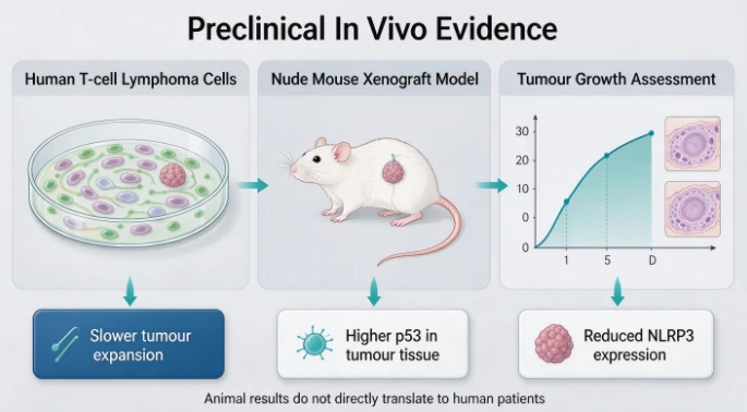
To move beyond isolated cell‑culture results, investigators built an in‑vivo tumour model by implanting human Hut78 malignant T‑cells subcutaneously into immunodeficient nude mice. After solid tumours had established, animals received daily intraperitoneal injections of either saline control, low‑dose CPhGs (20 mg/kg), or higher‑dose CPhGs (60 mg/kg), continuing for 12 consecutive days.
Study outcomes included:
Tumour‑volume measurements: Both CPhGs‑treated groups demonstrated slower tumour expansion versus saline controls; the 60 mg/kg high‑dose cohort produced the most prominent tumour‑growth suppression. Calculated tumour‑inhibition rates increased with dosage.
Body‑weight monitoring: Control‑group tumour‑bearing mice showed gradual weight loss. Mice receiving CPhGs maintained or gained body weight, suggesting reasonable animal tolerance to the tested dosages under experimental conditions.
Histopathology (HE‑staining): Tumour tissue sections from CPhGs‑treated animals exhibited increased tumour‑cell necrosis in a dose‑related fashion.
Immunohistochemistry staining of tumour tissue: Consistent with cell‑culture findings, tumour specimens from CPhGs‑treated mice displayed higher p53 protein signal intensity and reduced NLRP3 expression.
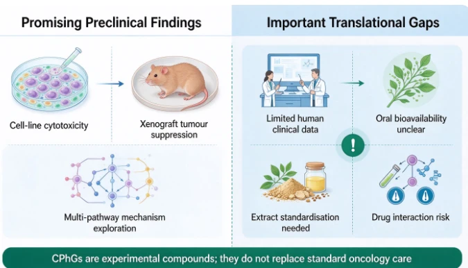
These animal‑model results corroborate the in‑vitro observations: systemically administered CPhGs could hinder human‑origin T‑cell‑lymphoma progression in living organisms. Nevertheless, critical caveats apply. Nude‑mouse models lack functional adaptive immune systems, and injection‑route dosing used in animal experiments cannot be replicated via oral human supplementation. Significant translational gaps separate mouse‑model results from clinical patient realities.
Broader Anti‑Tumour Context for Cistanche Phenylethanoid Glycosides
Apart from T‑cell lymphoma, existing pre‑clinical literature reports anti‑tumour potential for cistanche PhGs across diverse cancer models:
Liver cancer: PhGs modulate PI3K/AKT, TGF‑β/Smad pathways, induce hepatocellular‑carcinoma‑cell apoptosis and restrain migration.
Non‑small‑cell lung cancer: Echinacoside triggers mitochondria‑associated pyroptosis via Raf/MEK/ERK signalling modulation.
Breast cancer: Inhibits Wnt/β‑catenin and AKR1B10/ERK axes to reduce proliferation and metastatic potential.
Ovarian cancer: Suppresses PI3K‑AKT‑mTOR cascades, limiting tumour‑cell invasion and angiogenesis.
Colorectal, prostate and pancreatic tumour models: Multiple papers describe pro‑apoptotic, anti‑migratory pre‑clinical effects.
Most mechanistic themes recur across these different malignancies: PhGs tend to suppress oncogenic kinase pathways (PI3K‑AKT, MAPK), up‑regulate tumour suppressors such as p53, shift Bcl‑2/BAX balance toward apoptosis, modulate inflammatory‑inflammasome signalling and may interfere with tumour angiogenesis.
Even so, almost all datasets originate from cell‑culture and rodent assays. Human clinical trial data remain sparse. Herbal‑extract composition variability, bioavailability and metabolism in human bodies are poorly characterised for most cistanche PhG anti‑tumour applications. No established safe human dosage exists for anti‑cancer purposes.
Current Research Limitations & Future Directions
This line of pre‑clinical research yields fascinating mechanistic insights, yet multiple major hurdles must be crossed before any therapeutic translation can be contemplated:
Lack of human clinical evidence: All anti‑lymphoma data derive from cell‑lines and immunodeficient‑mouse xenografts. Animal models cannot perfectly mirror human T‑cell‑lymphoma complexity, patient immune status, drug metabolism, and tumour micro‑environment. There are no completed Phase I‑III human trials evaluating CPhGs in lymphoma patients.
Bioavailability and delivery challenges: Phenylethanoid glycosides' oral absorption characteristics in humans are incompletely understood. Successful tumour‑suppression in mice was achieved by intra‑peritoneal injection; oral ingestion may deliver far lower active‑compound exposure to lymphoid tumour tissue. Formulation research (e.g. improved extraction, nanoparticle delivery) will be required.
Extract standardisation issues: The described experiment applied four highly‑purified reference‑standard compounds mixed in defined proportions. Commercial cistanche extracts differ widely in PhG profiles, total PhG percentages, and contaminating secondary metabolites. Results cannot be extrapolated to consumer‑market cistanche products.
Mechanistic questions remaining: The exact molecular means by which CPhGs simultaneously activate caspase‑1‑GSDMD pyroptosis while repressing NLRP3 expression is incompletely decoded. Crosstalk networks connecting SIRT2/p53, PI3K/AKT, apoptosis and pyroptosis need deeper dissection. Cell‑line‑specific sensitivity differences (why pyroptosis manifests strongly in some malignant T‑cells but not others) also await clarification.
Safety and interaction data gaps: Long‑term safety profiles, potential herb‑drug interactions with chemotherapy or immunotherapy agents have barely been investigated. People living with haematological malignancies take complex medication regimens; unknown interactions could create clinical risk.
Future research priorities should include: optimised formulation and pharmacokinetic studies, additional immune‑competent animal‑model work, and eventually carefully‑designed early‑phase human safety‑oriented trials. Combination‑strategy studies exploring whether CPhG‑type compounds could synergise with existing anti‑lymphoma therapies also represent an interesting research direction.
Conclusion
Cistanche deserticola phenylethanoid glycosides (CPhGs), a mixture containing echinacoside, acteoside, cistanoside A and 2‑acetylacteoside, demonstrate potent pre‑clinical activity against human malignant T‑cells. Laboratory findings show CPhGs suppress malignant‑T‑cell proliferation through dual programmed‑cell‑death mechanisms:
Triggering both intrinsic and extrinsic apoptosis by tuning SIRT2/p53 tumour‑suppressor signalling and inhibiting oncogenic PI3K‑AKT survival pathways.
Provoking multi‑route pyroptosis (canonical, non‑canonical and GSDME‑dependent alternative pyroptosis), alongside the rare regulatory feature of inhibiting pro‑tumour NLRP3‑IL‑1β signalling while activating downstream pyroptosis effectors.
Xenograft nude‑mouse experiments confirmed that systemically administered CPhGs slowed in‑vivo human T‑cell‑lymphoma tumour growth, accompanied by elevated p53 and lowered NLRP3 within tumour tissue. Combined with existing literature covering liver, lung, breast and other tumour models, these results strengthen the scientific rationale for further investigating PhG‑type natural products as anti‑tumour lead compounds.
Crucially, these remain pre‑clinical laboratory discoveries. They do not constitute proof of efficacy for human lymphoma patients. Substantial translational barriers stand between cell‑culture / mouse outcomes and real‑world clinical utility. No person should utilise cistanche supplements as replacement for standard oncological treatment. What this body‑of‑work does accomplish is highlighting how bio‑active constituents from traditional medicinal plants can deliver multi‑target molecular effects worthy of continued basic‑science exploration, potentially inspiring future new‑drug‑discovery projects.
Health Disclaimer
This educational article summarises publicly‑available pre‑clinical laboratory and animal‑model research only. It is not medical advice. None of the statements have been evaluated by the FDA or any medical‑regulatory authority. Cistanche and its extracts are not approved for diagnosing, treating, curing or preventing cancer or T‑cell lymphoma. All information is for general science education purposes. If you have concerns over cancer or any medical condition, always consult a licensed healthcare oncologist or physician. Do not start, modify or discontinue any herbal supplement or medical therapy without professional medical supervision.