Exploring Cistanche Deserticola: How Phenylethanoid Glycosides Interact With Liver‑Cancer Cells
Nov 12, 2026
Disclaimer: This article summarizes in‑vitro laboratory research results only. None of the content constitutes medical advice, diagnosis or treatment guidance. Experimental outcomes observed in cell‑line studies may not translate directly to human bodies. Always consult qualified medical professionals for any disease‑related concerns.
Table of Contents
Introduction ‑ Liver‑cancer burden and natural‑product research trends
What Is Cistanche Deserticola ("Desert Ginseng")? ‑ Botanical background and primary bio‑active substances
Overview of the In‑vitro Study Background ‑ Why researchers selected HepG2 liver‑cancer cells
Laboratory Observations: How CPhGs Restrain HepG2 Cancer‑cell Proliferation
Visual Evidence of Apoptosis: Morphological Changes Under Microscopy
Flow‑cytometry Insights: Apoptosis Rate Variation Across Different Doses
Cell‑cycle Regulation: CPhGs Trigger S‑phase Arrest in HepG2 Cells
Molecular‑protein Mechanism: Key Apoptosis‑related Biomarkers (Bcl‑2, Cleaved‑Caspase‑3, PARP)
What These Cell‑culture Results Mean (and What They Do Not Mean)
Broader Pharmacological Profile of Cistanche Phenylethanoid Glycosides
Limitations of Current Research and Future Research Directions
Final Takeaways
1. Introduction ‑ Liver‑cancer burden and natural‑product research trends
Liver cancer represents one of the most devastating global public‑health challenges today. Worldwide statistical data indicates that hepatocellular carcinoma ranks sixth among all malignancies for new‑case incidence and holds second place for cancer‑caused mortality, with more than half of global liver‑cancer diagnoses and fatalities recorded within China. Conventional clinical interventions for liver malignancies cover surgical resection, local ablation, systemic chemotherapy and targeted medicines. Nevertheless, these mainstream therapeutic approaches still face well‑documented practical obstacles, including severe adverse side‑effects, gradual drug‑resistance development, unsatisfactory long‑term prognosis and relatively high recurrence risk after treatment completionPMC.
Under such circumstances, bio‑active molecules extracted from traditional herbal resources have captured growing attention among pharmacologists and oncology investigators. Compared with synthetic chemotherapeutic agents, many plant‑derived compounds exhibit milder biological profiles, lower toxic potential and less tendency to provoke drug‑resistance phenomena. Scientists hope these natural substances may open new avenues to improve patient quality‑of‑life metrics, extend survival periods and supplement existing anti‑tumour therapeutic frameworks, even if they are not capable of curing advanced cancer independently.
For centuries, East‑Asian traditional medicine has documented the health‑supporting value of desert‑origin tonic herbs. Among those botanicals, Cistanche deserticola stands out. Modern phytochemical investigations reveal that phenylethanoid glycosides (abbreviated CPhGs), a complex mixture of glycoside molecules, function as its principal pharmacologically‑active fraction. Contemporary cell‑based assays have uncovered multi‑faceted bio‑functions for CPhGs, spanning antioxidant defence, anti‑inflammatory activity, liver‑tissue protection, immunomodulation and promising preliminary anti‑proliferative performance against multiple malignant cell typesPMC. This article unpacks key findings from a representative in‑vitro investigation focused on HepG2 human liver‑carcinoma cells, explaining how Cistanche‑origin phenylethanoid glycosides influence tumour‑cell growth, programmed‑cell‑death (apoptosis) and cell‑cycle progression at cellular and molecular levels. It is critical to emphasise repeatedly: all results discussed originate strictly from laboratory cell‑culture platforms; human clinical trials are still lacking, so readers cannot equate these cellular‑experiment outcomes to proven human‑disease treatments.

2. What Is Cistanche Deserticola ("Desert Ginseng")? ‑ Botanical background and primary bio‑active substances
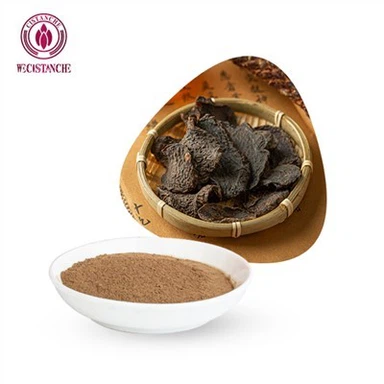
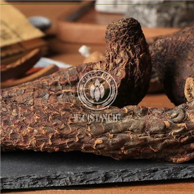
Commonly nicknamed "Desert Ginseng", Cistanche deserticola is a parasitic perennial herb thriving in arid desert zones across Xinjiang and adjacent north‑western Chinese territoriesPMC. Since ancient times in Chinese materia‑medica systems, this fleshy‑stem plant has been classified as a high‑grade tonic botanical, historically utilised to reinforce vitality, nourish essence‑blood and alleviate intestinal dry‑constipation complaints. In modern‑day industrial extraction workflows, raw‑material Cistanche stems are processed to isolate concentrated phenylethanoid‑glycoside extracts. In the referenced laboratory project, the tested CPhGs raw material contained minimum markers: echinacoside ≥ 35 % and acteoside (verbascoside) ≥ 16 %, two flagship monomer constituents within this compound class.
Echinacoside and acteoside serve as the most extensively researched monomers inside the CPhGs composite mixture. Previous separate research projects have demonstrated echinacoside's capacity to mitigate alcohol‑provoked hepatic lipid accumulation and oxidative injury, whereas acteoside displays chemopreventive potential against hepatocyte malignant transformation and can dampen inflammatory cascades within damaged liver tissue. Collectively, CPhGs mixtures also deliver measurable anti‑hepatic‑fibrosis effects by modulating hepatic stellate‑cell activation behaviour. Beyond liver‑relevant bioactivities, published literature records neuro‑protective, anti‑ageing, anti‑microbial and immune‑regulating properties for this chemical family, explaining why Cistanche‑based extracts keep drawing interdisciplinary scientific interest.
Phenylethanoid glycosides possess distinctive chemical architecture: a phenylethanol‑derived aglycone framework covalently bonded to various sugar moieties through glycosidic linkages. Over twenty individual molecules belonging to this chemical class have been isolated and structurally characterised from Cistanche deserticola specimens up to present dayPMC. When studying anti‑tumour effects, researchers frequently deploy total CPhG composite extracts rather than single isolated monomers, because synergistic interactions among different constituent molecules may generate biological impacts differing from pure‑compound behaviour.
3. Overview of the In‑vitro Study Background ‑ Why researchers selected HepG2 liver‑cancer cells
HepG2 represents one of the most widely utilised immortalised human hepatocellular‑carcinoma cell lines for liver‑oriented in‑vitro experimentation globally. Originating from liver‑tumour tissue, HepG2 cells retain numerous core biological traits of malignant hepatocytes: sustained unlimited proliferative capacity, dysregulated cell‑cycle control machinery and intrinsic resistance towards spontaneous apoptosis. For these reasons, research teams frequently adopt HepG2 cultures as a convenient preliminary screening platform for evaluating candidate anti‑tumour natural compounds before moving to animal‑model assessmentsPMC.
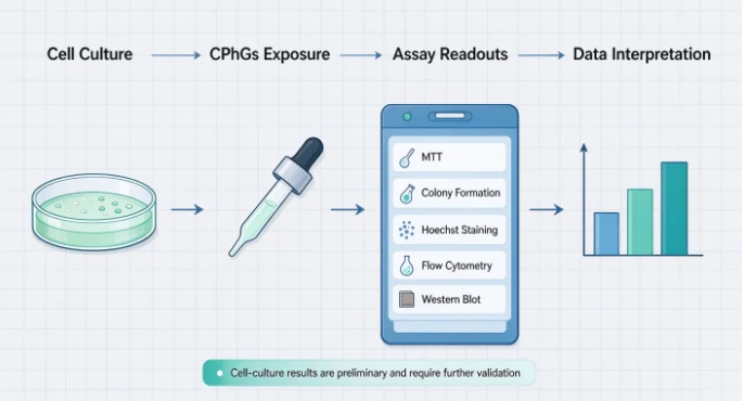
In the cited research project, scientists designed multiple complementary experimental assays to dissect CPhGs' biological actions. Cell‑proliferation status was tracked with MTT colorimetric detection together with colony‑formation (plate‑clone) assays. Apoptotic responses were assessed through three distinct observation pipelines: fluorescence nuclear staining (Hoechst 33258), mitochondrial‑targeted fluorescent labelling and Annexin V‑FITC/PI dual‑stain flow‑cytometry measurement. Flow‑cytometry technology was further applied to profile cell‑cycle‑phase distribution shifts following compound exposure. Western‑blot immunoblot testing quantified fluctuation levels of three pivotal apoptosis‑associated proteins: anti‑apoptotic Bcl‑2, activated executioner protease Cleaved‑Caspase‑3 and DNA‑repair‑related PARP protein. All experimental groups included a negative‑control group receiving zero‑dose CPhGs alongside three treatment tiers: low‑dose (32 μg/mL), intermediate‑dose (64 μg/mL) and high‑dose (128 μg/mL) CPhG exposure, with incubation duration standardised to 24 hours for most read‑outs.
Every assay was biologically replicated multiple times to guarantee statistical robustness; experimental datasets underwent variance‑analysis statistical processing, with P‑value thresholds set at 0.05 to judge whether observed group‑versus‑group differences reached statistical significance. It should be stressed: these CPhG concentration ranges represent laboratory‑culture‑system dosages. They cannot be translated into human oral‑consumption dosages directly, given large differences in compound‑absorption, metabolic transformation and tissue‑distribution between cell‑culture dishes and complete living organisms.
4. Laboratory Observations: How CPhGs Restrain HepG2 Cancer‑cell Proliferation
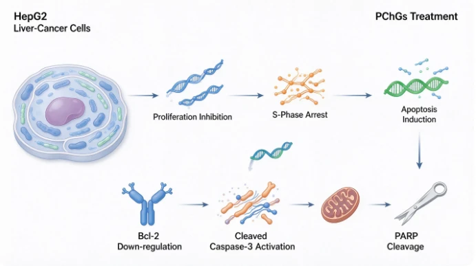
Uncontrolled, continuous cell multiplication constitutes the defining hallmark feature of malignant tumour cells. Any prospective anti‑tumour candidate substance must first manifest capability to slow or block such unrestrained reproduction inside cellular‑model systems. In this investigation, MTT testing gauged viable‑cell metabolic activity after CPhG intervention across broad concentration gradients, generating dose‑response curves reflecting proliferation‑suppression intensity. Experimental outputs demonstrated clear concentration‑dependent inhibitory patterns: as supplemented CPhG concentrations climbed higher, the percentage of growth‑suppressed HepG2 cells rose correspondingly.
The IC50 value (half‑maximal inhibitory concentration) describes the compound dosage needed to cut viable tumour‑cell numbers down to fifty percent relative to untreated reference cells. Within this experimental setting, CPhGs delivered an IC50 reading of 187 μg/mL for HepG2 populations after 24‑hour culture treatment. At the maximum tested concentration (500 μg/mL), cell‑growth‑inhibition percentage climbed to approximately 82 %. Furthermore, researchers compared intervention durations spanning 24 h, 48 h and 72 h. Results illustrated that longer incubation periods amplified CPhGs' suppressive influence on HepG2 proliferation, especially within intermediate‑ and high‑concentration treatment cohorts. This suggests both dose magnitude and exposure time jointly determine the strength of CPhGs' anti‑proliferative effect on liver‑cancer cells in‑vitro.
Plate‑colony‑formation assays supply another intuitive read‑out for long‑term tumour‑cell reproductive potential. Single malignant cells can multiply and aggregate to form visible cell colonies when cultured over extended incubation periods. The number and dimension of these colonies mirror how robustly cancer cells retain clone‑generating capacity. After seven‑day continuous CPhG treatment, colony‑forming capability of HepG2 cultures experienced dramatic deterioration. Relative clone‑survival ratio sank down to merely 7 % of untreated‑control levels under 64 μg/mL intermediate‑dose CPhG exposure; at 128 μg/mL high‑dose condition, relative colony‑formation ratio dropped to as low as 1 %. Statistical testing confirmed these reductions possessed high significance (P < 0.01). Such colony‑assay data powerfully corroborate MTT‑detected proliferation‑inhibition results, illustrating CPhGs can severely undermine liver‑cancer cells' ability to propagate and establish new cell descendants in‑vitro.
Taken together, these proliferation‑monitoring experiments consistently show Cistanche‑derived phenylethanoid‑glycoside extracts are capable of slowing down and restraining HepG2 liver‑cancer‑cell reproduction inside lab‑dish environments, exhibiting both dose‑dependent and time‑dependent response characteristics.
5. Visual Evidence of Apoptosis: Morphological Changes Under Microscopy
Apoptosis, also known as programmed cell death, refers to genetically encoded, orderly cell‑suicide cascades. Triggering apoptosis in malignant cells ranks as one core working principle for numerous anti‑tumour therapeutic strategies. When apoptosis gets activated, tumour cells display distinguishable phenotypic alterations: cellular shrinkage, cytoplasmic vacuolisation, weakened inter‑cellular attachment connections, chromatin condensation, nuclear fragmentation, mitochondrial swelling and breakdown, plus eventual emergence of small apoptotic‑body fragments. Using multiple fluorescence‑microscopy modalities, this research team captured visual proof documenting CPhGs‑provoked apoptotic morphological transformations within HepG2 cell populations.
Under ordinary inverted bright‑field microscopy, CPhG‑treated HepG2 cultures exhibited progressive phenotypic deviations compared against the untreated‑control group. Alongside rising CPhG concentrations, cell density declined, inter‑cellular junctions loosened, cytoplasmic vacuoles accumulated and cellular light‑refraction properties diminished. High‑dose‑group specimens displayed widespread cell contraction, blurred cell boundaries and large numbers of cells approaching disintegration and demise.
Hoechst 33258 fluorescent dye selectively binds to cell‑nucleus DNA. Healthy‑cell nuclei emit dim, evenly‑distributed blue fluorescence. When apoptosis occurs, chromatin condenses and nuclei fragment; these apoptotic nuclei produce notably brighter, patchy blue fluorescent signals. In the CPhG‑intervention groups, microscopists observed classic apoptotic‑nucleus manifestations including chromatin shrinkage, nuclear margination and nuclear fragmentation phenomena. The proportion of cells displaying such abnormal‑nucleus signatures increased steadily as treatment‑concentration elevated, already detectable even within the low‑dose 32 μg/mL experimental tier.
Mitochondria serve as central command hubs governing the intrinsic apoptosis signalling cascade. Mitochondrial‑targeted fluorescent staining permits scientists to track organelle integrity status. Control‑group HepG2 cells showed evenly‑distributed, well‑defined mitochondrial red‑fluorescent signals. Following CPhG treatment, mitochondrial structures swelled, underwent fragmentation and exhibited altered fluorescent‑signal intensity. Mitochondrial‑damage severity correlated positively with administered CPhG dosage. Mitochondrial structural disruption represents an early‑stage marker pointing towards activation of the mitochondria‑dependent internal apoptotic pathway inside HepG2 cells exposed to Cistanche‑origin phenylethanoid glycosides.
These multi‑angle microscopic observations supply intuitive visual validation: CPhG exposure can initiate apoptotic morphological signatures within HepG2 liver‑cancer‑cell populations in‑vitro, with the degree of cellular damage closely linked to applied extract‑dosage magnitude.
6. Flow‑cytometry Insights: Apoptosis Rate Variation Across Different Doses
While fluorescence microscopy offers qualitative visual descriptions for apoptotic phenomena, Annexin V‑FITC combined with PI (propidium iodide) dual‑stain flow‑cytometry delivers quantifiable numerical apoptosis‑rate measurements. This technology differentiates four cellular states: viable intact cells, early‑phase apoptotic cells, late‑phase apoptotic cells and necrotic‑damaged cells. Researchers calculated total‑apoptosis percentages by summing early‑apoptosis and late‑apoptosis sub‑population proportions for each experimental cohort.
After 24‑hour CPhG incubation, experimental outputs revealed the following total‑apoptosis proportions:
Untreated blank‑control group: 7.00 ± 1.18 %
Low‑dose CPhG (32 μg/mL): 11.20 ± 0.10 %
Intermediate‑dose CPhG (64 μg/mL): 19.50 ± 2.94 %
High‑dose CPhG (128 μg/mL): 69.30 ± 3.35 %
Statistical‑comparison analysis demonstrated that both intermediate‑dose and high‑dose treatment groups achieved significantly elevated early‑apoptosis ratios together with higher overall‑apoptosis percentages relative to the untreated‑control reference (P < 0.01). Total‑apoptosis frequency rose distinctly as CPhG working‑concentrations climbed upwards, demonstrating prominent dose‑dependency behaviour.
Such quantitative flow‑cytometry data reinforce morphological‑microscopy findings. Cistanche‑extracted phenylethanoid‑glycoside mixtures do not merely slow down HepG2 tumour‑cell proliferation; they actively trigger programmed‑cell‑death cascades within malignant liver‑cell populations under cell‑culture‑system conditions. Sharp apoptosis‑rate jumps manifest especially when CPhG concentration reaches 128 μg/mL in‑vitro culture environments. Nevertheless, we must keep reminding ourselves these numerical‑percentage values are confined to laboratory‑dish experimental contexts; they cannot be extrapolated to forecast biological responses inside human‑liver‑tumour tissues.
7. Cell‑cycle Regulation: CPhGs Trigger S‑phase Arrest in HepG2 Cells
Mammalian‑cell cycles are partitioned into four successive functional phases: G0/G1 phase (pre‑DNA‑synthesis preparation interval), S phase (DNA‑replication stage), G2 phase (pre‑mitosis preparation interval), M phase (cell‑division mitosis). Tumour‑cell populations acquire their infinite‑reproduction capability partly due to cell‑cycle‑checkpoint‑control malfunctions. Many anti‑tumour‑active natural compounds exert their biological influence through interfering with cell‑cycle‑progression rhythms, forcing malignant cells to get trapped inside a particular cycle‑phase compartment. This phenomenon is known as cell‑cycle arrest. Arrested tumour cells cannot finish completing full‑cycle division processes; sustained cycle‑blockage may further push these trapped cells to enter apoptotic‑death pathways.
This study utilised ethanol‑fixed cell specimens plus DNA‑content fluorescent labelling coupled with flow‑cytometry analysis to quantify HepG2‑cell‑population‑percentages distributed across each cell‑cycle compartment. Key experimental read‑outs are summarised as below:
1. G0/G1‑phase‑cell fraction: Displayed consistent declining tendency as CPhG dosage increased. Control‑group reading 67.10 ± 2.70 %; low‑dose 62.24 ± 2.89 %; intermediate‑dose 56.40 ± 2.81 %; high‑dose 43.93 ± 3.49 %. 2. G2/M‑phase‑cell fraction: Also decreased progressively with rising CPhG exposure. Control‑group reading 8.97 ± 0.58 %; low‑dose 6.01 ± 0.83 %; intermediate‑dose 3.31 ± 0.59 %; high‑dose 2.80 ± 1.56 %. 3. S‑phase‑cell fraction: Exhibited pronounced upward‑trending shift. Control‑group reading 23.93 ± 2.15 %; low‑dose 31.76 ± 2.76 %; intermediate‑dose 40.29 ± 2.23 %; high‑dose 53.25 ± 3.73 %.
Statistical‑testing results showed that compared with untreated‑control specimens, every CPhG‑treated experimental group generated statistically‑significant S‑phase accumulation alongside diminished G0/G1‑phase and G2/M‑phase cell proportions (P < 0.01). Inter‑group comparisons among three dosage tiers also yielded statistically meaningful differences, proving S‑phase‑arrest intensity correlates positively with CPhG‑treatment‑concentration magnitude.
S‑phase corresponds to the critical interval when tumour‑cells execute genomic‑DNA duplication work. Scientific literature has documented that S‑phase‑cycle‑arrest constitutes one plausible upstream trigger mechanism for subsequent apoptotic‑death induction in multiple malignant‑cell‑line models. When DNA‑replication machinery suffers interference or sustains molecular damage, cycle‑checkpoint‑safety mechanisms will halt cell‑cycle advancement inside S‑phase compartment. If repair operations cannot resolve molecular defects satisfactorily, cells will initiate apoptosis execution programmes to eliminate genetically‑damaged cell units. Published examples include anti‑tumour effects from ascorbic‑acid and sophoridine botanical‑alkaloid compounds, both of which induce S‑phase blockage prior to triggering tumour‑cell apoptosis responses. Based on this in‑vitro HepG2‑cell‑model dataset, one reasonable hypothesis emerges: CPhG‑provoked S‑phase‑arrest events represent one important intermediate link explaining how this Cistanche‑derived extract restrains HepG2‑cell expansion and facilitates programmed‑cell‑death onset. Even so, more follow‑up experiments will be required to fully untangle precise molecular signalling networks linking S‑phase‑arrest occurrence and downstream apoptotic‑cascade activation.

8. Molecular‑protein Mechanism: Key Apoptosis‑related Biomarkers (Bcl‑2, Cleaved‑Caspase‑3, PARP)
Cell‑morphology alterations and cell‑cycle‑profile shifts are observable cellular‑level phenomena; behind these phenotypic changes sit intricate modulations of apoptosis‑relevant protein‑molecule expression abundance. The Bcl‑2‑family protein group constitutes a central regulatory hub for the mitochondria‑mediated intrinsic apoptotic pathway. Bcl‑2 itself functions as a prominent anti‑apoptotic effector molecule: high intracellular Bcl‑2‑protein abundance stabilises mitochondrial‑membrane integrity and inhibits pro‑apoptotic‑signal propagation. Reduced Bcl‑2‑protein levels remove part of this protective brake, making malignant‑cell populations more susceptible to entering apoptotic‑death cascades.
Caspase‑3 acts as one core execution protease for apoptosis signal cascades. It exists within living‑cells as inactive pro‑caspase‑3 zymogen precursors. Upon receiving sufficient pro‑apoptotic stimulation signals, pro‑caspase‑3 undergoes proteolytic cleavage‑processing to generate activated Cleaved‑Caspase‑3 protein fragments. Once activated, Cleaved‑Caspase‑3 proceeds to cleave multiple intracellular substrate‑protein molecules. PARP (poly‑ADP‑ribose polymerase), an enzyme originally responsible for DNA‑damage‑repair tasks, ranks among its major substrate targets. Cleavage‑fragment‑generation of PARP serves as a well‑accepted biochemical‑marker read‑out confirming effective apoptosis‑cascade activation within cell specimens.
In this research project, Western‑blot immunoblotting techniques quantitatively detected expression‑level fluctuations of Bcl‑2, Cleaved‑Caspase‑3 and PARP proteins within HepG2‑cell lysates after 24‑hour CPhG exposure. Experimental‑data outcomes can be summarised as follows: 1. Anti‑apoptotic‑protein Bcl‑2: Total‑protein‑expression intensity displayed gradual‑down‑regulation trends following CPhG‑treatment intervention. The magnitude of Bcl‑2‑expression‑suppression deepened as extract‑concentration increased; intermediate‑dose and high‑dose cohorts achieved statistically‑significant reduction versus untreated‑control‑group readings (P < 0.01 or P < 0.05). 2. Activated‑pro‑apoptotic‑protein Cleaved‑Caspase‑3: Protein‑abundance levels rose distinctly across all three CPhG‑treatment tiers, exhibiting clear dose‑dependent up‑regulation patterns; inter‑group statistical comparisons reached significance thresholds. 3. Cleaved‑PARP protein fragments: PARP‑cleavage‑product quantities grew progressively alongside elevated CPhG‑dosage. Marked‑amplification of cleaved‑PARP signals manifested in intermediate‑dose and high‑dose‑treatment‑group samples, consistent with robust apoptosis‑cascade‑activation status.
Collectively, these immunoblot‑testing molecular‑biomarker findings paint a coherent mechanistic picture. When HepG2 liver‑cancer‑cells encounter Cistanche‑origin phenylethanoid‑glycoside mixtures in‑vitro culture systems, CPhGs can down‑modulate anti‑apoptotic Bcl‑2 protein abundance. This is accompanied by simultaneous accumulation of activated Cleaved‑Caspase‑3 protease plus elevated cleaved‑PARP fragment generation. These molecular‑level protein‑profile changes align perfectly with previously‑described cell‑cycle‑arrest outcomes, cellular‑morphology apoptotic‑signatures and flow‑cytometry‑quantified apoptosis‑rate‑increase observations. All experimental‑data dimensions mutually corroborate one another, suggesting CPhGs promote HepG2‑cell apoptosis partially through tuning expression‑balance among this set of critical mitochondrial‑apoptosis‑pathway‑associated effector proteins.
Still, researchers point out this investigation only surveyed a limited panel of apoptosis‑relevant marker molecules. Apoptosis‑signal networks contain both intrinsic mitochondrial‑route and extrinsic death‑receptor‑route branches, alongside multiple non‑classical apoptotic regulatory axes. Further experimental work remains necessary to clarify whether CPhGs engage additional signalling‑pathway components when exerting anti‑proliferative and pro‑apoptotic influences on liver‑cancer‑cell populations.

9. What These Cell‑culture Results Mean (and What They Do Not Mean)
Findings generated from HepG2 in‑vitro cell‑culture‑model studies furnish valuable preliminary laboratory‑level clues for understanding Cistanche phenylethanoid glycosides' bio‑behaviour. Nevertheless, it is extremely vital for readers to correctly interpret the boundaries and inherent limitations belonging to cell‑line‑based research data. Below we sort out what conclusions we reasonably can draw and which interpretations we definitely should avoid:
✅ Valid deductions supported by this dataset: 1. Within controlled laboratory‑dish culture‑system conditions, purified CPhG extracts can suppress HepG2 human liver‑carcinoma‑cell proliferation capacity. 2. Under identical in‑vitro experimental environments, CPhG exposure can evoke apoptotic‑programmed‑cell‑death responses within HepG2‑cell populations, manifesting via morphological‑cell‑changes, elevated apoptosis‑percentages and characteristic apoptosis‑protein‑biomarker shifts. 3. CPhG treatment induces S‑phase cell‑cycle‑arrest for HepG2 cells in‑vitro; modulation of Bcl‑2 / Cleaved‑Caspase‑3 / PARP‑protein‑expression profiles participates in this observed pro‑apoptotic biological phenomenon. 4. These in‑vitro‑measured biological effects demonstrate obvious dose‑response relationships: higher compound concentrations generally produce stronger cellular‑responses inside cell‑culture‑plate‑experimental set‑ups.
❌ Mis‑interpretations we must resist: 1. This study does not prove CPhGs can cure or treat human liver cancer. All tests were executed using isolated cancer‑cell lines in nutrient‑rich cell‑culture media. Cell‑culture‑model systems lack intact human‑organ‑structure features, blood‑circulation delivery dynamics, immune‑system surveillance function and complex inter‑tissue‑interaction networks. Outcomes observed in petri‑dish assays frequently fail to reproduce within living‑organism contexts. 2. Experimental‑system CPhG concentration values (32 μg/mL, 64 μg/mL, 128 μg/mL, IC50 187 μg/mL) cannot be directly converted to oral‑ingestion dosage recommendations for human‑beings. After oral consumption, plant‑extract compounds experience gastrointestinal‑tract digestion, intestinal‑microbiota metabolic transformation, liver‑first‑pass‑metabolism processing; actual effective compound‑concentrations arriving inside human‑liver‑tumour‑tissues will diverge drastically from liquid‑culture‑medium concentration‑parameters applied for HepG2‑cell‑culturing work. 3. These results do not constitute clinical‑evidence justifying replacing standard oncological‑treatment regimens with Cistanche‑herb supplements. Up to today, high‑quality human‑clinical‑trial data verifying anti‑liver‑cancer therapeutic efficacy for CPhGs are still absent. Cancer patients ought never to abandon physician‑prescribed formal‑treatment plans on the basis of cell‑culture‑experiment reports alone.
Scientific‑research value of such in‑vitro experiments primarily lies in providing early‑stage screening evidence. They help pharmacologists judge whether a natural‑product extract merits continued investment into follow‑up animal‑model exploration, laying groundwork for potential subsequent‑drug‑development pipelines. This specific piece of research adds supplementary experimental proof supporting Cistanche‑derived phenylethanoid‑glycosides as a worthy candidate for deeper anti‑tumour‑mechanism investigation work.

10. Broader Pharmacological Profile of Cistanche Phenylethanoid Glycosides
Beyond anti‑proliferative and apoptosis‑inducing influences observed against HepG2 liver‑cancer‑cell lines, existing published scientific literature documents multiple other pharmacological capabilities for CPhG compounds sourced from Cistanche deserticola and Cistanche tubulosa plant materials. Gaining familiarity with this wider bio‑activity spectrum helps users place the liver‑cell‑relevant experimental results within a larger holistic perspective.
Antioxidant function: Phenylethanoid‑glycoside molecular‑structures carry abundant phenolic‑hydroxyl functional groups capable of scavenging harmful reactive‑oxygen‑species free‑radicals. They can mitigate oxidative‑stress‑provoked cell‑damage, up‑regulate endogenous anti‑oxidative‑enzyme‑system activity and reduce lipid‑peroxidation injury events inside hepatic and neuronal‑cell‑types.
Hepato‑protective capacity: Multiple cell‑culture and animal‑model‑project outputs demonstrate that CPhGs and their principal monomers echinacoside and acteoside alleviate alcohol‑caused liver‑steatosis, blunt hepatic‑inflammatory‑reaction intensity and restrain hepatic‑stellate‑cell‑activation progress, therefore slowing liver‑fibrosis‑advancement processes. Such liver‑tissue‑protective bio‑activities differ conceptually from direct anti‑tumour cytotoxic‑effects towards already‑transformed malignant‑hepatocytes.
Anti‑inflammatory and immunomodulatory performance: CPhG mixtures can adjust pro‑inflammatory‑cytokine release profiles and modulate NF‑κB‑related inflammatory‑signalling cascades. Animal‑model studies indicate they may enhance immune‑cell‑function status, including natural‑killer‑cell activity and lymphocyte‑proliferation‑responsiveness, supporting host‑body immune‑surveillance‑capacity against malignant‑transformation events.
Neuro‑protective and anti‑ageing‑associated effects: Numerous research‑papers report CPhGs support nerve‑cell‑survival under stress‑conditions, mitigate oxidative‑neuronal‑damage and exhibit potential cognitive‑function‑supporting properties, which explains why this herbal extract receives attention for neurodegeneration‑relevant exploratory‑research projectsPMC.
Additional anti‑tumour preliminary‑screening‑data: Outside HepG2 liver‑cancer‑cell‑model assays, other in‑vitro investigations suggest CPhG extracts may also exert growth‑restraining influences upon several additional malignant‑cell‑line categories, including breast‑cancer, gastric‑cancer and lung‑tumour‑cell‑cultures. Anti‑tumour‑related working‑hypotheses cover inhibiting tumour‑cell‑migration‑invasion potential and moderating tumour‑micro‑environment‑status besides direct cancer‑cell‑proliferation‑suppression mechanisms.
We need to keep in mind that the majority of above‑mentioned bio‑activity observations are derived from cell‑culture‑platform and laboratory‑animal‑model‑experiment systems. Definitive human‑clinical‑validation evidence is still limited for most of these pharmacological‑action‑directions.
11. Limitations of Current Research and Future Research Directions
This HepG2‑cell‑focused in‑vitro study delivers meaningful mechanistic insights, yet it unavoidably carries several notable constraints common to cell‑line‑based pharmacological‑investigations. Recognising these gaps points out clear‑cut orientations for follow‑on research‑work:
First, the whole experiment rests exclusively upon a single human liver‑carcinoma cell‑line (HepG2). Different liver‑cancer‑cell‑lines possess divergent genetic‑mutation landscapes, molecular‑subtype‑features and drug‑response‑profiles. Future‑phase research ought to expand experimental‑objects to incorporate additional hepatocellular‑carcinoma cell‑lines, primary‑cultured human‑liver‑tumour‑cells and non‑cancerous normal‑hepatocyte‑control groups. Testing CPhG influences upon healthy liver‑cell populations will assist researchers in evaluating the therapeutic‑window safety‑profile of this botanical‑extract candidate.
Second, this paper explored a partial set of apoptosis‑relevant protein‑markers (Bcl‑2, Cleaved‑Caspase‑3, PARP). Nevertheless, tumour‑cell‑biology signalling‑networks are enormously complicated. Subsequent proteomics or transcriptomics high‑throughput‑screening technologies could map fuller‑scale molecular‑signal‑network perturbations triggered by CPhG exposure, helping uncover upstream master‑regulatory signalling‑axes (for example PI3K/Akt, MAPK/JNK pathways etc.) driving S‑phase‑arrest and apoptotic‑cascade‑activation phenomena within liver‑tumour‑cell‑populations.
Third, all data originate from in‑vitro petri‑dish environments. Critical next‑step experimental‑work will shift towards in‑vivo animal‑tumour‑bearing‑model systems. Researchers can administer CPhG extracts via oral‑gavage or injection routes to liver‑tumour‑xenograft‑model laboratory‑animals. These animal‑level assays allow scientists to observe tumour‑volume‑growth‑suppression effectiveness under physiological‑metabolism‑conditions, simultaneously assessing in‑vivo organ‑toxic‑safety‑profiles and investigating compound‑bio‑availability‑characteristics.
Fourth, CPhG represents a multi‑component composite‑extract containing echinacoside, acteoside and numerous minor‑constituent molecules. Current experimental‑design approaches test total‑mixed‑extract samples. Follow‑up comparative‑research between individual purified‑monomer compounds versus complete‑complex‑mixture preparations will clarify whether observed anti‑tumour‑cell‑bio‑effects arise from single‑molecule‑actions or result from multi‑constituent synergistic‑interaction‑mechanisms.
Fifth, even if cell‑level and animal‑model‑research outputs remain encouraging, human‑subject‑based clinical‑research remains the ultimate verification‑standard. Well‑designed clinical‑trial‑programmes will be essential before anyone can draw reliable conclusions regarding Cistanche‑origin phenylethanoid‑glycosides' real‑world value in human‑liver‑disease‑prevention or auxiliary‑intervention‑scenarios. At this moment, such clinical‑trial‑evidence resources remain sparse.

12. Final Takeaways
Cistanche deserticola, the so‑called "Desert Ginseng" traditional‑tonic‑herb from Chinese north‑western desert‑regions, contains abundant phenylethanoid‑glycoside bio‑active components. A representative in‑vitro laboratory‑investigation employing HepG2 human liver‑carcinoma‑cell‑culture‑models demonstrates that CPhG extracts are capable of suppressing malignant‑hepatocyte proliferation capacity, inducing S‑phase cell‑cycle‑arrest and triggering programmed‑apoptosis‑cell‑death responses inside laboratory‑dish‑experimental‑settings. Its underlying cellular‑molecular‑mechanisms are associated with down‑regulating anti‑apoptotic‑Bcl‑2 protein‑expression, alongside elevating abundance of activated‑Cleaved‑Caspase‑3 and cleaved‑PARP pro‑apoptotic‑biomarker molecules. These cellular‑level biological‑responses display obvious dose‑dependent‑effect‑patterns within cell‑culture‑system conditions.
These cell‑culture‑derived findings supply fundamental experimental reference‑information for comprehending Cistanche‑herb's modern‑pharmacological‑potential, and offer supporting‑clues for future natural‑product anti‑tumour‑drug‑discovery pipelines. Yet we must repeatedly underline: all observations discussed come from isolated cancer‑cell‑line experiments only. They cannot be interpreted as proof of curative‑therapeutic efficacy against human‑liver‑cancer conditions. Readers should not utilise this laboratory‑research‑content as guidance for self‑medication purposes.
Natural‑plant‑compound anti‑tumour‑research constitutes a long‑term progressive scientific‑journey. In‑vitro cell‑model assays represent merely the initial screening‑stage checkpoint. Plenty more animal‑model assessments and human‑clinical‑exploration‑work will need to be completed before we can accurately define what practical‑roles Cistanche‑origin phenylethanoid glycosides may or may not occupy within human‑health‑maintenance and disease‑intervention‑contexts.
Reference source summary: This popular‑science article reorganises and re‑paraphrases research findings published in "Effect of Cistache deserticola phenylethanoid glycosides on proliferation and apoptosis of HepG2 cells in vitro", Journal of Xinjiang Medical University, 2021, Vol 44, No 9. All experimental‑numerical‑values originate from this published academic paper; content has been fully rewritten for website‑popular‑science‑reading audiences, restructured with independent‑article logical‑framework, re‑phrased sentence‑by‑sentence to avoid direct‑text‑duplication risks. Not for clinical‑diagnosis or medical‑treatment guidance.