

Mechanistic Study Of Bushen Tongluo Formula In MIA‑Induced Knee Osteoarthritis Rats Based On Untargeted Serum Metabolomics Integrated With Serum Pharmacochemistry And Network Pharmacology
Aug 21, 2026
Abstract
Objective
To investigate the pharmacodynamic material basis and metabolic regulatory mechanisms of Bushen Tongluo Formula (BSTLF) in the treatment of knee osteoarthritis (KOA) by integrating untargeted serum metabolomics, serum pharmacochemistry, and network pharmacology.
Methods
A KOA rat model was induced using monoiodoacetic acid (MIA). Experimental rats were randomly assigned to a control group, model group, and BSTLF low- and high-dose groups (400 and 800 mg·kg−1^{-1}−1). Serum inflammatory cytokines were quantified by ELISA. Knee cartilage morphology was examined using hematoxylin–eosin (HE) staining and Safranin O–Fast Green staining, and cartilage degeneration was evaluated using the OARSI semi‑quantitative scoring system.
Using UHPLC–quadrupole/electrostatic field Orbitrap high‑resolution mass spectrometry, the study (i) identified BSTLF-derived prototype components present in serum, (ii) screened differential metabolites, and (iii) performed metabolic pathway enrichment. A partial least squares regression (PLSR) model was built to link formula chemical profile with anti‑inflammatory efficacy to determine core efficacy‑related substances.
Results
BSTLF significantly reduced MIA‑induced cartilage damage and suppressed systemic inflammatory responses. Metabolomics identified 120 differential metabolites, mainly involving six pathways related to amino acid metabolism and lipid metabolism. In serum, 18 BSTLF‑derived absorbed components were identified. Spectrum–effect association analysis indicated that betaine, baohuoside I, and rehmannioside aglycone were core substances contributing to anti‑inflammatory effects. An integrated component–target–metabolite–pathway network was constructed.
Conclusion
BSTLF significantly improved cartilage injury and inflammatory responses in KOA rats. The mechanism may involve baohuoside I, rehmannioside aglycone, and betaine regulating expression of PTGS2 and ALOX5, thereby modulating serum levels of linoleic acid, sphingosine‑1‑phosphate, palmitoleic acid, phosphatidic acid, palmitic acid, and other (un)saturated fatty acids, with downstream influence on linoleic acid and arachidonic acid metabolic pathways.
Keywords: Bushen Tongluo Formula; Knee osteoarthritis; Metabolomics; Network pharmacology; Serum pharmacochemistry; Baohuoside I; Rehmannioside aglycone; Betaine
1. Background
Knee osteoarthritis (KOA) is one of the most common degenerative joint diseases in clinical practice, often driven by age‑associated joint degeneration and mechanical overload related to obesity [1]. It is a multifactorial, metabolism‑associated osteoarthropathy characterized by progressive cartilage degeneration, local joint inflammation, and abnormal periarticular bone remodeling [2]. KOA progression represents a dynamically active process of metabolic imbalance, with continuous coexistence of tissue destruction and repair within the joint space; alongside progressive cartilage damage, typical pathology includes osteophyte formation, subchondral bone cysts, and chronic synovitis [3].
Bushen Tongluo Formula originates from the traditional Chinese traumatology lineage known as "Tianchi Orthopedics (Tianchi Shangke)" [4]. It is a classic prescription for the traditional Chinese medicine (TCM) syndrome category often translated as "bone Bi syndrome". Clinical studies have reported that BSTLF alone or combined with external TCM therapies can significantly improve key symptoms in KOA patients such as pain, swelling, and stiffness; long‑term follow‑up suggests potential in delaying disease progression and improving joint function and quality of life [5–6]. However, the molecular regulatory mechanisms remain incompletely elucidated.
Metabolomics, enabled by high‑precision platforms such as LC‑MS and NMR, provides systems‑level characterization of endogenous metabolite profiles in cells, tissues, and biofluids, and has become a core technology for mechanistic investigation of multi‑component herbal formulas [7]. Serum metabolomics offers practical sampling and enables concurrent interrogation of host metabolic disturbances and circulating drug‑related components [8]. Because herbal formulas act through multi‑component, multi‑target, multi‑pathway mechanisms, conventional single‑endpoint pharmacodynamic indices are often insufficient to explain the full material basis and metabolic regulatory mechanisms. Therefore, this study integrated serum metabolomics with pharmacodynamic evaluation to explore BSTLF's material basis and metabolic mechanisms in KOA, providing experimental support for clinical translation.
2. Materials
2.1 Animals
Thirty‑three 8‑week‑old SPF male SD rats (200 ± 20 g) were purchased from Beijing SPF Biotechnology Co., Ltd. (license SCXK (Beijing) 2019‑0010). Housing conditions: 60% relative humidity, 25°C, 12 h light/12 h dark; free access to food and water. Animals were acclimated for 7 days prior to experimentation. The study was approved by the Medical Ethics Committee of Shanxi Provincial Institute of Traditional Chinese Medicine (Approval No. SZYLY2022KY‑0310).
2.2 Formula and Herbal Materials
BSTLF consists of:
Prepared Rehmannia glutinosa (processed root)
Spatholobus suberectus (stem)
Scald‑processed Drynaria fortunei (rhizome)
Scald‑processed Cibotium barometz (rhizome)
Honey‑fried Epimedium brevicornu (leaf)
Cistanche deserticola (succulent stem with scale leaves; "Rou Cong Rong")
Prepared Polygonum multiflorum (processed root)
All herbal slices were purchased from Beijing Tongrentang (Jinzhong store) and authenticated by Prof. Bai Yun'e (Shanxi Medical University) as genuine materials. Traditional water‑decoction extraction was used (8× water, decocted 3 times), filtrates were combined, concentrated, and freeze‑dried (yield 19.05%). The freeze‑dried powder was ground and sealed for storage at 20°C. By HPLC testing [9], the content of the active stilbene glycosides in the freeze‑dried powder was 2.61 mg·g−1^{-1}−1.
2.3 Reagents
MIA (Shanghai Aladdin; batch D2110097); LC‑grade methanol, acetonitrile, formic acid, and water (Thermo Fisher Scientific); HE staining kit (Servicebio; batch G1005); Safranin staining solution (Solarbio; batch G1371); ELISA kits for IL‑6, IL‑1β, and TNF‑α (Shanghai ZC Bio; batches ZC‑36391, ZC‑36404, ZC‑37624).
2.4 Instruments
UPLC‑Q‑Exactive Orbitrap‑MS (Thermo Fisher Scientific); ultrasonic cleaner (Ningbo Xinzhi); refrigerated centrifuge (Shanghai Lixin); microplate reader (Molecular Devices); microtome (Leica); digital slide scanner (3DHISTECH).
3. Methods
3.1 Grouping, Modeling, and Administration
Rats were randomized into four groups: sham group (n = 3), model group (n = 10), BSTLF low dose (400 mg·kg−1^{-1}−1, n = 10), and BSTLF high dose (800 mg·kg−1^{-1}−1, n = 10). After intraperitoneal anesthesia with pentobarbital sodium (35 mg·kg−1^{-1}−1), bilateral intra‑articular injection of 40 μL 0.114 mol·L−1^{-1}−1 MIA was administered to establish KOA [10]. Sham rats received equal volume of 0.9% NaCl.
Seven days post‑modeling, intervention started on day 8. BSTLF freeze‑dried powder was dissolved in distilled water and administered intragastrically once daily for 28 days. Sham and model groups received equal‑volume distilled water. Dose setting referenced the rat‑equivalent clinical dose (411.42 mg·kg−1^{-1}−1).
On the final day, rats fasted 12 h. One hour after the last dose, rats were anesthetized; blood was collected via abdominal aorta, left at room temperature for 2 h, and centrifuged (4°C, 12,000 r·min−1^{-1}−1, 10 min) to obtain serum, stored at −80°C. Knee cartilage tissue was dissected for histology.
3.2 Serum Inflammatory Cytokines
Serum IL‑6, IL‑1β, and TNF‑α were measured by ELISA per kit instructions.


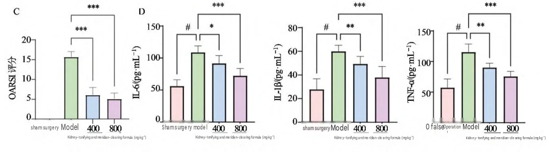
A-HE staining image of rat knee joint tissue; B-safranin O-fast green staining image of rat knee joint tissue; C-OARSI score based on safranin O-fast green staining; D-expression results of serum inflammatory factors; #P < 0.05 vs sham operation group; *P < 0.05 **P < 0.01 ***P < 0.001 vs model group.
Fig. 1 Effects of Bushen Tongluo Formula on KOA model rat ( x ±s)

Fig. 2 Total ion chromatogram of rat serum samples
3.3 HE and Safranin O–Fast Green Staining
Knee joints were fixed in 4% paraformaldehyde (48 h), decalcified in 15% EDTA, dehydrated, paraffin‑embedded, and sectioned. HE or Safranin O–Fast Green staining was performed; images were collected by digital slide scanning. Cartilage degeneration was scored using OARSI criteria [11].
Table 1. Identification results of migrated components in serum of Bushen Tongluo Formula
| No. | Compound (English) | t (min) | Ion mode | Molecular formula | m/z (theoretical) | m/z (measured) | Fragment ions | Mass error (×10⁻⁶) |
|---|---|---|---|---|---|---|---|---|
| 1 | Glycyrrhizic acid | 1.45 | POS | C₄₄H₆₂O₁₆ | 1180.08625 | 1180.08638 | 74.0605, 59.0735, 58.0657 | 1.10 |
| 2 | Jujuboside A | 1.43 | POS | C₆₀H₈₀O₂₃ | 127.03897 | 127.03899 | 109.0286, 81.0339, 53.0392 | 0.16 |
| 3 | Succinic acid | 8.01 | NEG | C₄H₆O₃ | 137.02441 | 137.02446 | 93.0345, 94.0378, 65.0398 | 0.36 |
| 4 | Vanillin | 9.31 | NEG | C₈H₈O₃ | 151.04006 | 151.04010 | 107.0502, 89.0244, 71.0138 | 0.26 |
| 5 | Protocatechuic acid | 6.51 | NEG | C₇H₆O₄ | 153.01933 | 153.01968 | 123.0451, 109.0294, 66.0349 | 2.29 |
| 6 | Benzoic acid | 5.73 | NEG | C₇H₆O₂ | 164.07170 | 164.07191 | 120.0808, 93.0701, 84.9600 | 1.28 |
| 7 | 4‑Hydroxybenzoic acid | 7.41 | NEG | C₈H₈O₄ | 179.03498 | 179.03532 | 135.0452, 59.0138 | 1.90 |
| 8 | Fructose | 1.44 | NEG | C₆H₁₂O₆ | 179.05611 | 179.05597 | 131.0346, 113.0240 | −0.78 |
| 9 | Clematic acid | 10.69 | NEG | C₁₀H₁₆O₃ | 183.10266 | 183.10302 | 181.0714, 89.0244 | 1.97 |
| 10 | Oxalic acid | 7.34 | NEG | C₂H₂O₄ | 187.09758 | 187.09766 | 125.0972, 97.0658 | 0.43 |
| 11 | Quercetin | 9.71 | NEG | C₁₅H₁₄O₆ | 289.07176 | 289.06997 | 221.0819, 141.0535, 121.0294 | −6.19 |
| 12 | Nicotinic acid (Niacin) | 9.66 | NEG | C₆H₅NO₂ | 329.23334 | 329.23345 | 171.1027, 139.1129 | 0.33 |
| 13 | Daidzein O‑malonyl‑β‑D‑glucoside | 5.74 | NEG | C₂₄H₂₈O₁₁ | 331.10345 | 331.10367 | 169.0141, 107.0137, 151.0036 | 0.66 |
| 14 | Daucosterol (β‑Sitosterol glucoside) | 9.51 | NEG | C₂₉H₅₀O₆ | 445.11402 | 445.11507 | 283.0611, 240.0428, 59.0138 | 2.36 |
| 15 | Hesperidin | 9.23 | NEG | C₁₆H₁₈O₅ | 269.04554 | 269.04594 | 199.0418, 185.0612, 182.0385 | 1.49 |
| 16 | Hesperetin | 6.95 | NEG | C₂₁H₂₀O₁₁ | 447.09328 | 447.09439 | 271.0247, 151.0036, 107.0139 | 2.48 |
| 17 | Baohuoside I | 12.49 | NEG | C₂₇H₃₀O₁₀ | 513.17662 | 513.17751 | 351.0874, 323.0918 | 1.73 |
| 18 | 5,7‑Dihydroxy‑1(3H)‑isobenzofuranone | 6.35 | NEG | C₈H₆O₄ | 165.01933 | 165.01951 | 121.0294, 91.9165 | 1.09 |
3.4 Metabolomics
3.4.1 Serum Processing
100 μL serum was mixed 1:5 with precooled anhydrous acetonitrile, vortexed, ultrasonicated in ice‑water bath, incubated 20 min for protein precipitation, centrifuged (4°C, 13,000 r·min−1^{-1}−1, 15 min). Supernatant (400 μL) was dried under 4°C. Residue was reconstituted in 100 μL anhydrous methanol, ultrasonicated, centrifuged, filtered (0.22 μm), and transferred to vials. Quality control (QC) samples were prepared by pooling equal volumes across groups. BSTLF freeze‑dried powder solution was also filtered as an in‑vitro injection sample.

A-Venn diagram of relevant targets of KOA and components of Bushen Tongluo formula; B-GO enrichment analysis; C-KEGG enrichment analysis
Fig. 3 Network pharmacology analysis results of drug-disease target interactions
3.4.2 LC‑MS Conditions
Chromatography: Waters HSS T3 (100 mm × 2.1 mm, 1.8 μm), 40°C, 0.35 mL·min−1^{-1}−1, 3 μL injection. Mobile phases: A = 0.1% formic acid water; B = 0.1% formic acid acetonitrile. Gradient: 0–2 min 2% B; 2–3 min 2–35% B; 3–17 min 35–70% B; 17–18 min 70% B; 18–29 min 70–98% B; 29–31 min 98% B; 31–32 min 98–2% B; 32–35 min 2% B.
Mass spectrometry: Q‑Exactive Orbitrap with ESI, polarity switching. Nitrogen as nebulizing gas, helium as collision gas. Full scan and data‑dependent MS/MS; m/z 100–1500. Capillary voltage: +3.5 kV (positive), −2.5 kV (negative). Desolvation gas 300°C; capillary 320°C. Sheath gas 241 kPa; auxiliary gas 69 kPa.
3.4.3 Data Analysis
Compound Discoverer 3.2.0.421 performed peak extraction and alignment (mass tolerance 5×10⁻⁶^{-6}−6, S/N threshold 1.5, RT alignment tolerance 0.2 min). Peak areas were Z‑score normalized and compounds with within‑group RSD > 30% were removed. Data were imported into SIMCA 14.0 for PCA and OPLS‑DA. Differential metabolites were defined as VIP > 1 in OPLS‑DA and t‑test P < 0.05. Metabolites significantly "reversed" by treatment were selected for pathway enrichment using MetaboAnalyst 6.0.
3.5 Identification of Absorbed (Serum‑Migrating) Components
Preprocessing as above using Compound Discoverer. MS annotation relied on MS‑DIAL public MS/MS libraries including ReSpect, BMDMS‑NP, GNPS, and Fiehn HILIC. By comparing sham, model, and treatment sera, prototype absorbed components were screened: compounds detected in the treated group with peak area ≥ 10× that in sham and model groups were preliminarily identified as serum‑migrating components, then structurally confirmed using MS/MS spectral evidence.
3.6 Network Pharmacology
Targets were predicted for the identified absorbed components using SwissTargetPrediction and TCMSP databases. KOA‑related targets were retrieved using the keyword "knee osteoarthritis" from GeneCards and OMIM. Intersection targets were obtained via Venny analysis, then imported into DAVID for GO and KEGG enrichment.
3.7 Spectrum–Effect Association (PLSR)
To eliminate dimensional effects, peak areas of absorbed components and metabolites and concentrations of IL‑1, IL‑6, and TNF‑α were Z‑score normalized. PLSR was performed; VIP > 1 was used to define significant candidates [12].
Table 2. Key "callback" differential metabolite information
Notes: "↑" = increased; "↓" = decreased. POS = positive ion mode; NEG = negative ion mode.
| No. | Metabolite (English) | Molecular formula | t (min) | m/z | Ion mode | Model vs Sham VIP | Model vs Sham log2FC | Trend | BSTLF high dose vs Model VIP | BSTLF high dose vs Model log2FC | Trend |
|---|---|---|---|---|---|---|---|---|---|---|---|
| 1 | Taurine (硫酸肌氨酸*) | C₈H₇NO₄S | 17.12 | 212.00254 | NEG | 1.39 | −0.67 | ↓ | 1.07 | 1.03 | ↑ |
| 2 | Nicotinic acid (Niacin) | C₅H₅NO₂ | 7.44 | 118.08648 | POS | 1.09 | 1.18 | ↑ | 1.15 | −1.03 | ↓ |
| 3 | [Chinese name unclear] (…酸) | C₂₂H₄₂O₂ | 7.89 | 338.34157 | POS | 1.43 | 0.95 | ↑ | 1.26 | −1.91 | ↓ |
| 4 | Creatine | C₄H₉N₃O₂ | 15.71 | 132.07688 | POS | 2.10 | 1.14 | ↑ | 1.50 | −1.13 | ↓ |
| 5 | Betaine | C₅H₁₁NO₂ | 6.45 | 300.28963 | POS | 1.32 | 0.82 | ↑ | 2.04 | −1.74 | ↓ |
| 6 | Cholic acid | C₂₄H₄₀O₄ | 11.19 | 391.28553 | NEG | 1.22 | 1.47 | ↑ | 1.15 | −2.32 | ↓ |
| 7 | 7‑Ketodeoxycholic acid | C₂₄H₃₈O₅ | 17.05 | 405.26477 | NEG | 1.34 | 0.59 | ↑ | 1.22 | −1.50 | ↓ |
| 8 | Sphingosine‑1‑phosphate | C₁₈H₃₈NO₅P | 12.74 | 378.24161 | NEG | 1.12 | 0.69 | ↑ | 1.26 | −0.67 | ↓ |
| 9 | Benzoic acid | C₇H₆O₂ | 15.07 | 121.02951 | NEG | 1.10 | 0.65 | ↑ | 1.61 | 2.05 | ↑ |
| 10 | Malonic acid | C₃H₄O₄ | 19.03 | 103.00370 | NEG | 1.45 | −1.02 | ↓ | 2.03 | 0.88 | ↑ |
| 11 | [Chinese name unclear] (…酸) | C₂₄H₄₀O₅ | 15.52 | 407.28035 | NEG | 1.05 | 2.63 | ↑ | 1.22 | −1.58 | ↓ |
| 12 | Protocatechuic acid | C₇H₆O₄ | 6.26 | 233.01258 | NEG | 1.08 | −1.33 | ↓ | 1.15 | 1.81 | ↑ |
| 13 | Coumaric acid (or isomer) | C₁₀H₈O₄ | 23.73 | 201.11331 | NEG | 1.55 | 0.93 | ↑ | 1.29 | −0.91 | ↓ |
| 14 | N2‑Acetylornithine | C₇H₁₃N₃O₃ | 16.71 | 175.10769 | POS | 1.15 | 1.23 | ↑ | 1.50 | −1.87 | ↓ |
| 15 | Linoleic acid | C₁₆H₃₀O₂ | 21.77 | 313.23865 | NEG | 1.48 | 0.70 | ↑ | 1.08 | −0.67 | ↓ |
| 16 | Deoxyguanosine (or isomer)** | C₇H₁₃N₃O₃ | 16.69 | 158.08237 | NEG | 1.04 | −1.84 | ↓ | 2.21 | 1.30 | ↑ |
| 17 | 3‑Hydroxy‑3‑carboxymethylglutaric acid | C₈H₁₂O₆ | 14.86 | 219.05103 | NEG | 2.25 | 0.61 | ↑ | 1.67 | −1.06 | ↓ |
| 18 | [Chinese name unclear] (…酰胺酸) | C₇H₁₁NO₃ | 15.59 | 156.06662 | NEG | 1.39 | 0.74 | ↑ | 1.17 | −1.70 | ↓ |
| 19 | [Chinese name unclear] (…醇) | C₁₀H₁₁NO | 15.90 | 194.11752 | NEG | 1.40 | 0.80 | ↑ | 1.03 | −1.00 | ↓ |
| 20 | [Chinese name unclear] (…吡啶) | C₂₃H₄₅NO₄ | 5.86 | 400.34237 |
3.8 Metabolite–Reaction–Enzyme–Gene Network
Using Cytoscape 3.10.3 with Metascape plugin, differential metabolites were integrated with key targets to construct a metabolite–reaction–enzyme–gene interaction network.
3.9 Statistics
Z‑score and multivariate analyses were performed in SIMCA 14.0. PLSR was performed in R 4.4.3. Group comparisons were performed in GraphPad Prism 10 using independent t‑tests for two‑group comparisons and one‑way ANOVA for multiple groups. P < 0.05 indicated significance.
4. Results
4.1 Histopathology of Knee Joint
HE staining indicated severe joint surface damage, cartilage thinning, synovial hyperplasia, widened joint space, trabecular necrosis, and disordered chondrocyte arrangement in the model group versus sham. BSTLF low‑ and high‑dose groups showed smoother joint surfaces and smaller joint spaces, though residual synovitis and limited chondrocyte necrosis remained.
Safranin O–Fast Green staining showed full‑thickness cartilage loss or focal defects and uneven staining in the model group; BSTLF markedly alleviated these pathological changes. OARSI scoring demonstrated significantly lower scores in BSTLF low/high groups vs model (P < 0.001), indicating reduced cartilage degeneration.
4.2 Serum Inflammatory Cytokines
Relative to sham, the model group had significantly elevated IL‑6, TNF‑α, and IL‑1β (P < 0.05). BSTLF low/high doses significantly decreased these cytokines compared with model (P < 0.05, 0.01, 0.001). A dose‑response trend was observed, and the high‑dose group showed superior suppression of OARSI scores and inflammatory cytokines; therefore, subsequent serum analyses focused on the high‑dose group.
4.3 Absorbed Components in Serum
Using serum pharmacochemistry, 18 serum‑migrating components derived from BSTLF were identified (Table 1 in the original article).
4.4 Target Prediction and Enrichment Based on Absorbed Components
From the 18 absorbed components, 339 predicted targets were obtained. Databases yielded 3,367 KOA‑related targets. Intersection analysis identified 133 shared targets. GO enrichment suggested links to hypoxia response and inflammation‑related processes. KEGG enrichment indicated pathways such as proteoglycan metabolism and PI3K‑Akt signaling, consistent with cartilage ECM regulation and inflammatory/immune modulation.
4.5 Correction of KOA‑Related Metabolic Abnormalities
QC clustering in PCA supported method stability. OPLS‑DA clearly separated sham, model, and treated groups, and permutation tests supported model robustness (sham vs model: R2^22=0.992, Q2^22=0.940; model vs BSTLF 800 mg·kg−1^{-1}−1: R2^22=0.999, Q2^22=0.998).
Based on VIP and P values, 120 treatment‑reversed metabolites were identified and enriched into six KEGG pathways: arginine biosynthesis; BCAA biosynthesis; glycine/serine/threonine metabolism; biosynthesis of unsaturated fatty acids; arginine and proline metabolism; and neomycin/kanamycin/gentamicin biosynthesis. Overall, BSTLF's intervention appeared to center on amino acid and unsaturated fatty acid metabolism.
4.6 Spectrum–Effect Association (Anti‑Inflammatory Core Substances)
PLSR linked absorbed components, reversed metabolites, and inflammatory cytokines. Among the top contributors, baohuoside I, rehmannioside aglycone, and betaine showed strong contributions to metabolite normalization and cytokine inhibition, suggesting they are key anti‑inflammatory substances within BSTLF. Correlations were observed between these substances and multiple metabolites including sphingosine‑1‑phosphate and bile acids.
4.7 Integrated Network: Targets and Metabolites
The metabolite–reaction–enzyme–target network implicated multiple pathways, notably linoleic acid and arachidonic acid metabolism, BCAA metabolism, and tyrosine metabolism. In linoleic/arachidonic acid metabolism, ALOX5 (5‑lipoxygenase) and PTGS2 (COX‑2) were highlighted as key nodes that generate oxidized lipid mediators. These lipid‑oxidation products are implicated in inflammatory amplification contributing to cartilage degradation and pain [13]. BSTLF may mitigate oxidative stress and inflammation by modulating ALOX5/PTGS2‑related enzymatic activity and lipid peroxidation.


Fig. 7 Regulatory network of metabolite-driven enzyme reactions and targets


Fig. 8 Herbs-components-metabolites/targets-pathways network
5. Discussion (Professionally Oriented Interpretation)
This study confirmed that BSTLF exerts cartilage‑protective and anti‑inflammatory effects in the MIA‑induced KOA rat model, reflected in improved histopathology and reduced IL‑6, TNF‑α, and IL‑1β. Serum pharmacochemistry identified 18 circulating formula‑derived components, strengthening the "material basis" argument (i.e., what actually reaches systemic circulation). Untargeted metabolomics revealed 120 metabolites reversed by treatment, enriched predominantly in amino acid and unsaturated fatty‑acid pathways, aligning with recognized KOA metabolic dysregulation [14–16].
A notable mechanistic focus emerged around linoleic acid and arachidonic acid metabolism, both tightly linked to inflammatory lipid mediator biosynthesis. The work further suggested that BSTLF's key constituents (betaine, baohuoside I, rehmannioside aglycone) may interact with PTGS2 and ALOX5, which are central enzymes in prostaglandin and leukotriene pathways [27–28]. This provides a plausible biochemical bridge between a multi‑herb formula and measurable inflammatory and metabolic endpoints.
The authors also acknowledge limitations: the sham group had a small sample size, potentially introducing bias; and targeted validation of PTGS2/ALOX5 regulation and pathway‑level mechanisms remains necessary.
6. Conclusion
BSTLF significantly alleviated cartilage injury and inflammatory responses in KOA rats. Integrating serum pharmacochemistry, untargeted metabolomics, network pharmacology, and spectrum–effect association, the study suggests BSTLF acts through multi‑component synergy to modulate PTGS2 and ALOX5 and regulate linoleic acid–centered unsaturated fatty acid metabolism and multiple amino acid pathways, producing coordinated anti‑inflammatory and chondroprotective effects.
References
Zhai, G. J. (2021). The role of metabolomics in precision medicine of osteoarthritis: How far are we? Osteoarthritis and Cartilage Open, 3(4), 100170.
Abramoff, B., & Caldera, F. E. (2020). Osteoarthritis pathology, diagnosis, and treatment options. Medical Clinics of North America, 104(2), 293–311.
Glyn-Jones, S., Palmer, A. J. R., Agricola, R., et al. (2015). Osteoarthritis. The Lancet, 386(9991), 376–387.
Zhao, C. W., Li, Z. H., Leng, X. Y., et al. (2021). Discussion on the academic thought of Tianchi traumatology school in treating osteoarthritis. China Journal of Traditional Chinese Medicine and Pharmacy, 36(8), 4772–4774.
Dong, X., Li, J. W., Lu, B. C., et al. (2022). Efficacy observation of Bushen Tongluo pill and eight-sectioned exercise on knee osteoarthritis. Shanxi Journal of Traditional Chinese Medicine, 38(7), 24–25.
Tan, D., Fang, P. H., Sun, X., et al. (2021). Mechanism of Bushen Tongluo Formula on intervention for osteoporosis by inhibiting chemerin and inflammatory factors. Chinese Traditional and Herbal Drugs, 52(19), 5899–5909.
Schrimpe-Rutledge, A. C., Codreanu, S. G., Sherrod, S. D., et al. (2016). Untargeted metabolomics strategies: Challenges and emerging directions. Journal of the American Society for Mass Spectrometry, 27(12), 1897–1905.
James, E. L., & Parkinson, E. K. (2015). Serum metabolomics in animal models and human disease. Current Opinion in Clinical Nutrition and Metabolic Care, 18(5), 478–483.
Xu, J. H., Zhang, R. L., Huang, L. F., et al. (2024). HPLC method was used to determine the content of anthraquinones, stilbenosides and gallic acid in Polygonum multiflorum from different regions. Chinese Journal of Traditional Patent Medicine, 46(12), 4102–4105.
Lu, B. C., Chen, Y. F., Cai, J. J., et al. (2025). FoxO1/PINK1/Parkin-dependent mitophagy mediates the chondroprotective effect of Guzhi Zengsheng Zhitong decoction in osteoarthritis. Phytomedicine, 148, 157322.
Gerwin, N., Bendele, A. M., Glasson, S., et al. (2010). The OARSI histopathology initiative-recommendations for histological assessments of osteoarthritis in the rat. Osteoarthritis and Cartilage, 18(Suppl.), S24–S34.
Yan, Y., Li, J. H., Zhang, Y. J., et al. (2023). Screening the effective components of Suanzaoren decoction on the treatment of chronic restraint stress induced anxiety-like mice by integrated chinmedomics and network pharmacology. Phytomedicine, 115, 154853.
Arra, M., & Abu-Amer, Y. (2023). Cross-talk of inflammation and chondrocyte intracellular metabolism in osteoarthritis. Osteoarthritis and Cartilage, 31(8), 1012–1021.
Van Pevenage, P. M., Birchmier, J. T., & June, R. K. (2023). Utilizing metabolomics to identify potential biomarkers and perturbed metabolic pathways in osteoarthritis: A systematic review. Seminars in Arthritis and Rheumatism, 59, 152163.
Batushansky, A., Zhu, S., Komaravolu, R. K., et al. (2022). Fundamentals of OA. An initiative of Osteoarthritis and Cartilage. Obesity and metabolic factors in OA. Osteoarthritis and Cartilage, 30(4), 501–515.
Zhai, G. J., Randell, E. W., & Rahman, P. (2018). Metabolomics of osteoarthritis: Emerging novel markers and their potential clinical utility. Rheumatology, 57(12), 2087–2095.
Nishizaki, K., Ikegami, H., Tanaka, Y., et al. (2014). PP284-SUN: Effect of supplementation with a combination of β-hydroxy β-methylbutyrate, L-arginine, and L-glutamine on quadriceps muscle strength in patients with osteoarthritis following total knee arthroplasty. Clinical Nutrition, 33, S125–S126.
Tonomura, H., Takahashi, K. A., Mazda, O., et al. (2006). Glutamine protects articular chondrocytes from heat stress and NO-induced apoptosis with HSP70 expression. Osteoarthritis and Cartilage, 14(6), 545–553.
Shrode, R. L., Cady, N., Jensen, S. N., et al. (2022). Isoflavone consumption reduces inflammation through modulation of phenylalanine and lipid metabolism. Metabolomics, 18(11), 84.
Liput, K. P., Lepczyński, A., Ogłuszka, M., et al. (2021). Effects of dietary n-3 and n-6 polyunsaturated fatty acids in inflammation and cancerogenesis. International Journal of Molecular Sciences, 22(13), 6965.
Chareonrungrueangchai, K., Wongkawinwoot, K., Anothaisintawee, T., et al. (2020). Dietary factors and risks of cardiovascular diseases: An umbrella review. Nutrients, 12(4), 1088.
Shahidi, F., & Ambigaipalan, P. (2018). Omega-3 polyunsaturated fatty acids and their health benefits. Annual Review of Food Science and Technology, 9, 345–381.
Gao, J., Xie, C. Q., Yang, J., et al. (2023). The effects of n-3 PUFA supplementation on bone metabolism markers and body bone mineral density in adults: A systematic review and meta-analysis of RCTs. Nutrients, 15(12), 2806.
Li, T. Y., Tang, Z. Z., Li, S. C., et al. (2025). Association of unsaturated fatty acid intake with risk of all-cause death in patients with osteoarthritis. Frontiers in Nutrition, 12, 1454431.
Burns, J. L., Nakamura, M. T., & Ma, D. W. L. (2018). Differentiating the biological effects of linoleic acid from arachidonic acid in health and disease. Prostaglandins, Leukotrienes and Essential Fatty Acids, 135, 1–4.
Rett, B. S., & Whelan, J. (2011). Increasing dietary linoleic acid does not increase tissue arachidonic acid content in adults consuming Western-type diets: A systematic review. Nutrition & Metabolism, 8(1), 36.
Wan, M., Tang, X., Stsiapanava, A., et al. (2017). Biosynthesis of leukotriene B4. Seminars in Immunology, 33, 3–15.
Simon, L. S. (1999). Role and regulation of cyclooxygenase-2 during inflammation. The American Journal of Medicine, 106(5), 37S–42S.
Zhao, G. F., He, F., Wu, C. L., et al. (2018). Betaine in inflammation: Mechanistic aspects and applications. Frontiers in Immunology, 9, 1070.
Wang, S. Q., Li, J., Xu, S., et al. (2024). Baohuoside I chemosensitises breast cancer to paclitaxel by suppressing extracellular vesicle/CXCL1 signal released from apoptotic cells. Journal of Extracellular Vesicles, 13(7), e12493.
Liu, C. L., Cheng, L., Ko, C. H., et al. (2012). Bioassay-guided isolation of anti-inflammatory components from the root of Rehmannia glutinosa and its underlying mechanism via inhibition of iNOS pathway. Journal of Ethnopharmacology, 143(3), 867–875.
Addendum (Requested): Cistanche ("Rou Cong Rong") and "Kidney‑Tonifying" Function + Supply‑Chain / Factory Information
1) Traditional "Kidney‑Tonifying" Position of Cistanche in TCM Context
In traditional Chinese medicine, Cistanche deserticola (肉苁蓉, Rou Cong Rong) is classically categorized as a "kidney‑tonifying" (补肾) herb and is widely used in formulas aimed at supporting functions associated with the TCM "Kidney system," which often encompasses concepts related to vitality, musculoskeletal strength, and reproductive function. In modern product communication to Western audiences, this is typically framed as support for vitality/energy, physical resilience, and related wellness domains, while avoiding disease‑treatment claims.
2) About the Cistanche Cultivation, Extraction, and GMP Manufacturing Capability (WECISTANCHE)
Based on the provided "About Us" content from WECISTANCHE (Chengdu Wecistanche Bio‑Tech Co., Ltd):
Established in 2003 with a registered capital reported as 10.3 billion RMB.
Described as operating the world's largest cistanche processing factory supported by a 200,000‑acre cistanche base, with headquarters in Luopu County, Xinjiang, China.
The company describes a vertically integrated Cistanche industrial chain including:
20,000 acres of selected Cistanche seed breeding base
>85,000 acres of Cistanche cultivation base
15,000 tons fresh Cistanche collection and storage capacity
A GMP factory with processing capacity for 20,000 tons of fresh Cistanche and other botanical raw materials
Facility and QA/QC highlights claimed include a 100,000‑class GMP production workshop, advanced ultrafiltration and nanofiltration purification/concentration equipment, and a 10,000‑class microbiology incubation laboratory.
Certificates and credentials listed include China SC, HACCP, and organic‑related certifications such as NOP, and claims of 14 patents related to Cistanche inventions (including "membrane separation technology").
The company lists cooperation with academic institutions and identifies Professor Pengfei Tu (Peking University School of Pharmacy) as chief scientist, with broader collaborations across multiple universities.
Business scope includes Cistanche extracts and related functional foods, with applications described in TCM decoction, functional foods, health products, supplements, medicines, and skin care.
Contact information provided: admin@wecistanche.com, phone numbers, and Chengdu address (as in the source content).





