The Glyoxalase System in Age-Related Diseases: Nutritional Intervention As Anti-Ageing Strategy Part 2
Jun 14, 2022
Please contact oscar.xiao@wecistanche.com for more information
3. Biology of the Glyoxalase System in Aging and Age-Related Diseases
3.1. The Involvement of Glyoxalase System in Aging and Age-Related Diseases
The aging process is characterized by a gradual impairment of the functional properties of cells, tissues, and whole organs. This may start with damage to cellular processes, such as mitochondrial function, proteostasis, and detoxifying systems. Among the insults or stresses associated with these compromises are an age-related decline of GLO1 activity, MG-derived AGEs, and age-related tissue dysfunction [109]. For example, Morcos et al. found a broad decline of GLO1 expression and activity in Caenorhabditis elegans with age and that overexpression of Glo1 increased median and maximum lifespan [110]. In addition, the loss of Glo1 decreased lifespan and demonstrated that the decrease in GLO1 activity increases mitochondrial ROS production, ultimately reducing lifespan. Sharma-Luthra et al. showed diminished GLO1 activity in mouse liver and spleen during the lifespan. However, GLO1 activity was increased in kidneys at 24 months[111]. GLO1 activity of rat liver tissue decreases with age, as well as under hypoxia in young rats [112]. GLO1 activity also declined during aging by approximately 50% in liver tissues of 80-week-old wild-type mice, as compared with 10-week-old wild-type mice [79]. This reduction in GLO1 was most pronounced in the liver. It was also observed in diabetic mice [79]. Interestingly, the observed decline of GLO1 activity was linked to a loss of GLO1 phosphorylation upon aging. GLO1 phosphorylation was not examined in other tissues. It remains to be clarified if tissue-dependent differences in the ratio of this post-transcriptional modification could be behind differences in GLO1 activity.
Please click here to know more
In humans, several studies have investigated the impact of aging on GLO1. These studies found a reduction in GLO1 activity in multiple tissues, such as arterial tissues, lenses, brain, and red blood cells with age [113-119]. However, systematic analysis to clarify in vivo changes in GLO1 activity associated with age has not been published to date. Obesity is a risk factor for chronic diseases during aging and there is increasing evidence supporting a genetic link between Glol and obesity [20,120,121]. A meta-analysis of quantitative trait loci in mice linked Glo1 to obesity-related phenotypes [122]. In humans, GLO1 was associated with upper-arm circumference and supra-iliac skinfold thickness, and anthropometric measurements were used as markers of hypertension [123]. In leptin-deficient ob/ob mice, a mouse overeating model of obesity, GLO1 protein was decreased in the liver [124]. Additionally, increased weight gain in mice lacking Glo fed with high-fat diets and decreased weight gain and adiposity in Glo1 overexpressing transgenic mice with a high-fat diet have been observed[125,126], supporting a functional role of GLO1 and dicarbonyl stress in obesity. Altogether, this evidence indicates that the downregulation of GLO1 might lead to cell and tissue dysfunction caused by dicarbonyl stress, which is a potential driver of obesity [20,121].
Given that the level of AGEs is dependent on blood glucose concentration, GLO1 expression and activity have also been investigated with regard to diabetes. Some studies showed that the overexpression of Glow in transgenic rats and mice could delay the development of microvascular complications in diabetes, such as nephropathy, retinopathy, and neuropathy [104,108,127]. In vitro studies showed that the reduction in GLO1 activity induced an accumulation of MG in endothelial cells under high glucose treatment [61,128,129]. In contrast, overexpression of GLO1 in these cells during glycation stress in vitro delayed the formation of AGEs[61]. Additionally, in vitro analysis showed that GLO1 overexpression reduced vascular complications under hyperglycemic conditions [107]. Several studies in both mouse and rat diabetic models showed that GLO1 protein or activity was decreased in different tissues, including kidney, sciatic nerve, liver, and extra-renal tissues [15-17,107,130].On the other hand, red blood cells of diabetic mice had higher GLO1 activity than non-diabetic mice [131]. Regarding human studies, GLO1 activity was increased in red blood cells in diabetic individuals compared to healthy subjects [116]. Additionally, patients with diabetes and microvascular complications had significantly higher activity of GLO1 in red blood cells compared to patients without complications, indicating a potential compensatory response to elevated dicarbonyl stress [116]. Together, these findings are consistent with glucose elevation, eliciting a hormetic effect [80].

Cistanche can anti-aging
All published epidemiologic studies indicate that people who consume lower glycemic index diets are protected against AMD, and even against the progression of AMD[132,133]. Consistent with these data, the more one subscribes to the Mediterranean, the lower the glycemic index diet, and the better the protection against early or late AMD[134]. The converse is also observed. These data imply that there are associations between our ability to process glucose and risk for AMD. They also suggest that protection against excess glucose or glucose metabolites, by GLO1 would be salutary. There is limited literature regarding the role of the glyoxalase system in eye disease. However, increasing evidence indicates a link between the impairment of the glyoxalase system and the development of DR. Expressions of GLO1 and GLO2 is downregulated in patients with DR, suggesting that dysfunction in this detoxifying system could be behind the development of retinopathy in humans [135,136]. Interestingly, a polymorphism that disrupts GLO1 promoter activity has been associated with retinopathy in diabetic subjects[137]. GLO1 activity was increased in retinal extracts from a mouse model that was protected from hyperglycemia-induced vasoregression (a hallmark of DR) by loss of TRPC(Transient Receptor Potential-Canonical)channels [138]. More directly, a transgenic rat model overexpressing GLO1 inhibited retinal AGE formation and prevented DR lesions [108]. Overall, we speculate that boosting GLO1 activity could reduce retinal AGEs in a diabetes context and prevent AGE-related pathologies [30].
There is robust literature indicating that consuming lower glycemic index diets limits the risk for cardiovascular diseases(CVD)[132]. Consistent with these findings, a large cohort study investigating genome-wide gene expression associations found a link between decreased GLO1 and CVD[139]. In hemodialysis patients, the GLO1 419A>C polymorphism was associated with an increased risk of CVD complications [140]. Further studies showed a high mortality rate in patients with homozygous GLO1 419CC mutation [141]. Additionally, a number of animal studies indicate the role of GLO1 in CVD. In Apolipoprotein E-deficient(ApoE-/-)mice, an established mouse model of atherosclerosis, GLO1 inhibition by bromobenzyl-glutathione cyclopentyl diester increased vascular adhesion and augment atherogenesis [142]. In addition, GLO1 overexpression seemed to preserve cardiac function post-myocardial infarction by increasing vascularity [143]. Furthermore, GLO1 is downregulated in hypoxia linked to tissue ischemia [75,144]. Anti aging cistanche Together these studies implicate an important role for GLO1 in CVD.
Several studies suggest a role for GLO1 in neurodegenerative diseases, such as Alzheimer's (AD) and Parkinson's disease (PD) and it has been proposed that dicarbonyl stress may be a hallmark of AD[145].GLO1 expression and activity declined in both advancing stages of AD and with increasing age in comparison to age-matched controls[117,118]. GLO1 levels decreased in old-aged human cortices compared to young brains. Elevated GLO1 levels were found in Alzheimer's patients and GLO1 amount also decreased with age[119]. However, several animal models suggest that GLO1 expression is increased in neurodegenerative disease. GLO1 expression was increased in the brains of P301L mutant tau transgenic mice which develop neurofibrillary tangles, a histopathologic hallmark of AD and dementia [118]. Additionally, Ciavardelli et al. found increased GLO1 expression in the cerebellum of triple-transgenic AD mouse, which expresses mutant presinilin1 (M146V), amyloid precursor protein (see), and tau(P301L)transgenes, and developed neuropathological progression of AD[146]. Experimental α-synuclein deficient PD mice had increased GLO1 expression in brain tissue compared to wild-type controls suggesting that α-synuclein may have a role in regulating MG-mediated dicarbonyl stress and thereby stress responsiveness increased in GLO1 expression [147].
Different studies have investigated a relationship between GLO1, brain function, behavior, and psychosocial status. GLO1 has been associated with anxiety although the literature remains controversial. GLO1 duplications were associated with anxiety-like behavior in strains of laboratory mice [148]. Transgenic mice overexpressing Glol displayed an anxiety phenotype. cistanche benefícios A decline in MG, a GABAA receptor agonist, in the brain by overexpression of GLOl was proposed to explain the link to the anxiety state [105]Kollmannsberger et al. observed that the insertion of Glow duplication into a mouse model did not induce the anxiety phenotype in these mice[149], suggesting potential gene dosage thresholds. Previous literature pointed to both an association between increased GLO1 expression and anxiety; and the opposite, anxiety linked to decreased GLOl expres-sion[150,151]. GLOl has also been linked to mood-affective disorders. An unusual clinical GLO1 deficiency, due to a rare frameshift mutation of GLO1, was associated with a high risk of severe schizophrenia [26]. Another study showed that GLOl expression of peripheral blood leukocytes declined in bipolar disorder patients [152]. A recent study showed that MG treatment decreased dopamine levels in the prefrontal cortex of mice along with a~25% decrease in the GLOl protein, resulting in memory deficits and depressive-like behavior [153]. Additionally, GLO1 protein and activity in brain tissues were reduced in the D-galactose-induced aging mouse model [154]. All of these studies suggest a relevant role of the glyoxalase system in brain function.

GLOl has been investigated and linked to studies of tumor growth and cancer therapy (reviewed in [155]). The human GLO1 gene is a hotspot for copy number variation, as confirmed in human population studies [156,157], and increasing GLO1 expression associated with human tumors[158]. The glyoxalase system appears to have a dual role in cancer: a tumor-suppressor function in a healthy population and a mediator of multidrug resistance in established cancer. Briefly, tumor cells require a high rate of detoxification of MG to counteract the high glycolytic rate. Cistanche Extract Anti Radiation GLO1 activity and expression are generally increased in many tumor cells. This reduces intracellular MG levels and avoids MG-induced apoptosis [20]Indeed, experimental GLO1 overexpression reduces intracellular MG levels, which decreases the activation of the p38 MAPK-NFKB pathway, inhibiting the proapoptotic Bax and p53 proteins, and, ultimately, enhancing the anti-apoptotic Bcl-2 protein expression [159,160]Moreover, overexpression of GLOl in human tumors is commonly linked to Glol gene amplification [158]. GLO1 overexpression can also be acquired by oncogene-linked malignant transformation [161] and chronic treatment with antitumor agents [162]. One of the first developed GLOl-inhibiting agents in tumor cells was the S-p-bromobenzyl glutathione cyclopentyl diester that leads to apoptosis in GLO1-overexpressing cancer cells[164].
3.2.Genetically Modified Models for the Study of Glyoxalase System Biology
A growing literature indicates that the accumulation of toxic AGEs impacts the cellular function in all tissues, playing a pathogenic role in the progression of molecular and cellular aging [109]. However, to date, suitable pharmacological tools to reduce the formation of AGEs or accelerate the AGEs degradation in a clinical context are lacking. For these reasons, there has been a significant effort to develop genetic models for the study of glyoxalase system function in vivo. Both cellular and animal models have been used to prove the causal involvement of MG and AGEs in multiple pathological disorders. In Table 1, relevant studies focused on the association between Glol genetically modified cells and animal models are summarized (Glow knockdown, KD; Glol knockout, KO; Glol overexpressed), accumulation of MG and toxic AGEs in tissues, modulation of lifespan in organisms and the prevention or development of age-related disease.
Regarding Glo1 KD studies, there are several studies showing that the lack of GLO1 accelerates the accumulation of MGand MG-derived AGEs. The intracellular accumulation of MGby GLO1 KD was reported to disrupt collagen homeostasis in L6 myoblasts [165]Nigro et al. also found increased levels of MGand AGEs in GLO1 KD mouse aortic endothelial cells [166]. GLO1 KD in non-diabetic mice induced an alteration in glomerular proteins by MG, causing alterations in kidney morphology similar to diabetic nephropathy [127]
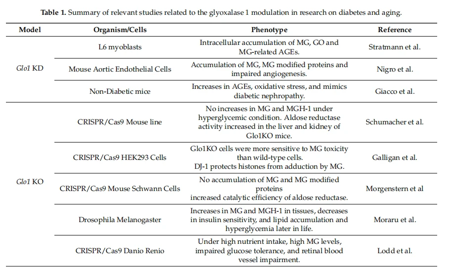
Recently, CRISPR-Cas technology allowed for the creation of viable Glo1 KO organisms [167]. Curiously, some data in both KO cells and animal models showed no increased levels of MG in tissues, indicating that alternative routes, such as aldose reductase and DJ-1 might be able to increase catalytic efficiency and protect proteins from glycation [86,92,94,168]. Galligan et al.found that the lack of GLO1 in HEK293 cells may induce deglycase activity of DJ-1, reducing MG-H1 deposition on chromatin [86] and Shu-macher et al.observed increased aldose reductase activity in the liver and kidney of Glo1 KO mice [94]. Increases in Minn tissues, as well as MG-derived hydroimidazolones and AGE-derived proteins, were observed in animals from which Glol was KO[93,169]. Lipid accumulation and hyperglycemia later in life were also observed [169]. Similarly, overfeeding Danio rerio Glo1 KO resulted in increased MG levels in tissue inducing hyperglycemia, insulin resistance, and alteration in the retinal blood vessels [93].
Conversely, multiple studies using Glo1 gene overexpression showed protective effects. Morcoset al. showed that the overexpression of Glow enhanced lifespan in Caenorhabditis elegans and, reduced glyoxal and MG-derived hydroimidazolones, mitochondrial MG-derived protein modification as well as oxidative stress biomarkers [110]. In diabetes models, Glo1 overexpression reduced glomerular protein modifications by MG, decreased oxidative stress markers, and prevented the development of diabetic kidney pathology [127]. cistanche herba Consistent with these observations, mice with Glol overexpression had lower levels of MG in the brain [105]. Vulesevic et al. found a reduction of circulating biomarkers of inflammation, such as VCAM-1(vascular cell adhesion molecule 1) and E-selectin (endothelial-leukocyte adhesion molecule 1), and diminished RAGE expression in diabetic Glo1 overexpress-ing mice [170]. Other studies with rat models overexpressing GLO1 had similar results, showing a decreased concentration of glyoxal and MG-derived hydroimidazolones in tissues, reduction in AGE formation, and prevention of renal and endothelial dysfunction in response to induced diabetes [103,104,108]. Reduced damage was also observed in the diabetic retinal neuroglia and vascular cells with Glo1 overexpression [108]. In cells overexpressing Glo1, MG levels decreased and delayed MG-induced protein modifications [171,172]. Finally, the upregulation of GLO1 and aldose reductase were protective against diabetic nephropathy alterations in diabetic patients [173]suggesting that boosting this system could be a possible therapeutic approach to delay the development of age-related diseases.
4. Nutritional Intervention to Enhance the Glyoxalase System and Decrease Accumulation of AGEs
There is an increasing interest in the discovery of small-molecule regulators with the capacity to modulate glyoxalase systems. He et al.recently summarized high-throughput microplate assays to identify novel regulators, chemical structures, and the structure-activity relationship between GLO1 protein and modulators (reviewed in [174]). cistanche penis growth These studies are useful for drug design and the optimization of therapeutic doses for these compounds will require human clinical trials. However, dietary interventions based on supplements or changes in diet may be an alternative source of multiple glyoxalase activity enhancers to prevent or alleviate glycation-derived damage with age. In this section, we summarize the current status of knowledge about the antiglycation activities of different nutritional compounds.
4.1.Isothiocyanates
Isothiocyanates are natural compounds formed by enzymatic conversion of glucosinolates, metabolites highly enriched in cruciferous vegetables. Sulforaphane and allyl isothiocyanate are two dietary isothiocyanates proven to enhance the activity of GLO1 in an NRF2-dependent manner and prevent glycation stress-induced DNA damage [65,71,175]. Proteomic analysis identified GLO1 as a protein differentially expressed in cells treated with sulforaphane [176]. Sulforaphane counteracts glycation-induced damage by lowering the phosphorylation of MAPK signaling pathways (ERK1/2, JNK, and p38) and, consequently, enhancing the glyoxalase system in primary neonatal rat cardiomyocytes and SH-SY5Y neuroblastoma cells [71,176]. Of note, the effect of sulforaphane could be cell-dependent, as the incubation of these isothiocyanates did not affect GLO1 activity in isolated peripheral blood mononuclear cells from healthy individuals [72].
4.2.Polyphenols
Polyphenols are natural compounds found in plant-based foods. They include flavonoids, phenolic acid, polyphenolic amides, and other polyphenols. Although a vast literature informs about the action of polyphenols on glyoxalase function, the role of polyphenols remains controversial. On one hand, studies report that some polyphenols can inhibit glyoxalase activity in vitro. Curcumin, a polyphenol commonly used as a food flavoring, as well as other flavonoids such as baicalein showed biological activity inhibiting GLO1 [177-180]. Oral administration of naringenin, a flavonoid present in citrus fruits, diminished the GLO1 enzymatic activity and increased AGEs intermediates in two mouse skin tumor models [181]. On the other hand, increasing literature reports that many of these micronutrients enhance GLO1 activity. Resveratrol, found in grapes, berries, and peanuts, upregulates the glyoxalase system through the upregulation of the ERK pathway and the nuclear translocation of NRF2. The depletion of NRF2 abrogates the resveratrol-induced upregulation of glyoxalase expression [182]. The effectiveness of resveratrol was demonstrated both in vivo upon oral treatment in streptozotocin-nicotinamide-induced diabetic rats and in vitro in several cell types [17,183,184]. A recent report adds new molecular clues and indicates that SIRTl plays a role in the effective response of resveratrol against glycation-derived cytotoxicity [185]. Other polyphenols were also found to be bioactive in a glyoxalase-dependent manner. A polyphenol isolated from the edible seaweed Ishige oka-mural, diphlorethohy droxycarmalol, inhibits glycation stress in vitro in an NRF2-dependent manner [186] and polyphenols from Chilean native berries also increased GLO1 activity and were cytoprotective in human gastric epithelial cells[187]. Recent reports have focused on the therapeutic potential of polyphenols in neurodegenerative disorders. Resveratrol, curcumin, capsaicin, and epigallocatechin gallate induce neuroprotective effects through NRF2-induction in experimental models of Alzheimer's and Parkinson's disease (reviewed in [188,189]).
Flavonoids, the most common class of polyphenolic compounds in the human diet, are found in tea, citrus fruit, citrus fruit juices, berries, red wine, apples, and legumes. Flavonoids have attracted attention in the last decade, in part, due to their efficacy against glycation stress. Flavonoids are classified in different subgroups according to their chemical structure (anthocyanidins, anthoxanthins, flavanones, flavanonols, and flavans) and members of almost every subclass have been reported to modulate GLO1.
Cyanidin belongs to the class of anthocyanidin and is found in many red berries and other fruits. Cyanidin exerts a protective effect against glycation stress by increasing the activity of GLO1 but with no changes in Glo1 mRNA expression in rat pancreatic β-cells [190]. Anthocyanin has been reported to induce therapeutic activities in a wide range of disorders in the nervous systems, such as cerebral ischemia, Alzheimer's disease, and Parkinson's disease [191]. Anthocyanins prevent neurotoxicity, but there is limited information about the impact of anthocyanins on the glyoxalase system.
Within the group of flavonoids, the class of anthoxanthins is likely the best characterized regarding antiglycation properties. However, the protective mechanism against glycation stress does not always correlate with the modulation of the glyoxalase system. Fisetin, a plant polyphenol found in many fruits and vegetables, promotes the expression and activity of GLO1, ameliorating major complications of diabetes in Akita mice, a model of type 1 diabetes [192]. Although reduced levels of glycated protein were found in different tissues of fisetin-treated animals, no significant change was detected in blood sugar. The impact of fisetin seems to be pleiotropic given that it also induced the expression of glutamate-cysteine ligase(GCL), the rate-limiting enzyme in GSH production, and reduced levels of RAGE receptor [192]. Quercetin also has anti-glycation properties by enhancing the expression of GLO1 and GLO2 proteins and increasing cellular viability against MG-induced proteotoxicity in primary cultures of cerebellar neurons [193]. Other in vitro analyses showed that the protective effect of quercetin against high glucose-derived glycation stress is via NRF2/GLO1 pathway in neuroblastoma SH-SY5Y cells [175].
In vivo analyses corroborate in vitro experiments. OQuercertin enhanced GLO1 in the brain of streptozotocin-induced diabetic rats [194] and lowered plasma MG levels in a randomized, double-blind, placebo-controlled crossover trial [195]. However, neither quercetin nor epicatechin significantly changed the expression of GLO1 in peripheral blood mononuclear cells [195]. Compounds derived from the flavonol morin were recently shown to be protective in cultured mouse primary cerebellar neurons and Caenorhabditis elegans treated with MG. Increased NRF2, GLO1, and GLO2 expression, glyoxalase activity, and GSH concentration were found in morin-treated models [196].
Flavanones comprise another type of flavonoid that modulates glyoxalase activity. Hesperetin, the main flavonoid in lemons and sweet oranges, activated the NRF2 pathway, enhanced GLO1, and ameliorated renal changes in streptozotocin-induced diabetic rats [197]. Of note, trans-resveratrol and hesperetin synergize to enhance the expression and activity of GLO1 and induce a decrease in glycated protein[198,199]. A trans-resveratrol and hesperetin formulation was shown to enhance angiogenesis and wound closure in diabetic mice [171]. Increased GLO1 activity in peripheral blood mononuclear cells was found in a randomized, placebo-controlled crossover clinical trial [198].
Genistein is an isoflavone found in soy-based foods, coffee, and other different plant-based foods. Dietary genistein enhanced the expression of GLO1 and GLO2 and signifi-cantly decreased MG and AGEs concentrations in two mouse models, fed with a high-fat diet alone or in combination with MG [200].
In the subgroup of flavans, catechin, also enhanced the glyoxalase system activity and improved the detoxification of AGEs intermediates [193].
Xanthohumol, a prenylated flavonoid found in hops and beer, attenuated glycation stress in osteoblastic MC3T3-E1 cells by increasing NRF2 and GLO1 activity[201]. Different reports have shown neuroprotective properties of xanthohumol in murine neuroblastoma N2a cells stably expressing human Swedish mutant amyloid precursor protein and in ischemic stroke animal model [202]. However, it was not explored if the upregulation of GLO1 activity contributes to the neuroprotective effect. The xanthonoid mangiferin is another natural phenolic compound that upregulates NRF2 signaling, elevates glyoxalase activity, prevents the formation of AGEs, and ameliorates diabetes-associated cognitive decline in rats [203-205].
4.3. Vitamins
Pyridoxamine, a form of vitamin B, has been shown to reduce glycation stress, by preventing the formation of MG-derived AGEs [206]. Enhanced erythrocyte GLO1 activity was found in streptozotocin-induced diabetic Sprague Dawley rats treated with pyridoxamine, which showed the reduced formation of AGEs in plasma [206]. However, pyridoxamine treatment failed to counteract AGE formation, did not lead to differences in GLO1 activity, or ameliorate disease progression in a model of experimental autoimmune encephalitis [207]J. Changes in GLO1 expression were also not found in pyridoxamine-treated mice overexpressing inducible nitric oxide synthase in pancreatic β-cells )208]. The effect of pyridoxamine might be tissue-dependent, as GLO1 expression was induced in visceral fat but not in perivascular fat tissues in Sprague Dawley rats fed a high-fat diet [209],
Vitamin D was also reported to exert a protective role against glycation stress [210,211]. Vitamin D supplementation in type-2 diabetes participants showed a trend in increasing GLO1 expression, a transcriptional downregulation of RAGE receptor, and decreased AGE serum in a double-blind randomized placebo-controlled [211]. Nevertheless, the action of vitamins remains controversial because vitamin treatment had no significant impact in other studies. For example, dietary vitamin E intake did not change mRNA Glo1 levels and glutathione reductase 1 in the brain of inbred strains of mice [212]. In addition, vitamin D seemed to compensate for the loss of GLO1 in other diabetic contexts [213].
4.4. Other Dietary Compounds
Ursolic acid, found in the peels of fruits along with herbs and spices, showed an antiglycation effect in the kidneys of diabetic mice. Enhanced renal Glol mRNA expression and glyoxalase activity were found in treated animals along with lower levels of plasma AGEs [214]. Some plant extracts used in traditional medicine in Asia have been evaluated. Monastic is a metabolite fermented by Monascus purpureus, a fungus used in the production of fermented food in Asia. Monastic is a natural PPARy agonist that enhances GLO1 expression through activation of NRF2 and protects from glycation stress in rats orally administrated MG[215]. Extracts from Psoralea corylifolia seeds increased the expression of hepatic GLO1 in MG-treated mice [216].Indole-4-carboxaldehyde isolated from the edible seaweed Sargas-sum thunbergia was also able to enhance mRNA and the expression of GLO1 in vitro [217].
Finally, the oral administration of glycine has been shown to promote the nuclear translocation of NRF2, thus protecting against glycation stress in different tissues. Glycine had a suppressive effect on glycation stress, manifested by increased activity and the expression of aortic GLO1 and reduced levels of AGEs [218]. Additionally, glycine enhanced the glyoxalase system function and protects against renal glycation stress in streptozotocin-induced diabetic rats [219].
5. Concluding Remarks and Pending Questions
AGEs are a common pathogenic factor for multiple age-related diseases, such diverse as diabetes, AMD, CVD, neurocognitive and neurologic disorders, etc. The glyoxalase system is among the most robust protective capacities against the formation of AGEs. Other mechanisms also seem to complement—or compensate for impaired—GLO1 activity. Although different protective mechanisms counteract the accumulation of these cytotoxic compounds, the activity of these cellular defenses declines with age, and AGEs are gradually deposited, compromising aged tissues. Metabolic conditions such as diabetes and the consumption of Western, high glycemic diets exacerbate the AGE accumulation and represent a serious burden on public healthcare.
In order to diminish the detrimental effect of glycation stress on health, strategies extending the capacity of anti-AGE pathways with age should be salutary. The glyoxalase system is a primary mechanism that limits the synthesis of AGEs and alterations in its activity are linked to multiple age-related diseases. Pharmacological interventions have not been successful in clinical settings so far. Dietary management or nutritional supplements may represent safe and low-cost alternatives to prolong the functionality of detoxifying routes such as the glyoxalase system. Further research is required to define nutritional approaches to overcome the progression of age-related diseases associated with glycation stress.
This article is extracted from Cells 2021, 10, 1852. https://doi.org/10.3390/cells10081852 https://www.mdpi.com/journal/cells