Total Glycosides Of Cistanche: Brain Protection Against Intermittent Hypoxia Driven Oxidative Damage
Nov 16, 2026
Introduction: When Repeated Low Oxygen Cycles Hurt Brain Function
Millions of people worldwide experience repeated drops in blood oxygen levels while asleep. Obstructive sleep apnea (OSA), the most common condition causing this pattern, forces the human body into repeating cycles: oxygen falls sharply, then rebounds rapidly when breathing resumes. This back and forth shift, known medically as intermittent hypoxia, does more than make people feel tired during daytime hours. Over months and years, recurring hypoxia reoxygenation cycles can trigger persistent oxidative stress inside brain tissue, particularly within the hippocampus, the critical brain region responsible for learning, spatial memory and information consolidation.
Many OSA patients receive continuous positive airway pressure (CPAP) devices as primary clinical intervention. CPAP physically prevents airway collapse and largely resolves intermittent hypoxia for compliant users. Still, a notable percentage of individuals struggle with mask discomfort, nasal irritation or poor nightly adherence, which leaves chronic intermittent hypoxia and its downstream brain related consequences unaddressed. To date, pharmaceutical antioxidant agents tested for easing OSA linked neuronal damage have delivered limited clinical success, creating demand for exploring plant derived antioxidant candidates as complementary research subjects.
For decades, extracts from Cistanche deserticola, a desert parasitic herb widely documented within traditional Asian herbal practice, have drawn scientific interest. Its primary bioactive fraction, total glycosides of Cistanche deserticola (TG CD), is dominated by phenylethanoid glycosides such as echinacoside and acteoside, compounds well known for free radical scavenging, anti inflammatory and neuro protective properties in multiple rodent disease models, including dementia, cerebral ischemia and accelerated aging rodents. Existing published animal data confirm TG CD can improve learning memory deficits triggered by oxidative insults, yet few research projects had specifically examined whether these glycosides could counteract brain harm produced by long term intermittent hypoxia until a 2023 peer reviewed animal study published in Chinese Journal of Respiratory and Critical Care Medicine.
This article translates that pre clinical rodent research into plain language educational content. It explains what intermittent hypoxia does to hippocampal tissue, how scientists model sleep apnea like oxygen fluctuation in laboratory rats, what behavioral memory and oxidative stress biomarkers they measured, what measurable changes total cistanche glycosides generated in this controlled setting, and, crucially, where the boundaries of this animal based evidence lie. Nothing inside this piece constitutes medical advice for human sleep apnea patients; all observations originate from laboratory rat experiments only.

Understanding Intermittent Hypoxia, Hippocampus and Oxidative Stress Biomarkers
Before diving into experiment details, readers need clarity about three core concepts: intermittent hypoxia's unique injury pattern, why the hippocampus suffers disproportionately, and what SOD and MDA represent as widely accepted laboratory markers of oxidative burden.
Intermittent Hypoxia: Worse Than Steady Low Oxygen
People often imagine hypoxia simply means "not enough oxygen". Intermittent hypoxia works differently: oxygen concentration swings continuously between low oxygen status and normal room air oxygen levels, hundreds of times each day inside experimental chambers or inside a sleep apnea sufferer's body. Each swing resembles repeated ischemia reperfusion events. Every time oxygen returns after a low oxygen episode, brain mitochondria and cellular enzyme systems produce large surges of reactive oxygen species (ROS). ROS attack lipid molecules within neuron cell membranes, distort protein folding and even harm nuclear genetic material if antioxidant defenses cannot neutralize them fast enough.
Chronic steady hypoxia triggers damage too, yet research repeatedly notes intermittent hypoxia creates far heavier oxidative stress loads for nervous tissue, largely because of this repetitive re oxygenation shock cycle. Damage accumulates week by week instead of appearing overnight. In rodent models, measurable memory impairment and biochemical brain changes progressively worsen across two week, four week, six week and eight week exposure timelines, mirroring slow onset cognitive complaints seen among long untreated OSA human patients.

The Hippocampus: Memory's Vulnerable Command Center
The hippocampus sits deep inside the temporal lobe. Every time we navigate unfamiliar locations, learn new facts or store recent events into long term memory, hippocampal neurons perform central signal processing work. Unfortunately, hippocampal nerve cells rank among the most oxygen sensitive cell types across the whole mammalian brain. When ROS concentrations rise persistently, hippocampal neurons degrade gradually: signal transmitting synapses weaken, cell membrane integrity degrades and eventually neuronal function declines, translating directly into poorer spatial learning and memory retention performance observable in behavioral testing paradigms like the Morris water maze.
SOD and MDA: Two Opposite Sides Biomarkers for Oxidative Status
Scientists rely on paired biomarkers to quantify oxidative stress balance within tissue samples:
Superoxide dismutase (SOD): A primary endogenous antioxidant enzyme produced naturally inside cells. Higher SOD activity means stronger internal free radical neutralizing capacity. When tissues face heavy oxidative assault, SOD gets consumed, so measured SOD activity values drop downward.
Malondialdehyde (MDA): A stable chemical by product formed when free radicals oxidize polyunsaturated fats within cell membranes. Elevated MDA levels act as a "damage footprint": higher MDA readings signal more extensive lipid peroxidation injury to neuron membranes.
In simple terms: Good protective status = higher SOD + lower MDA. When oxidative stress wins out: SOD values fall, MDA values climb upward. This dual marker testing framework is standard practice across neurological pre clinical research for assessing plant extract neuro protective potential.
Total Glycosides of Cistanche Deserticola: Where This Botanical Candidate Comes From
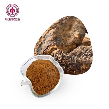
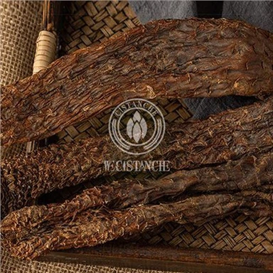
Cistanche deserticola is a root less desert parasitic plant growing across arid zones of northwest China. It attaches itself onto root systems of drought tolerant desert shrubs and absorbs water and nutrition from host plants rather than photosynthesizing sunlight. Its fleshy stems are harvested and processed into extracts for modern laboratory investigations as well as traditional herbal preparations.
Total glycosides of Cistanche deserticola (TG CD) represent a purified glycoside rich fraction isolated via solvent extraction workflows. Its dominant bioactive substances are phenylethanoid glycosides: echinacoside and acteoside (verbascoside) make up the largest proportion of this mixture, alongside minor companion iridoid glycosides and other secondary plant metabolites.
Published pre clinical literature from multiple independent labs has documented TG CD's biological profiles:
Scavenges excess reactive oxygen species molecules
Raises antioxidant enzyme activity inside nervous tissue
Reduces lipid peroxidation marker accumulation
Supports neuron survival under oxygen deprivation toxicity
Improves spatial learning and memory performance in dementia model rodents, vascular dementia animals and senescence accelerated mouse strains
Nevertheless, prior to the 2023 study, researchers lacked direct experimental evidence answering one critical question: Would oral TG CD administration mitigate hippocampal oxidative harm and associated memory deficits specifically brought on by weeks long intermittent hypoxia exposure? That exact research gap became the motivation for this rat model investigation.
How the Intermittent Hypoxia Rodent Study Was Designed
This section breaks down the experimental setup of the 2023 peer reviewed study into easy to follow segments: animal subjects, grouping logic, intermittent hypoxia chamber operating workflow, TG CD dosage administration, behavioral memory assessment methodology, tissue biochemical testing steps and statistical analysis rules.
Animal Subjects & Group Allocation
Seventy two healthy adult male Wistar laboratory rats with starting body weight 170 ± 10 g took part in the full trial. Animals were randomly sorted via digital random number procedure into three major experimental cohorts:
Blank control group: kept breathing normal 21% oxygen ambient air without hypoxia exposure or herbal extract dosing
Intermittent hypoxia only group (IH group): placed inside custom built intermittent hypoxia chambers, receiving no plant extract intervention
TG CD intervention group: receiving identical intermittent hypoxia chamber exposure plus daily oral gavage feeding of total glycosides of Cistanche deserticola at dosage 50 mg/kg body weight per day
Each main group was further subdivided into four time point subgroups with six rats for every time condition. Subgroups underwent experimental procedures lasting for 2 weeks, 4 weeks, 6 weeks and 8 weeks respectively. This staggered sacrifice arrangement allowed researchers to track progressive changes as intermittent hypoxia injury accumulated over extended durations. All animal handling protocols obtained formal ethical committee approval from the local university animal ethics review board before starting laboratory operations.

Intermittent Hypoxia Chamber Operating Protocol
For both hypoxia exposed groups, daily hypoxia chamber treatment ran 8:30 AM until 4:30 PM each experimental day. Inside custom fabricated closed chambers, gas flow controllers cycled nitrogen gas and compressed room air in repeating 120 second cycles:
First 30 seconds: nitrogen gas flows in, pulling oxygen concentration gradually down to 5 %
Next 40 seconds: high volume compressed air input pushes oxygen levels rapidly back up to normal 21 %
Final 50 seconds: low flow air maintains chamber oxygen stable near 21 %
Digital oxygen sensing instruments continuously monitored chamber atmosphere, keeping oxygen swing tightly constrained within ±0.5 % target boundaries. Rats inside blank control cages stayed in identical style chambers receiving constant 21 % oxygen without gas cycling manipulations. After finishing daily chamber sessions, all rats returned to standard housing cages with unrestricted food and drinking water access, consistent temperature humidity housing conditions shared across all cohorts.
TG CD Dosing Implementation
Thirty minutes before each day's hypoxia chamber session, rats belonging to the TG CD intervention group received oral gavage delivery of dissolved total cistanche glycosides (purity: 85 %, sourced from commercial laboratory reagent supplier). Compound powder dissolved in sterile injectable water, and liquid administration volume was standardized to 10 mL per kilogram rat body weight. Hypoxia only group rats received identical volume sterile water gavage without active ingredient content to match procedural stress conditions across groups. Dosing continued uninterrupted for respective 2, 4, 6 and 8 week experimental durations for each subgroup animal set.
Behavioral Memory Testing: Morris Water Maze Assay
The Morris water maze system represents the gold standard behavioral test for evaluating rodent spatial learning and hippocampus dependent memory in pre clinical neuroscience research. A large circular water filled tank holds opaque water, with one hidden submerged escape platform sitting beneath the liquid surface in a fixed quadrant position. Rats must use visual spatial cues positioned around the tank's outer perimeter to locate this invisible safety platform. Two primary read out metrics get recorded:
Escape latency time: How many seconds pass before each rat successfully climbs onto the hidden escape platform. Longer escape latency signals poorer spatial learning capability; shorter latency demonstrates better acquisition of spatial navigation memory.
Target quadrant crossing duration: After removing the escape platform entirely, researchers record how many total seconds each rat spends swimming within the original platform location quadrant. Animals with intact memory will gravitate back toward the remembered quadrant, spending more time swimming there. Shorter time within target quadrant serves as marker of degraded spatial memory retention.
For each scheduled time point (after completing 2, 4, 6 and 8 weeks of experimental exposure), rats completed multi day maze training cycles followed by formal recorded testing sessions to capture these two behavioral endpoints for statistical comparison across cohorts.
Tissue Biomarker Laboratory Measurement
Once behavioral testing wrapped for a given time point subgroup, laboratory rats received intra peritoneal anesthesia injection, followed by rapid brain tissue dissection. Fresh hippocampal tissue was separated, flash frozen and stored at −80 C deep freezer conditions until biochemical assay processing. Using commercially available test kit protocols, lab technicians measured hippocampal tissue SOD enzymatic activity values (WST 8 testing method) and MDA tissue concentration levels (TBA color reaction assay method). Every assay followed kit manufacturer specified procedural steps strictly to guarantee measurement result reproducibility.

Statistical Analysis Rules
All numerical results were expressed as mean value plus minus standard deviation. SPSS 19.0 statistical software processed datasets. One way analysis of variance followed by LSD post hoc multiple comparison tests handled inter group comparisons. Scientists defined P value less than 0.05 as threshold marking statistically significant differences between measured outcome datasets. Important reminder: statistical significance denotes low probability observed differences arise purely by random chance; it does not automatically equate to large magnitude biological effects, and these outcomes remain confined within rat model experiment boundaries only.
Key Experimental Outcomes: Behavioral and Biochemical Observations
This section presents interpreted study findings, avoiding direct verbatim copying from original Chinese language journal text, summarizing progressive changes across 2 week through 8 week time series data.
Spatial Learning & Memory Behavior Results from Morris Water Maze
Compared against blank control animals, both the hypoxia only group and TG CD treated hypoxia exposed group showed significantly prolonged escape latency durations at every measured time point (2 wk, 4 wk, 6 wk, 8 wk). As intermittent hypoxia exposure extended week by week, escape latency kept growing longer, reaching maximum values at the 8 week mark. This demonstrates chronic intermittent hypoxia progressively impairs spatial learning performance in rats, as expected from prior neuroscience literature.
When researchers directly compared TG CD intervention rats against hypoxia only animals: at every matching time interval, TG CD treated animals achieved statistically shorter escape latency values. While TG CD treated rats still performed worse than normoxic blank control rats overall, their learning deficits were consistently milder than animals receiving hypoxia exposure with zero botanical intervention.
For target quadrant dwell time metric: hypoxia only group rats spent significantly fewer seconds swimming within original platform quadrant relative to blank controls across all four time points; dwell time kept shrinking as hypoxia weeks accumulated. However, TG CD intervention cohort registered significantly extended target quadrant residence time compared with hypoxia only counterparts at every time point.
Collectively, maze behavior data tell this story: total cistanche glycoside oral treatment could not fully erase intermittent hypoxia provoked memory related behavioral deficits, yet it measurably softened the severity of both learning acquisition impairment and spatial memory retention loss inside this laboratory rat experimental setting.
Hippocampal Tissue Oxidative Stress Biomarker Results (SOD & MDA)
Biochemical assay measurements from dissected hippocampus tissue mirrored behavioral test trends step by step:
Relative to blank control rats, hypoxia only group hippocampal samples displayed consistently lower SOD antioxidant enzyme activity and markedly elevated MDA lipid peroxidation damage marker levels for 2, 4, 6 and 8 week time points. Over successive weeks of intermittent hypoxia treatment, SOD activity kept dropping lower and MDA concentrations climbed steadily upward, confirming cumulative progressive oxidative stress injury building up inside hippocampal tissue over time.
Comparing TG CD intervention group with hypoxia only group: at every single time point, TG CD treated hypoxia exposed rats demonstrated significantly higher hippocampal SOD activity readings, paired with statistically reduced hippocampal MDA concentrations. Again, TG CD animals still showed worse oxidative stress biomarker status than normoxic blank controls, meaning the botanical glycoside intervention produced partial protective effects rather than complete reversal of hypoxia driven oxidative damage signatures within this animal model setup.
Core Mechanistic Conclusion Suggested by This Dataset
Within controlled laboratory rat experimental conditions, TG CD intervention partially preserved hippocampal antioxidant enzyme SOD activity, lowered MDA lipid peroxidation damage marker levels, and correspondingly alleviated some portion of intermittent hypoxia induced spatial learning and memory deficits as measured by Morris water maze behavior assays. Study authors hypothesized that the neuro protective outcomes primarily stem from suppression of excessive hippocampal oxidative stress.

Critical Limitations of This Pre Clinical Rodent Study
Readers researching botanical supplements must develop healthy critical reading habits toward animal model research. This experiment delivers valuable mechanistic clues, but it carries several well defined limitations that must never be overlooked when extrapolating toward human health contexts:
All data come exclusively from male laboratory rats. No female animal subgroups existed within this trial design. Rodent metabolic rate, hormone status and brain oxidative stress responses differ by biological sex; outcomes observed here cannot automatically extend to female animal models, let alone human male or female subjects.
Only one fixed TG CD dosage (50 mg/kg/day) was tested. This investigation lacked multiple dose gradient arms, so scientists cannot establish dose response curves: we do not know whether lower dosages would produce weaker protective effects or whether higher dosage administration might deliver stronger benefits or trigger toxicity related outcomes. Future multi dose experimental work remains required.
The project measured functional behavior endpoints plus two core oxidative stress biomarkers only. It did not carry out detailed histopathological neuronal cell morphology inspection, nor did it map precise intracellular signaling pathway activation changes. Proposed mechanistic explanation ("suppress hippocampal oxidative stress") represents reasonable inference derived from measured biomarker shifts, not direct proof pinpointing exact molecular signaling cascades.
Intermittent hypoxia chamber environment is highly controlled laboratory artificial model. Real world human obstructive sleep apnea pathophysiology adds numerous confounding variables: sleep fragmentation stress, systemic inflammation, metabolic comorbidities, aging related brain changes, genetic variability, lifestyle factors and concurrent medication intake. The simplified rat model cannot replicate this full complexity human disease picture.
No human clinical trial data available. Positive pre clinical rodent study outcomes never guarantee identical functional results translate over into human bodies. Differences in gastrointestinal absorption, compound metabolic transformation, tissue distribution and target tissue bioavailability separate rodents from human beings. Results cannot be interpreted as proof that consuming cistanche glycoside supplements will improve memory in sleep apnea human patients.
This research project did not examine safety toxicology profiling for repeated long term TG CD oral administration. Long duration safety profiles for extended period high glycoside intake remain incompletely characterized in vivo.

Contextual Broader Research: Other Cistanche Glycoside Pre Clinical Neuro Protection Literature
This intermittent hypoxia rat study does not stand alone. Multiple independent pre clinical teams have published separate rodent model papers describing neuro protective properties for total glycoside / phenylethanoid glycoside fractions derived from Cistanche deserticola or related species raw material, which helps contextualize this 2023 dataset:
In Alzheimer disease model rodents, cistanche total glycoside treatment raises brain tissue SOD activity, cuts MDA lipid peroxidation levels and partially rescues Morris water maze spatial memory deficits, consistent with antioxidant mediated neuro protective mode of action.
Vascular dementia rat model work also records learning memory behavior improvements coupled with antioxidant biomarker modulation after cistanche glycoside intervention.
Cerebral ischemia reperfusion injury rodent studies report TG CD driven antioxidant pathway (Nrf 2 / Keap 1 axis) activation, lowered MDA, elevated SOD activity, reduced brain infarction volume and improved neurological function scores in stroke simulated animals.
Accelerated senescence mouse strain (SAMP8) studies note cistanche glycoside administration can mitigate age related memory decline via boosting antioxidant enzyme status and supporting synaptic plasticity inside hippocampal brain regions.
While experimental models differ across these projects (neurodegeneration, stroke, aging versus intermittent hypoxia insult), a recurring theme emerges across publications: phenylethanoid rich cistanche glycoside extracts consistently modulate oxidative stress related biomarkers and partially preserve memory related behavioral outcomes across diverse oxidative insult rodent paradigms. Still, every single one of these investigations stays confined to pre clinical animal lab settings, awaiting human clinical validation work.
Practical Guidance for Supplement Sourcing: What Buyers Should Check for Cistanche Glycoside Raw Materials
If you are a supplement brand developer, formulator, procurement specialist or informed consumer researching cistanche glycoside containing products, the wide variability of botanical extract quality represents a major real world challenge. Two different commercial products labelled "Cistanche extract / total cistanche glycosides" can show drastically different bio active compound concentrations, based on plant origin species, harvest region, extraction technology and post processing workflows. Below are key points for quality verification checklist:
Verify marker compound quantification via HPLC laboratory report: Do not rely only upon marketing language like "high strength cistanche extract". Request Certificate of Analysis (CoA) documentation explicitly listing percentage content for echinacoside and acteoside (verbascoside), the signature phenylethanoid glycoside marker substances. These two compounds form the major active fraction behind most documented antioxidant neuro protective pre clinical observations. Avoid suppliers only supplying generic ratio extract claims without HPLC quantitative marker readings.
Confirm raw material botanical species identity: Cistanche deserticola and Cistanche tubulosa are two primary commercial source species, and their glycoside profiles differ; make sure documentation clarifies exact botanical source species.
Review extraction process descriptions: Solvent types, solid liquid ratio, extraction temperature time parameters directly shape final glycoside composition profile. Reputable suppliers can describe their full extraction workflow transparently.
Contaminant safety screening data: Heavy metal residual testing, pesticide residue panel and microbial limit testing reports for each batch are non negotiable for human consumption oriented botanical extract materials.
Stability testing evidence: Phenylethanoid glycoside molecules can degrade under elevated temperature or high humidity storage circumstances. Accelerated stability study data demonstrate whether marker compound content remains within specification across intended product shelf life duration. Without stability validation, initial batch lab values cannot guarantee potency retention by end of shelf life.
Frequently Asked Questions
Q1: Does this rat study prove cistanche glycoside supplements can fix memory issues for human sleep apnea patients?
Answer: Definitely not. This is animal pre clinical research only. Positive rodent experiment results do not constitute clinical proof of effectiveness for human beings. Obstructive sleep apnea in humans is complex multi factor illness. CPAP therapy remains gold standard clinical intervention for OSA patients. Any botanical supplement related ideas should always be discussed with licensed health care providers and never replace established medical treatments.
Q2: What exactly do SOD and MDA tell us when reading botanical neuroscience papers?
Answer: They act as tissue level oxidative stress readouts. SOD = endogenous antioxidant enzyme capacity; MDA = chemical footprint recording lipid membrane oxidative damage. When treatment group shows higher SOD plus lower MDA versus damage model group, it suggests test substance may exert antioxidant related biological effects inside tested tissue. This represents biomarker correlative evidence; it does not alone prove full causal mechanisms.
Q3: Can I calculate human supplement dosage directly from the rat 50 mg/kg day experimental dose?
Answer: Not safely for self dosing purposes. Rodent to human dose conversion involves allometric scaling calculations, plus unknown human oral bioavailability factors for cistanche glycosides. The 50 mg/kg day dosage is purely laboratory experimental dosage for Wistar rats; it is not recommended human intake guidance.
Q4: Are total glycosides of cistanche the same thing as ordinary cistanche herb powder?
Answer: No. Total glycosides extract represents concentrated purified fraction isolated via solvent extraction processes. Plain crushed herb powder contains full spectrum plant material with far lower concentrations of phenylethanoid glycoside marker compounds. Biological responses observed using purified glycoside extract cannot be expected to appear at equal magnitude when consuming simple raw herb powder products.
Final Summary
Chronic intermittent hypoxia, the core physiological insult behind untreated obstructive sleep apnea syndrome, triggers progressive hippocampal oxidative stress injury, which contributes toward learning and memory functional decline within laboratory animal models. The 2023 peer reviewed rat model study explored whether orally administered total glycosides from Cistanche deserticola might counter these harmful consequences.
Within controlled laboratory rat experimental conditions, TG CD intervention partially preserved hippocampal antioxidant enzyme SOD activity, lowered MDA lipid peroxidation damage marker levels, and correspondingly alleviated some portion of intermittent hypoxia induced spatial learning and memory deficits as measured by Morris water maze behavior assays. Study authors hypothesized that the neuro protective outcomes primarily stem from suppression of excessive hippocampal oxidative stress.
Nevertheless, important research limitations apply: this is single dose male rat pre clinical experiment without human clinical trial validation data. It cannot serve as evidence for human therapeutic claims for sleep apnea or memory impairment conditions. It adds one more piece of pre clinical evidence supporting cistanche phenylethanoid glycosides as promising neuro protective botanical research candidate worthy of further investigation.
Source reference note for article footer Pre clinical animal study source: Zhang P, et al. Effect of total glucosides of Cistanche deserticola on oxidative stress and cognitive function in rats with intermittent hypoxia. Chinese Journal of Respiratory and Critical Care Medicine, 2023, 22(4):269‑273.
Disclaimer: Pre clinical animal study educational content only
All information presented in this article relates exclusively to laboratory rat pre clinical research observations. It is for general educational understanding of botanical pharmacology research literature only. This content does not constitute medical advice, diagnosis or treatment recommendation for any human disease including obstructive sleep apnea, memory loss or neurodegenerative conditions. Rodent study findings do not guarantee identical biological responses occur inside human bodies. Never disregard professional medical guidance or delay seeking medical care because of information you have read here. Always consult qualified health care practitioners before starting any new botanical supplement regimen, especially if you live with chronic illness, take prescription medications, are pregnant or nursing.