Useful Knowledge For Acute Kidney Disease
Mar 12, 2022
Contact: joanna.jia@wecistanche.com / WhatsApp: 008618081934791
Kidney Injury, Acute
SUSAN GARWOOD
Department of Anesthesiology, Yale University School of Medicine, New Haven, CT, USA
Synonyms
Acute renal failure; Acute tubular necrosis (ATN); Ischemic renal failure; Prerenal azotemia; Prerenal ischemia

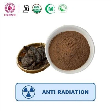
cistanche is good for acute kidney injury
Definition
Acute kidney injury (AKI) was coined by the American Society of Nephrology Renal Research Group to unify definitional terms of acute kidney disease and is used to reflect the entire spectrum from minimal elevations in serum creatinine to anuric renal failure. A consensus definition of AKI (acute kidney injury) was proposed by the Acute Dialysis Quality Initiative (ADQI) Group created the RIFLE classification (Risk, Injury, Failure, Loss, End-stage kidney disease) based on increases in serum creatinine compared to baseline. AKI (acute kidney injury) comprises the first three classes of this system (Risk, Injury, and Failure) and is categorized as a 1.5-fold increase (Risk), twofold increase (Injury), and threefold increase (Failure) in serum creatinine in [1]. Less than 20% of patients with AKI (acute kidney injury) as defined by the RIFLE classification will be identified as having acute renal failure by ICD-9-CM code.
Prerenal azotemia (prerenal failure) is caused by an absolute or relative reduction in renal perfusion, resulting in a modest reduction in glomerular filtration rate (GFR) and increased serum creatinine and blood urea. It is usually, but not always, accompanied by oliguria. It occurs as a physiological response to a lowered renal perfusion that may result from a globally or regionally reduced blood flow to the kidney, but regardless of the cause, the initial compensatory mechanisms correct renal blood flow (RBF) and bring GFR toward normal. During prerenal azotemia, renal tubules and microvasculature remain structurally and functionally intact. It is by definition reversible, is frequently a precursor to acute kidney injury, but is not included as part of the acute kidney injury spectrum.
Acute tubular necrosis (ATN) is frequently used synonymously with acute kidney injury since the early phases of the pathophysiology are dominated by structural and functional changes to the proximal tubule epithelial cells. However, the term itself comes from animal experiments of renal ischemia where glomerular filtration rate (GFR) falls secondary to luminal obstruction and back-leak of the filtrate by gross necrosis and sloughing of proximal tubule epithelial cells. In the clinical situation, less than 1% of epithelial cells show signs of necrosis and sloughing but there is a partial or total loss of the apical brush border in most of the proximal tubule epithelial cells with consequent functional derangements including reduced GFR.
Ischemic acute renal failure (ARF) occurs secondary to an absolute or relative reduction in renal perfusion which may be global or regional. The key sign of clinical ischemic ARF is a rapidly progressive and profound reduction in glomerular filtration rate (GFR), which continues and even progresses after the return of renal perfusion to baseline. This has led to the addition of an extension phase to the traditional model of renal injury resulting in the new paradigm of initiation, extension, maintenance, and recovery [2]. In ischemic ARF, there are marked pathophysiological changes that occur in both the tubules and the renal vasculature.
In the early phase of ischemic AKI (initiation phase), autoregulatory mechanisms that were effective in prerenal azotemia begin to fail and RBF declines with GFR while adenosine triphosphate (ATP) stores become depleted. Inappropriate and selective renal vasoconstriction occurs in response to the sympathetic nervous system and renin-angiotensin stimulation due to heightened sensitivity of the vascular endothelium to these and a number of other vasoconstrictors such as thromboxane, endothelin, and leukotrienes. Furthermore, vasorelaxation in response to stimuli that normally generate endothelium-dependent vasodilators is also inhibited which may be attributable to increased cytosolic and mitochondrial calcium concentrations noted at this stage.

cistanche ingredients are good for acute kidney injury
Treatment
Traditional treatment of AKI is predicated on the restoration of renal perfusion and GFR and the provision of adequate cardiac output is the keystone to medical management of this problem. Currently, there is no practical method or monitoring of renal blood flow or renal perfusion measurement and optimization relies on providing adequate filling pressures and cardiac output. Pharmacological therapy to enhance either renal perfusion or GFR other than by increasing cardiac performance has proven to be disappointing in the clinical setting and therapies which functioned well in animal models such as dopaminergic agents and atrial natriuretic peptide (ANP) have not been reproducible in the clinical arena.
The future of therapeutic agents for the treatment of AKI (acute kidney injury) has taken a different approach and relies on an understanding of the pathophysiological events that occur during AKI (acute kidney injury)[3]. The concept of the different phases of AKI and an understanding of the cellular events which
take place during these phases [4] has allowed the development of novel therapeutic agents which are now making their way into late-phase clinical trials.
Tubular Epithelial and Vascular Endothelial Pathophysiology in Ischemic ARF
Depletion of adenosine triphosphate (ATP) is a key event in the early stages of ischemic AKI (acute kidney injury) initiating changes in the cytoskeleton of both the tubular epithelium and vascular endothelium and disruption of tight junctions. This allows loss of tubular filtrate and plasma, decreasing glomerular filtration rate and renal blood flow even further. Ischemic tubular epithelial cells express cytokines and adhesion molecules, which attract and upregulate leukocytes. Vascular endothelial cells become activated and express adhesion molecules, trapping red cells and leukocytes particularly in the vessels of the corticomedullary junction. Because of this inflammatory reaction, the process of cellular hypoxia and injury may continue and even progress in the corticomedullary region after perfusion has been restored and repair and regeneration have begun in other regions.
A number of cascades are set in motion by the rapid depletion of ATP, the key events being the induction of nitric oxide synthase, resulting in oxidant injury and activation of caspases: the key event in the apoptosis/necrosis cascade. Nucleotide depletion culminates in the accumulation of hypoxanthine that contributes to the generation of reactive oxygen molecules and impaired calcium sequestration resulting in the well-documented rise in free intracellular calcium with subsequent activation of proteases and phospholipases. This pathophysiological approach to AKI (acute kidney injury) has led to the development of a number of classes of drugs that may be active in the prevention and or treatment of clinical AKI (acute kidney injury). These are anti-apoptotic agents, anti-inflammatory, and antioxidant drugs.

cistanche ingredients are good for acute kidney injury
Novel Therapeutic Agents in the Treatment of AKI
The key step in apoptosis/necrosis is the activation of caspases (cysteine aspartate-specific proteinases), which are highly operative during programmed cell death: initiating and executing the final stages of cell death. Activation of caspases occurs either by an intrinsic or extrinsic pathway involving members of the Bcl-2 superfamily of genes and proteins that govern mitochondrial outer membrane permeability and can be pro-apoptotic anti-apoptotic. A number of regulatory mechanisms exist that affect the balance of pro-and anti-apoptosis, the most important of which during renal ischemia appears to be the pro-apoptotic transcription factor p53. Expression of p53 is triggered by hypoxia and loss of cell-cell adhesion and is itself regulated by PARP (Poly ADP-Ribose Polymerase). Because of their key roles in cell apoptosis and necrosis, caspases, p53 and PARP have been targeted as novel therapeutic intervention sites in several disease states and are currently undergoing a number of clinical trials. Caspase inhibitors were identified as potentially useful agents in AKI (acute kidney injury) by the Acute Kidney Injury Network (http://www.akinet.org/) since the catalytic domain of most caspases processes the proforms of interleukin-1b and interleukin-18 and are thus active in the inflammatory process as well as apoptosis. PARP is similarly active in the inflammatory process so these two entities make attractive targets for renal protection strategies. Clinical trials of these agents in AKI (acute kidney injury) registered with Clinicaltrials.gov can be found at http://clinicaltrials.gov/.
Of greater interest are two older drugs with anti-apoptotic and anti-inflammatory activities, which circumvent the need for preliminary safety trial in man; these are tetracyclines and human recombinant erythropoietin (hr-EPO).
Tetracyclines are well known to possess a wide repertoire of anti-inflammatory and immunomodulatory actions in rheumatologic and dermatologic diseases. In animal studies, tetracyclines have been demonstrated to decrease the activity of p53. In rat models of ischemic renal injury, minocycline reduces tubular cell necrosis, microvascular permeability, and inflammation with a salutary effect on renal function. The mechanism of action is thought to be complex and includes inhibition of free radical and cytokine formation, modulation of matrix metalloproteinase activity by transcriptional and posttranslational regulation, inhibition of caspase expression, and direct blockade of cytochrome c release from mitochondria. Minocycline is currently undergoing clinical trials to prevent AKI (acute kidney injury) after cardiac surgery.
HrEPO has multiple effects on cellular energetics during kidney injury and has been demonstrated to have antiapoptotic effects, anti-inflammatory effects, growth factor effects, and regulate kidney homing circulating stem cells during the repair/recovery phase. Of these novel renoprotective agents, HrEPO penetrated furthest into clinical trials. Promising preliminary data in patients undergoing contrast-enhanced imaging studies, kidney transplants, and cardiac surgery have been presented at the conference level but have not made it yet into mainstream publication.
A review of the current paradigm of AKI (acute kidney injury) reveals an important phase in the later stages: repair and differentiation. Since many cases of AKI (acute kidney injury) are diagnosed late, this is an attractive phrase to target for therapy. While most therapeutic options have focused on the injury phase of AKI, there is now a growing interest in targeting repair and regeneration. Observations in human renal transplantation reveal that extrarenal progenitor cells participate in the regeneration phase of injured renal tissue; male recipients of female kidneys that went on to develop AKI have been shown to have tubule cells that contain Y chromosomes. The existence of renotropic progenitor cells opens the door for a completely new therapeutic strategy. Two possible methodologies have been envisaged. First, progenitor cells can be engineered to deliver antiinflammatory molecules to injured renal tissue. Bone marrow-derived mesenchymal stem cells (MSCs) have been differentiated ex vivo to express ligands for adhesion molecules so that they can be recruited to areas of inflammation and deliver cytokines. Mesenchymal stem cells have also been modified genetically and reintroduced to mice prior to differentiation so that they retain the potential for self-renewal and are capable of contributing to the formation of kidney cells, including mesangial cells, tubular epithelial cells, and podocytes. These cells home and traffic to kidney tissue, engraft into injured tubules and fuse with tubular cells displaying paracrine and endocrine activity. Furthermore, MSCs themselves possess immunomodulatory properties that make them attractive for potential use in AKI which is dominated by inflammatory and immune mechanisms. The potential utility of these MSCs in the treatment of AKI is impressive. Preoperative harvest and expansion of bone marrow-derived MSCs allow for pre-donation in surgical patients stratified as high risk for AKI for either preemptive therapy or early treatment within hours of injury if early biomarkers of AKI are employed. However, of greater interest, there is an enormous potential supply of human MSCs from lipoaspirates and amniocentesis specimens, which can be induced toward kidney epithelial lineage and banked for either autologous therapy or histocompatible recipients. Allure is the first company to develop viable cell therapies for the treatment of AKI and kidney transplantation and has funded a Phase I Clinical Trial for AKI which is currently recruiting. (http://clinicaltrials.gov/). No preliminary data are readily available yet.

cistanche ingredients are good for acute kidney injury
Evaluation/Assessment
The evaluation of ischemic AKI (acute kidney injury) begins with risk profile assessment and there are a number of well-documented independent risk factors for ischemic AKI (acute kidney injury). Some of these are not modifiable (age, gender, race) but there are factors that can be modified/optimized if not eradicated (low cardiac output, hypotension, hypovolemia). Further confounding factors should be sought and again removed or modified (exposure to nephrotoxins, sepsis, anemia). Traditionally, any degree of renal dysfunction is detected by the occurrence of an increase in serum creatinine or the development of oliguria. Both of these are late-occurring events in the history of an ischemic renal injury and are furthermore nonspecific. Hence, there has been much research effort into finding early renal biomarkers that will tag the initiation of AKI in high-risk patients. Current candidates that have performed well in clinical trials are a urinary panel (neutrophil gelatinase-associated lipocalin, NGAL; interleukin-18, IL-18) and a blood panel (kidney injury molecule-1, KIM-1; NGAL; cystatin C).
Regardless of whether the diagnosis is made early or late in the injury, the initial part of patient assessment usually involves the differentiation of prerenal, renal, and postrenal causes. Typically, postrenal causes can be eliminated by physical findings of obstructive uropathy or by ultrasonography. The differentiation between prerenal and renal causes may be more difficult to make because they often merge from one into the other and frequently coexist. Nevertheless, an evaluation and correction of fluid balance and cardiac output at this point may prevent further overt injury. Therapy with diuretic agents has not been proven to alter outcomes in terms of morbidity (a requirement for renal replacement therapy) or mortality but is useful for fluid balance management if blood products or intravenous nutrition is required. Management of the patient with AKI (acute kidney injury) is thereafter supportive and hinges on the prevention of other organ dysfunction and comorbid events but may ultimately require renal replacement therapy.
After-care
The aftercare of patients with AKI (acute kidney injury) continues to be supportive and will be dominated in most patients by the treatment of other complications, which tend to develop. In studies of AKI after cardiac surgery, significantly more patients who develop AKI will require discharge to tertiary care or extended care facility than those who do not develop AKI (acute kidney injury).
Prognosis
The incidence of AKI (acute kidney injury) in all hospitalized patients is approximately 7% and most of these cases are ischemic AKI. When intensive care patients are considered the event rate can increase to 25% with an associated short-term mortality rate of 50–80%.
In a large study of over 10,000 patients without preoperative chronic kidney disease who went on to develop AKI (acute kidney injury) after major surgery, both short-term and long-term survival rates were significantly curtailed compared to those who did not develop AKI (acute kidney injury) [5]. The early survivors
of AKI (acute kidney injury) had yearly mortality that is tenfold higher than the general population. Even mild forms of AKI (Risk and Injury, by the RIFLE classification) were independently associated with increased mortality rates. Overall, 97% of patients with AKI had either partial or complete recovery of renal function at the time of hospital discharge. Nevertheless, even those patients with complete renal recovery after AKI still had an increased adjusted hazard ratio for long-term death.
Approximately one-third of patients developing AKI (acute kidney injury) that requires renal replacement therapy will progress to chronic kidney disease; approximately 10% will progress to end-stage kidney disease.

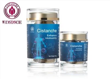
Cistanche products are good for acute kidney injury
From: ' Kidney Injury, Acute ' by SUSAN GARWOOD
----Jean-Louis Vincent & Jesse B. Hall (eds.), Encyclopedia of Intensive Care Medicine, DOI 10.1007/978-3-642-00418-6, # Springer-Verlag Berlin Heidelberg 2012
References
1. Bellomo R, Ronco C, Kellum JA, Mehta RL, Palevsky P (2004) Acute renal failure – definition, outcome measures, animal models, fluid therapy and information technology needs: the second international consensus conference of the Acute Dialysis Quality Initiative (ADQI)
Group. Crit Care 8:R204–R212, The ADQI workgroup
2. Sutton TA, Fisher CJ, Molitoris BA (2002) Microvascular endothelial injury and dysfunction during ischemic acute renal failure. Kidney Int 62:1539–1549
3. Chatterjee PK (2007) Novel pharmacological approaches to the treatment of renal ischemia-reperfusion injury: a comprehensive review. Naunyn-Schmiedebergs Arch Pharmacol 376:1–43
4. Devarajan P (2006) Update on mechanisms of ischemic acute kidney injury. J Am Soc Nephrol 17:1503–1520
5. Bihorac A, Yavas S, Subbiah S, Hobson CE, Schold JD, Gabrielli A, Layon AJ, Segal MS (2009) Long-term risk of mortality and acute kidney injury during hospitalization after major surgery. Ann Surg 5:851–858