Biomarkers Of Acute Kidney Injury And Pathophysiological Mechanisms Of Progression From Acute Kidney Injury To Acute Kidney Disease To Chronic Kidney Disease
Oct 11, 2024
Biomarkers of acute kidney injury
Extensive research is currently underway to explore novel biomarkers for the early detection and prognosis of AKI. As outlined by the Acute Illness Quality Initiative AKI Biomarker Consensus Conference, these biomarkers fall into three broad categories: stress markers, injury markers, and functional markers [18]. Stress markers serve as early indicators of cellular stress and are used to predict AKI. In contrast, injury markers indicate structural damage, which may or may not lead to a decline in renal function. Finally, functional markers are associated with changes in glomerular filtration rate (GFR), providing a measure of changes in renal function. Subclinical AKI is defined as the presence of at least one positive novel biomarker in the absence of elevated serum creatinine levels, indicating early renal stress and injury before functional loss. The added value of these novel biomarkers is that they enable early diagnosis and detection of renal injury, even in the absence of overt functional impairment [19-21].

click to cistanche for kidney disease
Stress markers
Urinary Dickkopf-3 (DKK3) is a glycoprotein derived from renal tubular epithelial cells (TECs) that is used for risk assessment and prediction of AKI. Preoperative urinary DKK3 levels have been identified as an independent predictor of postoperative AKI [22]. Urinary levels of TIMP-2 and IGFBP-7 can be used as markers to indicate G1 cell cycle arrest. These markers may increase rapidly after cellular stress, usually within 4 to 12 hours, even before injury occurs [23,24]. In patients who progress to AKI stage 2-3, urinary [TIMP-2]•[IGFBP7] concentrations surge on the day of exposure, showing a clear pattern of rise and subsequent decline around most exposures [25]. Therefore, biomarkers such as TIMP-2 and IGFBP-7 can help detect subclinical forms of AKI that are not recognized using traditional testing methods.
Injury markers
Alanine aminopeptidase, alkaline phosphatase, and gamma-glutamyl transpeptidase are enzymes located on the brush border villi of proximal tubular cells [24]. When these cells are damaged, these enzymes are released into the urine and can be detected and used to indicate tubular damage in patients [26].
The renin-angiotensin-aldosterone system (RAAS) is activated in AKI, resulting in elevated levels of angiotensin II (Ang II) in the kidney. This Ang II triggers proinflammatory and profibrotic pathways, contributing to the progression of AKI [27]. Angiotensinogen is a precursor peptide of angiotensin peptide that is stable in urine, making it a practical marker for assessing RAAS activity and providing more specific insights than direct measurement of Ang II in urine. Urinary angiotensinogen can serve as a marker of intrarenal RAAS activity and predict the progression of CKD. In patients with severe AKI, angiotensinogen effectively predicted the combined outcome of worsening AKI [area under the ROC curve (AUC) = 0.77] and the need for renal replacement therapy (RRT) or mortality (AUC = 0.73) [28].
Meprin A, composed of α- and β-subunits, is a membrane-associated neutral metalloendopeptidase in the zinc endopeptidase astaxanthin family that is primarily localized to the brush edge membrane of the proximal tubule and intestine [29]. Extensive studies have highlighted its role in the pathogenesis of AKI caused by ischemia-reperfusion (IR) injury and cisplatin nephrotoxicity. Notably, after renal vascular IR, a significant change in the distribution of alpha-fetoprotein a was observed from its regular linear pattern along the brush border membrane to the underlying basement membrane. It is believed that the redistribution of alpha-fetoprotein a during insulin resistance leads to cellular damage and triggers an inflammatory response. Furthermore, the presence of medroxyprogesterone in the urine during AKI suggests that it is shed under this pathological condition [30-32].

Calprotectin is a cytoplasmic calcium-binding complex derived from neutrophils and monocytes. In the setting of endogenous AKI, urinary calprotectin levels are significantly increased [33,34]. C-C motif chemokine ligand 14 (CCL14) is a proinflammatory chemokine that is released into the urine when renal tubular cells are stressed or injured. According to the results of the RUBY study, elevated CCL14 levels can be used as a predictive marker for persistent AKI in critically ill patients, especially those with severe AKI [35]. NGAL exists in three different forms: a monomeric glycoprotein form derived from neutrophils and TECs, a homodimeric protein derived from neutrophils, and a heterodimeric protein produced by renal tubular cells. These forms of NGAL can be detected in serum and urine during the development of AKI, especially after ischemic or toxic-induced renal injury [26,33,36], with NGAL having the best predictive accuracy for the development of AKI [37].
Urinary KIM-1 is a transmembrane glycoprotein produced by proximal tubular cells and released into the urine after tubular injury. It has been identified as a reliable marker of AKI in adults [15,36]. In fact, additional biomarkers are released upon tubular injury, including L-FABP, a protein located in the cytoplasm of the renal proximal tubules. Interleukin (IL)-18, a proinflammatory cytokine, is also one of the biomarkers associated with tubular injury. Monitoring the levels of these biomarkers can help in the assessment and diagnosis of AKI [36,38]. These molecules consist of constitutive proteins released by the damaged kidney, substances that are upregulated by the injury, and non-renal tissue products that are filtered, reabsorbed, or secreted by the kidney [18].
Functional markers
Levels of cystatin C, a cysteine protease inhibitor produced by nucleated human cells, increase within 12 to 24 hours after renal injury [39]. Cystatin C is thought to have better accuracy than serum creatinine in identifying individuals with reduced GFR [40]. First, serum creatinine does not rapidly reflect changes in GFR, especially in cases of unstable GFR [41]. In addition, serum creatinine is not eliminated from the body solely through glomerular filtration; it also involves partial secretion by the renal tubules. This widely recognized process can result in a significant overestimation of GFR. Therefore, cystatin C is particularly useful for detecting mild decreases in GFR [42]. Proenkephalin A is an endogenous polypeptide hormone found in various tissues, such as the adrenal medulla, nervous system, immune system, and renal tissue [43]. Proenkephalin A has been reported to be a useful biomarker for early detection of AKI and prediction of shorter duration and successful release of RRT [44,45]. Both cystatin C and proenkephalin A are considered injury biomarkers as well as functional biomarkers. When used in combination, they facilitate a more comprehensive assessment and accurate diagnosis of AKI [24].
Limitations and possibilities for future research on novel AKI biomarkers
Unlike cardiac troponins, which are specifically released by damaged cardiomyocytes [46], renal injury biomarkers and serum creatinine may interact with inflammatory responses. Such cross-reactivity may complicate their ability to accurately recapitulate renal injury, particularly in conditions such as sepsis [47]. Peak concentrations of different biomarkers vary depending on the time since the initial injury, which poses a significant challenge to accurately interpreting these findings in the clinical setting. Future research should emphasize exploring the mechanisms of injury to deepen our understanding of the various AKI phenotypes based on their pathophysiological characteristics. Exploring the biological transition from initial renal stress to the emergence of subclinical or clinical AKI could open up new targets for therapeutic intervention. These advances may include targeted therapies or strategic approaches, all with the ultimate goal of improving patient outcomes.
Pathophysiology of AKI
It originates from a variety of insults, such as renal hypoperfusion, sepsis, major surgery, immune diseases affecting the renal parenchyma, the use of radiocontrast agents or nephrotoxic drugs, and postrenal causes [69]. The pathophysiology of AKI varies depending on the situation, and the etiology of AKI can be classified as prerenal, intrarenal, or postrenal [70]. This article briefly summarizes this pathophysiology.
Prerenal causes
① In the case of renal hypoperfusion caused by hypovolemia, autoregulatory and neurohumoral mechanisms are triggered to maintain GFR. However, persistent renal hypoperfusion leads to persistent oxygen insufficiency and adenosine triphosphate (ATP) depletion, resulting in epithelial cell damage [71]. This subsequently activates an inflammatory response, inducing endothelial damage, and ultimately renal injury [72,73].
② In sepsis, inflammatory cytokines can induce leukocyte activation, recruit neutrophils, and trigger endothelial damage and coagulation. In addition, these inflammatory mediators may bind to specific receptors expressed by renal endothelial and tubular epithelial cells, leading to direct injury [74]. Damaged cells release damage-associated molecular patterns (DAMPs), which further contribute to vasodilation, increased vascular permeability, and a prothrombotic environment [75]. In addition, disordered DAMPs and pathogen-associated molecular patterns (PAMPs) may activate Toll-like receptor 2 (TLR2) and Toll-like receptor 4 (TLR4) on the proximal tubules, subsequently initiating interstitial inflammation. Vascular dysfunction, endothelial injury, immune dysregulation, and abnormal cellular responses to injury together contribute to the development of AKI in sepsis [76].
③ AKI caused by major surgery can be attributed to fluid depletion, including blood loss and extravasation of fluid into the third space [77]. In addition, anesthetics may cause peripheral vasodilation and myocardial depression, thereby affecting renal perfusion. In the case of AKI associated with cardiac surgery, extracorporeal circulation may cause IR injury, leading to cell damage and death by increasing mitochondrial permeability [77,78]. Renal IR injury is the main cause of AKI, leading to perioperative tubular epithelial cell apoptosis, necrosis, and inflammation [79].
Intrarenal causes
① Nephrotoxic drugs are transferred and concentrated in the nephron, which may cause damage to renal tubular epithelial cells (TEC) through direct cytotoxic effects. In addition, these toxins can cause mesangial cell contraction by impairing intrarenal hemodynamics [72,80]. Some nephrotoxic drugs may induce acute tubular injury. For example, polycationic aminoglycosides, which have a significant effect on anionic phospholipid membranes, may interact with megalin-cubosome receptors located on the apical surface. This interaction promotes the endocytosis of aminoglycosides, resulting in their subsequent internalization into cells and ultimate transport to lysosomes [81]. Lysosomal injury with osteoblast formation and mitochondrial damage leads to tubular cell apoptosis and/or necrosis. In addition, some drugs, such as β-lactam antibiotics, proton pump inhibitors, and immunotherapy, can induce acute interstitial nephritis [82]. These drugs or their metabolites may trigger immune responses through different mechanisms. They can attach to the tubular basement membrane as haptens or prehaptens, triggering an immune response against this antigen. Dendritic cells and tubular cells present the antigen to naive CD41 T helper cells, leading to the formation of various T helper cell subsets. These cells release cytokines, such as interleukins and interferons, which attract macrophages, eosinophils, CD8 T cells, and mast cells/basophils to the tubulointerstitium, ultimately leading to acute interstitial nephritis [83].
② In individuals genetically susceptible to autoimmune activation, renal consequences may involve glomerular inflammation and damage, such as rapidly progressive glomerulonephritis [84].
Postrenal causes
Extrarenal or intrarenal obstruction may increase intratubular pressure, impair renal blood flow, and initiate an inflammatory process that ultimately leads to AKI [85].
Summary
The classification of renal diseases into AKI, AKD, and CKD highlights the risk of progression and the causal relationship between AKI/AKD and CKD. It addresses the need for better biomarkers for early AKI detection beyond traditional serum creatinine, and the complex molecular pathways involved in the transition from AKI to CKD. Biomarkers for early detection of AKI are divided into stress, injury, and functional markers. AKI arises following multiple insults, leading to complex pathophysiological events such as cellular hypoxia, inflammation, and nephrotoxicity, and subsequent renal damage.
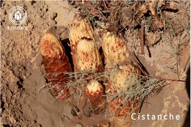
How Does Cistanche Treat Kidney Disease?
Cistanche is a traditional Chinese herbal medicine used for centuries to treat various health conditions, including kidney disease. It is derived from the dried stems of Cistanche deserticola, a plant native to the deserts of China and Mongolia. The main active components of cistanche are phenylethanoid glycosides, echinacoside, and acteoside, which have been found to have beneficial effects on kidney health.
Kidney disease, also known as renal disease, refers to a condition in which the kidneys are not functioning properly. This can result in a buildup of waste products and toxins in the body, leading to various symptoms and complications. Cistanche may help treat kidney disease ase through several mechanisms.
Firstly, cistanche has been found to have diuretic properties, meaning it can increase urine production and help eliminate waste products from the body. This can help relieve the burden on the kidneys and prevent the buildup of toxins. By promoting diuresis, cistanche may also help Reduce high blood pressure, a common complication of kidney disease.
Moreover, cistanche has been shown to have antioxidant effects. Oxidative stress, caused by an imbalance between the production of free radicals and the body's antioxidant defenses, plays a key role in the progression of kidney disease. ies help neutralize free radicals and reduce Oxidative stress, thereby protecting the kidneys from damage. The phenylethanoid glycosides found in cistanche have been particularly effective in scavenging free radicals and inhibiting lipid peroxidation.
Additionally, cistanche has been found to have anti-inflammatory effects. Inflammation is another key factor in the development and progression of kidney disease. Cistanche's anti-inflammatory properties help reduce the production of pro-inflammatory cytokines and inhibit the activation of mandatory pathways for inflammation, thus alleviating inflammation in the kidneys.

Furthermore, cistanche has been shown to have immunomodulatory effects. In kidney disease, the immune system can be dysregulated, leading to excessive inflammation and tissue damage. Cistanche helps regulate the immune response by modulating the production and activity of immune cells, such as T cells and macrophages. This immune regulation helps reduce inflammation and prevent further damage to the kidneys.
Moreover, cistanche has been found to improve renal function by promoting the regeneration of renal tubes with cells. Renal tubular epithelial cells play a crucial role in the filtration and reabsorption of waste products and electrolytes. In kidney disease, these cells can be damaged, leading to damaged renal function. Cistanche's ability to promote the regeneration of these cells helps restore proper renal function and improve overall kidney health.
In addition to these direct effects on the kidneys, cistanche has been found to have beneficial effects on other organs and systems in the body. This holistic approach to health is particularly important in kidney disease, as the condition often affects multiple organs and systems. che has been shown to have protective effects on the liver, heart, and blood vessels, which are commonly affected by kidney disease. By promoting the health of these organs, cistanche helps improve overall kidney function and prevent further complications.
In conclusion, cistanche is a traditional Chinese herbal medicine used for centuries to treat kidney disease. Its active components have diuretic, antioxidant, anti-inflammatory, immunomodulatory, and regenerative effects, which help improve renal function and protect the kidneys from further damage. , cistanche has beneficial effects on other organs and systems, making it a holistic approach to treating kidney disease.