Cistanche Deserticola Polysaccharides: Pre‑Clinical Insights Into Lung Inflammation, Macrophage Polarisation And The Gut‑Lung Axis
Nov 09, 2026
Disclaimer: This article covers pre‑clinical animal‑ and cell‑based research only. It is not medical advice. Cistanche‑derived compounds have not been approved for treating acute respiratory distress syndrome (ARDS) in humans. Dietary supplements cannot diagnose, cure or prevent disease. Always consult a qualified clinician for severe breathing issues, lung conditions or before starting any new supplement regimen. Meta data for your CMS (do not publish inside article body): Title tag: Cistanche Deserticola Polysaccharides: Lung Inflammation, Macrophages and Gut‑Lung‑Axis Research Meta description: What pre‑clinical lab studies tell us about Cistanche deserticola polysaccharides, macrophage polarisation, STING/NF‑κB pathways, gut‑lung axis and acute inflammatory lung injury. URL slug: /cistanche‑polysaccharides‑lung‑inflammation‑gut‑lung‑axis Primary keyword: Cistanche deserticola polysaccharides Secondary keywords: Cistanche polysaccharides, gut‑lung axis, macrophage M2 polarisation, STING/NF‑κB, acute respiratory distress syndrome ARDS, Akkermansia, herbal immunomodulator

Introduction
Acute respiratory distress syndrome (ARDS) represents one of the most dangerous forms of severe lung injury seen in modern critical‑care medicine. Triggered by sepsis, bacterial infection, trauma or inflammatory insults, ARDS manifests as runaway systemic and pulmonary inflammation, damage to the delicate alveolar‑capillary barrier, pulmonary oedema and severely impaired oxygen exchangePMC. Reported mortality rates can reach 40‑46 %, and clinical care today relies heavily on mechanical ventilation and supportive strategies; specific drug therapies to halt the core inflammatory cascade remain limitedPMC. This gap drives researchers worldwide to look for new molecular targets and bioactive natural compounds.
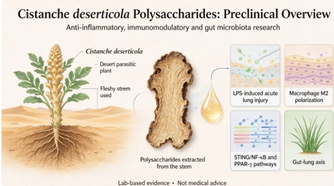
Cistanche deserticola, commonly nicknamed "desert ginseng", is a well‑documented tonic herb in traditional Chinese materia medica. Its fleshy stem hosts multiple bioactive fractions: phenylethanoid glycosides, iridoid substances, amino acids, and polysaccharides. Cistanche deserticola polysaccharides (CDP) are water‑soluble carbohydrate polymers extracted from this desert parasitic plant. A growing body of pre‑clinical work shows CDP exerts anti‑inflammatory, antioxidant and gut‑microbiota‑regulating properties across multiple disease models, including brain injury, intestinal disorders and age‑related impairment. A recent animal‑ and cell‑based research project explored how CDP may act against lipopolysaccharide‑triggered acute lung injury, focusing on macrophage reprogramming, inflammatory signalling cascades and cross‑talk between gut and lung tissue, also known as the gut‑lung axis. This article unpacks that lab‑based evidence in plain language, clarifying what has been observed in the laboratory and where knowledge gaps still remain.
1. Understanding ARDS, Macrophage Plasticity and the Gut‑Lung‑Axis Framework
1.1 The immune chaos behind ARDS
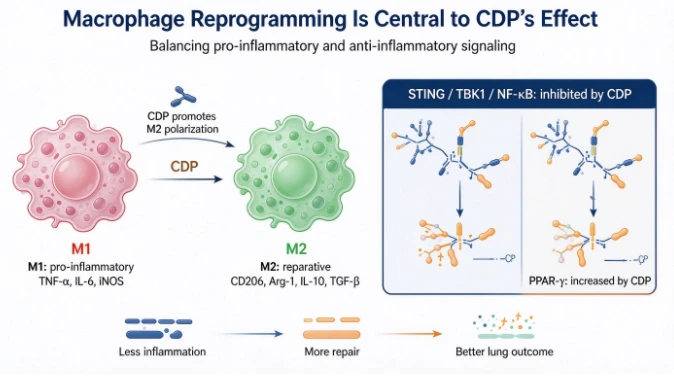
At the heart of ARDS pathology lies dysregulated innate immunity. Macrophages, versatile sentinel immune cells, play a decisive role. These cells show remarkable functional flexibility, described in research terms as "polarisation". Broadly speaking, macrophages can shift towards two opposing functional profiles: - M1‑type (pro‑inflammatory): Activated by bacterial toxins such as lipopolysaccharide (LPS). They secrete large quantities of TNF‑α, IL‑6, iNOS and other pro‑inflammatory mediators, kick‑starting defensive responses but also driving excessive tissue destruction when over‑activated. - M2‑type (repair‑oriented): Produce anti‑inflammatory cytokines like IL‑10 and TGF‑β, express surface markers such as CD206 and Arg‑1. Their main roles include clearing cellular debris, dampening inflammation and supporting tissue reconstruction after injury.
During ARDS, immune homeostasis breaks down: M1‑dominant responses persist too long, while the shift toward reparative M2 macrophages stalls. Uncontrolled release of inflammatory mediators floods lung tissue, ruptures alveolar barriers and fills lung spaces with fluid, making gas exchange nearly impossible. Therefore, encouraging macrophages to transition toward the M2 reparative phenotype has become an attractive target for experimental anti‑inflammatory drug development.
1.2 The gut‑lung‑axis: organs communicating far apart
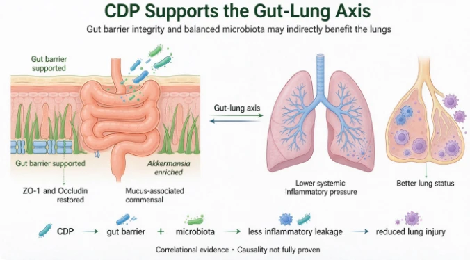
Modern immunology has confirmed robust bidirectional signalling between intestinal tract and respiratory organs, termed the gut‑lung axis. Even when injury originates in the lungs, gut health often deteriorates simultaneously. Severe pulmonary inflammation can compromise intestinal tight‑junction proteins (ZO‑1, Occludin), weaken the gut physical barrier and trigger gut‑microbiota dysbiosis. Conversely, a leaky gut and imbalanced microbial communities release inflammatory signals and metabolites into systemic circulation, further amplifying distant lung injury. This cross‑organ communication helps explain why many critically‑ill ARDS patients concurrently suffer intestinal complications. It also opens a thought‑provoking possibility: interventions that stabilise gut microbiota and gut barrier integrity may indirectly mitigate inflammatory damage in the lungs.
1.3 STING / NF‑κB and PPAR‑γ: two key signalling switches
Two molecular signalling modules feature prominently in this pre‑clinical research. First, the STING‑TBK1‑NF‑κB inflammatory cascade: STING is an intracellular DNA‑sensor protein. When activated by misplaced cytoplasmic DNA from damaged or dying cells, it recruits TBK1 kinase and subsequently triggers NF‑κB translocation into cell nuclei, turning on hundreds of pro‑inflammatory‑related genes and cytokine production. Excessive STING/NF‑κB activation is repeatedly linked to aggravated acute lung injury.
Second, PPAR‑γ, a nuclear‑receptor protein highly expressed in macrophages. PPAR‑γ acts as an inflammation‑brake; its activation promotes M2‑type macrophage maturation, curbs NF‑κB‑driven transcription and boosts anti‑inflammatory mediator release. Balancing STING/NF‑κB and PPAR‑γ activity represents a promising strategy for calming hyper‑active innate immunity. This study set out to test whether CDP could modulate these two axes, alongside gut‑lung‑axis events, to ease LPS‑provoked ARDS‑like pathology.
2. Overview of the pre‑clinical experimental setup
This pre‑clinical work combined in‑vitro cell culture experiments and in‑vivo mouse‑model investigations. Two core experimental systems were used: 1. Cell‑level assays: Kasumi‑1 monocyte‑derived cells (to study macrophage polarisation), and A549 human alveolar epithelial cells to model lung‑epithelial inflammatory responses. LPS was deployed to simulate bacterial‑toxin‑driven inflammatory insult. 2. Animal‑level assays: 6‑8‑week‑old male C57BL/6J mice, widely used for immunology research. ARDS‑like acute lung injury was induced via intratracheal LPS instillation, a standard pre‑clinical approach that reproduces major hallmarks of human ARDS, including pulmonary oedema, immune‑cell infiltration and elevated circulating pro‑inflammatory cytokinesPMC. CDP was administered by oral gavage at low (200 mg/kg/day) and high (400 mg/kg/day) dosages for seven consecutive days before LPS‑challenge.
Critical validation experiments included macrophage‑depletion testing: researchers injected clodronate‑loaded liposomes to largely eliminate macrophages in some animal groups. This experimental trick answers a vital causal question: if CDP's protective effects vanish when macrophages are removed, it demonstrates macrophages are essential mediators of CDP's biological activity, rather than CDP working through some entirely separate pathway.
Multiple read‑out techniques were deployed: histopathological H&E tissue staining for lung and colon tissue; immunohistochemistry for gut‑barrier tight‑junction proteins; flow cytometry for immune‑cell phenotyping; RT‑qPCR for gene‑expression profiling; Western‑blot protein analysis for signalling‑pathway activity; ELISA for cytokine quantification; and 16S rDNA high‑throughput sequencing to profile compositional shifts in gut‑microbiota communities.
Important caveat: Animal‑study doses cannot be directly translated to safe human‑supplement‑dosage equivalents. These lab‑grade CDP preparations are carefully characterised experimental materials, not commercial off‑the‑shelf supplement powders.
3. Major pre‑clinical findings: CDP effects on lung inflammation and macrophage reprogramming
3.1 CDP mitigates LPS‑triggered lung tissue damage in mice
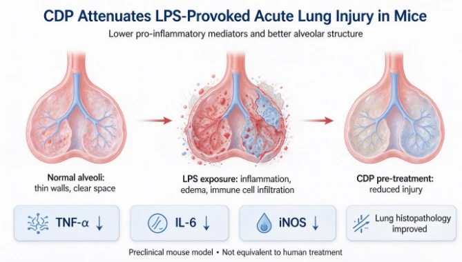
After LPS‑challenge, the untreated ARDS‑model mice exhibited clear signs of severe pulmonary injury: marked body‑weight loss, thickened alveolar septa, widespread inflammatory‑cell infiltration, alveolar‑space collapse and tissue oedema. By contrast, animals pre‑treated with CDP displayed dose‑dependent improvements. High‑dose CDP substantially alleviated histological lung damage and partially prevented LPS‑induced weight decline. Consistent with tissue‑section observations, molecular testing showed CDP treatment lowered pulmonary and serum concentrations of key pro‑inflammatory mediators including TNF‑α, IL‑6 and iNOS.
3.2 CDP drives M2‑type reparative macrophage polarisation
In cell‑culture experiments, CDP treatment increased CD206‑positive M2‑phenotype cell populations in Kasumi‑1 cells. M2‑marker genes CD206, Arg‑1, together with anti‑inflammatory‑cytokine transcripts IL‑10, TGF‑β and chemokine CCL22 were significantly up‑regulated under CDP intervention. When researchers transferred supernatants from CDP‑treated Kasumi‑1 cells onto LPS‑stressed A549 alveolar epithelial cells, the epithelial‑cell inflammatory‑gene activation (TNF‑α, IL‑6, iNOS) was strongly suppressed. This indicated CDP‑reprogrammed macrophages release soluble protective factors that calm lung‑epithelial‑cell inflammation.
In living mice, flow‑cytometry analysis confirmed CDP‑treated groups had elevated CD206‑positive M2‑macrophage abundance within spleen tissue. Lung‑tissue RT‑qPCR and serum ELISA results also confirmed higher levels of M2‑associated anti‑inflammatory cytokines IL‑4, IL‑10 and TGF‑β.
3.3 Macrophage‑depletion cancels CDP's lung‑protective benefit
When researchers eliminated macrophages with clodronate liposomes, CDP no longer conferred measurable protection. Even with CDP dosing, macrophage‑depleted animals still displayed severe lung histopathology, high TNF‑α and IL‑6 levels, and significant body‑weight loss. This experiment delivers a key mechanistic take‑away from this study: CDP's protective effect against LPS‑provoked acute lung injury is largely macrophage‑dependent. Without functional macrophages present, CDP cannot produce the observed anti‑inflammatory outcomes in this mouse‑model.
3.4 CDP modulates STING/NF‑κB and PPAR‑γ signalling pathways
Western‑blot protein measurements of lung tissue illuminated how CDP reshapes intracellular signalling: LPS‑exposed model mice showed strong activation of the STING‑TBK1‑NF‑κB axis, with elevated STING protein, increased phosphorylation of TBK1 and NF‑κB. CDP‑pre‑treatment effectively reduced STING abundance and lowered phosphorylation of TBK1 and NF‑κB, meaning it dampened this major pro‑inflammatory cascade. Simultaneously, CDP markedly raised PPAR‑γ protein expression in lung tissue. Put together, pre‑clinical data suggests CDP works through dual‑directional signalling regulation: it inhibits the STING/NF‑κB pro‑inflammatory cascade, while boosting the anti‑inflammatory PPAR‑γ axis, jointly pushing macrophage cells toward the reparative M2 phenotype.
4. CDP reshapes intestinal barrier integrity and gut‑microbiota composition (gut‑lung‑axis evidence)
Beyond lung immunity, this research also explored CDP's influence on intestinal health in the context of LPS‑induced ARDS‑like injury.
4.1 CDP restores gut tight‑junction protein expression
LPS‑triggered systemic inflammation damaged colon tissue in model mice, lowering expression of critical tight‑junction proteins ZO‑1 and Occludin, weakening intestinal physical barrier function. Immunohistochemistry results demonstrated CDP supplementation significantly restored ZO‑1 and Occludin protein abundance, helping to preserve intestinal epithelial‑barrier integrity. A robust gut barrier limits microbial‑product leakage into blood circulation and thereby reduces secondary systemic inflammatory pressure that can worsen remote‑organ lung injury.
4.2 CDP reshapes gut‑microbiota community composition
16S rDNA sequencing of mouse caecal gut‑microbiota samples yielded several notable observations. While CDP did not produce statistically significant shifts in alpha‑diversity (within‑sample species richness), beta‑diversity analysis revealed large compositional differences between groups: LPS‑model mice had distinct gut‑microbiota profiles compared to healthy controls, and CDP‑treated animals diverged clearly from untreated ARDS‑model animals, moving closer toward the healthy‑control microbial configuration.
At the phylum level, LPS‑challenge drove elevated relative abundance of Firmicutes, Deferribacterota and Proteobacteria (groups containing multiple opportunistic pathogenic bacterial taxa), alongside reduced Bacteroidota fractions. CDP intervention reversed many of these shifts: it brought down Firmicutes, Deferribacterota and Proteobacteria abundance and strongly enriched Akkermansia (phylum Verrucomicrobia). Akkermansia muciniphila is a well‑studied mucus‑dwelling commensal bacterium known to support intestinal‑mucus‑layer maintenance, strengthen gut‑barrier function and exert immunomodulatory activity. LEfSe biomarker‑analysis further identified Akkermansia as the signature‑discriminatory microbial taxon in CDP‑receiving animal groups. By contrast, Lachnospiraceae_NK4A136_group bloomed in untreated LPS‑model animals and was suppressed after CDP treatment.
These pre‑clinical findings illustrate that under acute systemic inflammatory stress mimicking ARDS, CDP simultaneously protects gut physical barrier function and remodels gut‑microbiota community structure, enriching beneficial mucus‑associated commensal microbes while suppressing over‑expansion of potential pathobionts. Consistent with the gut‑lung‑axis hypothesis, these gut‑system improvements likely contribute indirectly to reduced pulmonary‑inflammatory burden in this animal‑model, although further research is needed to fully untangle cause‑and‑effect relationships between microbiota changes, macrophage polarisation and lung outcomes.
5. Synthesising the proposed pre‑clinical working‑mechanism model
Based on the combined cell‑culture and mouse‑model findings, researchers put forward a multi‑layer mechanistic model for CDP action in LPS‑provoked acute inflammatory lung injury: 1. Orally‑administered CDP reaches the gastrointestinal tract, supporting intestinal tight‑junction‑protein expression and remodelling gut‑microbiota composition, enriching beneficial taxa including Akkermansia, restraining opportunistic‑pathogen overgrowth and preserving gut‑barrier integrity. This reduces microbial‑product translocation‑driven systemic inflammatory pressure. 2. CDP modulates innate‑immune‑cell behaviour, promoting monocyte‑macrophage transition toward reparative M2‑polarisation. At molecular level, this occurs partly via suppression of the STING/NF‑κB pro‑inflammatory signalling cascade and concurrent up‑regulation of PPAR‑γ anti‑inflammatory nuclear‑receptor signalling. 3. Reprogrammed M2‑type macrophages secrete anti‑inflammatory cytokines IL‑10, TGF‑β and IL‑4, suppressing excessive pro‑inflammatory‑cytokine storms (TNF‑α, IL‑6, iNOS). This alleviates pulmonary‑tissue oedema, immune‑cell infiltration and alveolar‑structural damage. 4. Macrophage‑depletion experiments confirm macrophages are a necessary central mediator for CDP‑conferred lung‑tissue‑protection in this pre‑clinical setting.
Crucially, this model originates exclusively from pre‑clinical lab data. Human clinical trials are absent, and causal chains between gut‑microbiota shifts and macrophage‑polarisation are not yet definitively proven.
6. Important limitations of current pre‑clinical research
Readers exploring natural‑compound research need clear awareness of study constraints: 1. Animal‑model gap: This work uses LPS‑induced mouse acute‑lung‑injury as a proxy for human ARDS. Human ARDS arises from highly heterogeneous clinical triggers (viral pneumonia, bacterial sepsis, trauma etc). Mouse‑model findings do not guarantee identical biological responses will occur in human patients. 2. Dose and formulation constraints: Oral dosages (200 mg/kg and 400 mg/kg) in mice cannot be directly scaled for human‑supplement‑use. The CDP preparation used in‑house for experiments is a lab‑extracted characterised polysaccharide sample; commercial Cistanche supplements on the market vary widely in polysaccharide content, purity, extraction‑processing methods and species source (Cistanche deserticola vs Cistanche tubulosa). Lab‑study material is not interchangeable with retail‑product material. 3. Causal‑proof boundaries: The study observed simultaneous changes in gut‑microbiota, gut‑barrier function, macrophage‑polarisation and lung‑inflammation. However, it has not performed faecal‑microbiota‑transplantation experiments, the gold‑standard assay for proving gut‑microbiota changes directly cause the observed lung‑protective effects. Therefore, gut‑microbiota shifts are strongly correlated, but definitive causal evidence remains pending. 4. Cell‑model imperfections: A549 cancer‑derived alveolar‑epithelial cell line is frequently‑used surrogate for human primary alveolar epithelium but carries inherent tumour‑cell metabolic differences. Future work using primary lung‑epithelial cells or organoid‑systems would strengthen mechanistic conclusions. 5. No human‑trial data: There are no published human‑clinical‑trial results evaluating CDP for ARDS or acute‑lung‑injury treatment. We cannot draw conclusions on human efficacy, safety‑profile or optimal human‑dosing.
7. Real‑world take‑aways for supplement shoppers and health‑enthusiasts
1. This is pre‑clinical basic‑science research only: Cistanche polysaccharides are not an ARDS treatment. If you or someone you experience acute severe breathing difficulty, this is a medical emergency requiring immediate hospital‑based clinical care; natural‑product supplements are no substitute. 2. Do not extrapolate mouse‑study dosages for personal‑supplement‑dosing: Animal experimental dosages are for laboratory‑research purposes only. Retail‑supplement‑labels should be followed, or guidance sought from a healthcare‑provider. 3. Scrutinise supplement‑product specifications: Check which Cistanche species is used, test‑certified polysaccharide content, third‑party‑tested purity for heavy‑metals, microbial contamination and residual‑solvents. Extraction ratios (e.g. 10:1) tell you nothing about absolute active‑polysaccharide concentration. 4. Potential‑interaction awareness: Cistanche‑derived polysaccharides possess immunomodulatory properties. People taking immunosuppressant‑medications, diabetes‑medication, pregnant or nursing individuals should consult their clinician before adding new herbal‑based supplements.
8. Future‑research directions
This body of pre‑clinical work opens several promising avenues for follow‑up investigation: - Faecal‑microbiota‑transplantation experiments to establish causal links between CDP‑reshaped gut‑microbiota communities and macrophage‑polarisation / lung‑inflammatory outcomes. - Use of specific‑molecular‑pathway inhibitors or gene‑knock‑out animal‑models to further dissect STING/NF‑κB and PPAR‑γ pathway contributions. - Testing CDP effects in alternative ARDS‑relevant animal‑models beyond LPS‑instillation (e.g. viral‑infection‑triggered lung‑injury). - Purification of individual polysaccharide‑sub‑fractions from crude CDP to identify which molecular‑weight‑sub‑populations drive immunomodulatory and gut‑microbiota‑regulating activity. - Ultimately, well‑designed human‑clinical‑trials are required to move beyond pre‑clinical observations toward real‑world‑therapeutic application.

Conclusion
Pre‑clinical cell‑culture and mouse‑model research demonstrates that Cistanche deserticola polysaccharides (CDP) can mitigate lipopolysaccharide‑provoked acute inflammatory lung injury. Its proposed multi‑modal‑mechanism includes driving reparative M2‑macrophage polarisation via suppression of STING/NF‑κB pro‑inflammatory signalling and activation of PPAR‑γ pathways, alongside maintenance of intestinal‑barrier integrity and rebalancing of gut‑microbiota communities, enriching beneficial Akkermansia. Macrophage‑depletion assays underline macrophages as an essential mediator for CDP‑derived lung‑protection in this experimental‑system.
Nevertheless, all conclusions are confined to laboratory‑pre‑clinical data. Human clinical evidence is absent. These findings highlight Cistanche polysaccharides as an interesting natural immunomodulatory candidate worthy of deeper investigation, but they should not be misinterpreted as proof of human‑therapeutic efficacy for ARDS or other severe lung‑conditions. Further mechanistic validation and human‑clinical‑research will be required to translate these promising lab‑observations into practical health‑applications.
Reference source: Adapted from the master‑degree thesis: Liu Jingyun. Study on the Effects and Mechanisms of Cistanche deserticola Polysaccharide on Macrophage Polarization and Gut Microbiota in Acute Respiratory Distress Syndrome, Southwest Medical University, 2025. All material is substantially re‑phrased, restructured for general‑audience educational blog format. This is secondary interpretive popular‑science writing, not original academic‑paper reproduction.