Echinacoside Modulates The CGAS–STING Pathway To Alleviate LPS‑Induced Inflammatory Injury In Alveolar Epithelial Cells (A549)
Aug 20, 2026
Authors: Jianfeng Luo; Tao Bai
Affiliation: Ward 1, Department of Infectious Diseases, Wuhan Jinyintan Hospital, Wuhan 430000, Hubei, China
Key Takeaways (for natural‑ingredient & supplement R&D readers)
LPS exposure injured A549 alveolar epithelial cells, reducing proliferation markers and increasing apoptosis and inflammatory cytokines.
Echinacoside (ECH) showed dose‑dependent protection: improved cell viability/proliferation and reduced apoptosis and inflammatory mediators.
Mechanistically, the benefits were associated with down‑regulation of the cGAS–STING–TBK1–IRF3 signaling axis at both mRNA and protein levels.
A cGAS–STING pathway activator (RocA) weakened/antagonized ECH's protective effects, supporting pathway involvement.
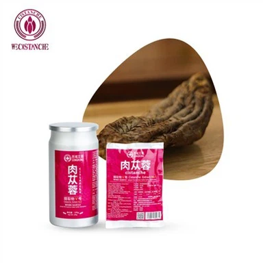
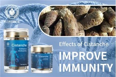
Relevance to botanical development: Cistanche (肉苁蓉) is widely recognized as a botanical rich in echinacoside, making it a practical upstream material to consider for product R&D-while remembering this study is in vitro and uses purified ECH.
Table 1. Effects of different concentrations of ECH and RocA on A549 cells (OD570_{570}570, xˉ±s\bar{x}\pm sxˉ±s, n=6n=6n=6)
| Concentration (μmol/L) | ECH (OD570_{570}570) | RocA (OD570_{570}570) |
|---|---|---|
| 0 | 0.45 ± 0.03 | 0.43 ± 0.04 |
| 3 | 0.42 ± 0.05 | 0.45 ± 0.06 |
| 6 | 0.43 ± 0.04 | 0.42 ± 0.05 |
| 9 | 0.46 ± 0.06 | 0.46 ± 0.07 |
| 12 | 0.44 ± 0.05 | 0.44 ± 0.05 |
| 24 | 0.47 ± 0.06 | 0.20 ± 0.03¹ |
F / P value: ECH: 0.857 / 0.521; RocA: 22.050 / <0.001
Note: Compared with 0 μmol/L: ¹ P<0.05P<0.05P<0.05.
Abstract (rewritten in English, point‑by‑point)
Objective
To evaluate whether echinacoside (ECH) protects human alveolar epithelial cells from lipopolysaccharide (LPS)‑induced inflammatory injury, and whether this protection is linked to regulation of the cGAS–STING signaling pathway.

Methods (study design)
Cell model: Human alveolar epithelial cell line A549.
Cytotoxicity/viability screening: MTT assay to test ECH and a cGAS–STING pathway activator (RocA) for toxicity.
Grouping:
Control (standard culture)
LPS (30 mg/L)
LPS + ECH low/medium/high dose (9, 12, 24 μmol/L)
LPS + ECH (24 μmol/L) + RocA (12 nmol/L)
Endpoints:
Proliferation: EdU staining and PCNA (Western blot)
Apoptosis: Flow cytometry (Annexin V‑FITC/PI); Bax/Bcl‑2 (Western blot)
Inflammation: TNF‑α, IL‑6 in supernatant (ELISA)
Pathway readouts: cGAS, STING mRNA (qRT‑PCR); cGAS, STING, TBK1, IRF3 proteins (Western blot)
Table 2. Comparison of EdU-positive rate, PCNA expression, apoptosis rate, Bax/Bcl-2 expression, and TNF-α/IL-6 levels in A549 cells among groups (xˉ±s\bar{x}\pm sxˉ±s, n=6n=6n=6)
| Group | EdU-positive rate | PCNA/GAPDH | Apoptosis rate (%) | Bax/GAPDH | Bcl-2/GAPDH | TNF-α (μg/ml) | IL-6 (ng/ml) |
|---|---|---|---|---|---|---|---|
| Control | 57.38 ± 3.24 | 1.67 ± 0.15 | 4.38 ± 1.09 | 0.37 ± 0.06 | 1.63 ± 0.12 | 4.65 ± 1.08 | 18.47 ± 1.19 |
| LPS | 27.51 ± 1.68¹ | 0.49 ± 0.08¹ | 36.75 ± 2.31¹ | 1.59 ± 0.13¹ | 0.28 ± 0.06¹ | 30.71 ± 1.68¹ | 49.23 ± 2.75¹ |
| ECH-L | 33.72 ± 1.96² | 0.86 ± 0.10² | 29.16 ± 2.27² | 1.24 ± 0.11² | 0.67 ± 0.08² | 22.58 ± 1.52² | 41.78 ± 2.46² |
| ECH-M | 40.59 ± 2.40²,³ | 1.27 ± 0.13²,³ | 21.93 ± 1.64²,³ | 0.85 ± 0.09²,³ | 1.08 ± 0.10²,³ | 15.83 ± 1.16²,³ | 32.64 ± 2.18²,³ |
| ECH-H | 48.21 ± 2.57²,³,⁴ | 1.58 ± 0.14²,³,⁴ | 13.86 ± 1.45²,³,⁴ | 0.49 ± 0.08²,³,⁴ | 1.35 ± 0.11²,³,⁴ | 7.91 ± 1.13²,³,⁴ | 23.59 ± 1.57²,³,⁴ |
| RocA | 35.79 ± 2.38⁵ | 0.92 ± 0.11⁵ | 25.74 ± 1.69⁵ | 0.93 ± 0.10⁵ | 0.74 ± 0.09⁵ | 19.76 ± 1.20⁵ | 36.78 ± 2.61⁵ |
F value: 118.927; 85.600; 245.724; 131.552; 159.598; 321.852; 161.353
P value: all <0.001
Notes (multiple comparisons):
Compared with Control: ¹ P<0.05P<0.05P<0.05
Compared with LPS: ² P<0.05P<0.05P<0.05
Compared with ECH-L: ³ P<0.05P<0.05P<0.05
Compared with ECH-M: ⁴ P<0.05P<0.05P<0.05
Compared with ECH-H: ⁵ P<0.05P<0.05P<0.05
Results (main findings)
Compared with Control, LPS:
Decreased EdU‑positive rate, PCNA, and Bcl‑2
Increased apoptosis rate, TNF‑α, IL‑6
Upregulated cGAS and STING at mRNA level
Increased Bax and cGAS–STING pathway proteins (cGAS, STING, TBK1, IRF3)
(All statistically significant, P<0.05P<0.05P<0.05.)
Compared with LPS alone, ECH (9→12→24 μmol/L) dose‑dependently:
Restored proliferation (higher EdU‑positive rate and PCNA)
Reduced apoptosis (lower apoptosis rate, lower Bax, higher Bcl‑2)
Suppressed inflammation (lower TNF‑α and IL‑6)
Downregulated cGAS–STING signaling (lower cGAS/STING mRNA; lower cGAS, STING, TBK1, IRF3 proteins)
(Dose‑dependent, P<0.05P<0.05P<0.05.)
RocA counteracted ECH's protection under LPS injury conditions (P<0.05P<0.05P<0.05), consistent with cGAS–STING pathway involvement.
Conclusion
ECH enhanced viability and proliferative activity of LPS‑challenged alveolar epithelial cells and suppressed apoptosis and inflammatory responses. The protective mechanism is likely related to inhibition/blockade of over‑activated cGAS–STING signaling.
Background (why this matters)
In patients exposed to or infected by LPS (a major Gram‑negative bacterial component), acute lung injury (ALI) and its severe form ARDS are key drivers of morbidity and mortality. Characteristic pathology involves diffuse injury to alveolar epithelium and capillary endothelium, leading to barrier leakage and pulmonary edema.
Despite progress in supportive care, effective targeted therapies remain limited, so identifying new drug candidates and targets is important.
Echinacoside (ECH) is a phenylethanoid glycoside with reported activities including antioxidant, anti‑inflammatory, cytoprotective, and anti‑apoptotic effects.
For natural‑product developers: Cistanche (肉苁蓉) is a traditional botanical often highlighted for being rich in echinacoside, making it a practical source material when exploring ECH‑oriented formulations-while also requiring careful standardization and translation work beyond cell assays.
The cGAS–STING pathway is a cytosolic DNA‑sensing axis that promotes type I interferon and inflammatory mediator production. Evidence suggests that excessive activation of cGAS–STING contributes to inflammatory and autoimmune pathology, and that suppressing this over‑activation may help attenuate inflammatory lung injury.
Materials and Methods (R&D‑friendly summary)
Cell line and culture
A549 cells were cultured in low‑glucose DMEM with 10% FBS and 1% antibiotics, with medium changes every 2 days. Experiments were performed at ~80–90% confluence.
Reagents (high level)
ECH (vendor information listed in the original paper)
LPS
RocA (used here as a cGAS–STING pathway activator)
Assay kits: MTT, EdU, apoptosis kit (Annexin V‑FITC/PI), ELISA kits for TNF‑α and IL‑6
Molecular assays: RNA extraction, reverse transcription, qRT‑PCR reagents; antibodies for Western blot (PCNA, Bax, Bcl‑2, cGAS, STING, TBK1, IRF3, GAPDH)
Dose selection (viability screening)
ECH (0–24 μmol/L) showed no significant cytotoxicity in A549 cells and therefore 9/12/24 μmol/L were used.
RocA was non‑toxic within 0–12 nmol/L, while higher concentration reduced viability; 12 nmol/L was selected.
LPS injury induction and grouping
LPS injury was induced with 30 mg/L LPS.
Treatment arms were as described in the Abstract (Control; LPS; LPS+ECH low/medium/high; LPS+ECH high+RocA).
Outcome measurements
Proliferation: EdU‑positive cells by fluorescence microscopy; PCNA by Western blot.
Apoptosis: Flow cytometry (Annexin V‑FITC/PI); Bax and Bcl‑2 protein levels.
Inflammatory mediators: TNF‑α and IL‑6 in culture supernatant by ELISA.
Pathway expression: cGAS and STING mRNA by qRT‑PCR using GAPDH as internal control; proteins (cGAS, STING, TBK1, IRF3) by Western blot.
Statistics: ANOVA with SNK‑q post‑hoc testing (GraphPad Prism 9.0), significance set at P<0.05P<0.05P<0.05.
Results (what changed with LPS, ECH, and RocA)
1) Cell viability / toxicity window
ECH was well tolerated up to the tested range used for mechanistic studies.
RocA required cautious dosing; a non‑toxic working concentration was selected.

Figure 1. A549 cell proliferation in each group (EDU staining, ×200)
2) Proliferation was suppressed by LPS and restored by ECH
LPS reduced EdU incorporation and PCNA expression, indicating decreased proliferative activity.
ECH increased EdU‑positive rates and PCNA in a dose‑dependent manner.
Adding RocA reduced these gains, suggesting the pathway contributes to ECH's pro‑survival/pro‑proliferation effect.
3) Apoptosis increased with LPS and was inhibited by ECH
LPS increased apoptosis rate, increased Bax, and decreased Bcl‑2.
ECH reversed these changes dose‑dependently (lower apoptosis rate and Bax, higher Bcl‑2).
RocA shifted the phenotype back toward apoptosis, weakening ECH protection.

Figure 2. Western blot analysis of PCNA, Bax, and Bcl-2 expression in each group
4) Inflammatory cytokines (TNF‑α, IL‑6) were reduced by ECH
LPS elevated TNF‑α and IL‑6 in supernatants.
ECH reduced both cytokines dose‑dependently.
RocA increased them relative to ECH high dose alone, consistent with pathway re‑activation amplifying inflammation.
5) cGAS and STING transcription was induced by LPS and suppressed by ECH
LPS increased cGAS and STING mRNA.
ECH lowered their expression (dose‑dependent).
RocA increased expression compared with ECH high dose.

Figure 3. Comparison of A549 cell apoptosis in each group
6) cGAS–STING pathway proteins were downregulated by ECH
LPS increased cGAS, STING, TBK1, and IRF3 protein expression.
ECH reduced these proteins dose‑dependently.
RocA increased pathway protein levels versus ECH high dose alone.
Table 3. Comparison of cGAS/STING mRNA and cGAS–STING pathway protein expression in A549 cells among groups (xˉ±s\bar{x}\pm sxˉ±s, n=6n=6n=6)
| Group | cGAS mRNA | STING mRNA | cGAS/GAPDH | STING/GAPDH | TBK1/GAPDH | IRF3/GAPDH |
|---|---|---|---|---|---|---|
| Control | 1.00 ± 0.12 | 1.00 ± 0.11 | 0.32 ± 0.07 | 0.36 ± 0.08 | 0.29 ± 0.05 | 0.25 ± 0.06 |
| LPS | 2.15 ± 0.16¹ | 2.28 ± 0.17¹ | 1.68 ± 0.13¹ | 1.85 ± 0.16¹ | 1.57 ± 0.13¹ | 1.62 ± 0.14¹ |
| ECH-L | 1.74 ± 0.13² | 1.83 ± 0.14² | 1.34 ± 0.12² | 1.42 ± 0.13² | 1.24 ± 0.11² | 1.23 ± 0.12² |
| ECH-M | 1.36 ± 0.11²,³ | 1.31 ± 0.12²,³ | 0.82 ± 0.09²,³ | 1.03 ± 0.11²,³ | 0.86 ± 0.09²,³ | 0.78 ± 0.09²,³ |
| ECH-H | 0.73 ± 0.09²,³,⁴ | 0.62 ± 0.08²,³,⁴ | 0.45 ± 0.08²,³,⁴ | 0.57 ± 0.09²,³,⁴ | 0.47 ± 0.08²,³,⁴ | 0.38 ± 0.07²,³,⁴ |
| RocA | 1.58 ± 0.12⁵ | 1.69 ± 0.15⁵ | 1.17 ± 0.11⁵ | 1.28 ± 0.14⁵ | 1.12 ± 0.10⁵ | 0.97 ± 0.10⁵ |
F / P values:
cGAS mRNA: 103.612 / <0.001
STING mRNA: 124.857 / <0.001
cGAS/GAPDH: 160.066 / <0.001
STING/GAPDH: 124.141 / <0.001
TBK1/GAPDH: 150.139 / <0.001
IRF3/GAPDH: 158.576 / <0.001

Figure 4. cGAS-STING pathway protein expression in cells of each group
Discussion (interpretation for product and mechanism readers)
LPS is commonly used to model inflammatory lung injury. Here, LPS directly impaired alveolar epithelial cell proliferative capacity and promoted apoptosis, accompanied by elevated TNF‑α and IL‑6-two cytokines widely linked to inflammatory lung damage.
ECH acted as a cytoprotective and anti‑inflammatory agent in this in‑vitro setting, consistent with prior reports that ECH can mitigate oxidative stress, inflammation, and apoptosis in lung‑injury contexts.
The cGAS–STING axis (and downstream TBK1/IRF3) appears to be a mechanistic node: ECH lowered cGAS–STING pathway markers, and RocA blunted ECH's benefits, supporting the hypothesis that ECH protects cells at least partly by blocking excessive cGAS–STING activation.
What this suggests for natural botanical development (Cistanche context)
Because Cistanche (肉苁蓉) is known for high echinacoside content, it is a logical botanical candidate when exploring ECH‑centered formulations aimed at inflammatory balance and epithelial protection themes.
However, translating this to supplements requires caution: this work is cell‑line based, uses LPS injury and purified ECH, and does not establish human efficacy, oral bioavailability, metabolism, or clinically relevant dosing.
Limitations and next steps (as implied by the authors)
The findings need validation in in vivo ALI/ARDS models to confirm efficacy and mechanistic linkage to cGAS–STING under physiological conditions.
Further work would be required to map upstream triggers and confirm whether effects are direct pathway inhibition versus secondary to reduced cellular stress.
References (kept complete, as provided)
Li N, Liu B, Xiong R, et al. HDAC3 deficiency protects against acute lung injury by maintaining epithelial barrier integrity through preserving mitochondrial quality control. Redox Biol, 2023;63(1):102746-61.
Long ME, Mallampalli RK, Horowitz JC. Pathogenesis of pneumonia and acute lung injury. Clin Sci (Lond), 2022;136(10):747-69.
Mokrý AD. Acute lung injury-from pathophysiology to treatment. Physiol Res, 2020;69(Suppl 3):S353-66.
Wang W, Jiang S, Zhao Y, et al. Echinacoside: a promising active natural products and pharmacological agents. Pharmacol Res, 2023;197(1):106951-66.
Pan Y, Liu Y. Echinacoside alleviates airway remodeling and inflammation in an ovalbumin-induced neonatal mouse model of asthma by modulating the SIRT1-NF-κB pathway. Allergol Immunopathol (Madr), 2023;51(4):71-7.
Cui YH, Li KF, Jin B, et al. Echinacoside inhibits bleomycin-induced lung epithelial cell injury and inflammatory response. Chinese Journal of Pathophysiology, 2021;37(11):2024-30.
Xie W, Deng L, Lin M, et al. Sirtuin1 mediates the protective effects of echinacoside against sepsis-induced acute lung injury via regulating the NOX4-Nrf2 axis. Antioxidants (Basel), 2023;12(11):1925-45.
Alonso Paiva IM, Santos AR, Brito CB, et al. Role of the cGAS/STING pathway in the control of Brucella abortus infection acquired through the respiratory route. Front Immunol, 2023;14:1116811.
Zhao J, Zhen N, Zhou Q, et al. NETs promote inflammatory injury by activating cGAS-STING pathway in acute lung injury. Int J Mol Sci, 2023;24(6):5125-45.
Wu X, Jiang Y, Li R, et al. Ficolin B secreted by alveolar macrophage exosomes exacerbates bleomycin-induced lung injury via ferroptosis through the cGAS-STING signaling pathway. Cell Death Dis, 2023;14(8):577-92.
Zhang SW, Cheng SL, Xing QF. Pachymic acid alleviates colon epithelial cell injury in ulcerative colitis rats by inhibiting the cGAS-STING signaling pathway. Journal of Immunology (China), 2023;39(8):672-80.
Zhang JJ, Li YY, Qin J. lncRNA LUCAT1 targets miR-203a-3p to affect apoptosis and inflammatory response in LPS-induced alveolar type II epithelial cells. Journal of Immunology (China), 2023;39(8):689-95.
Li J, Deng SH, Li J, et al. Obacunone alleviates ferroptosis during lipopolysaccharide-induced acute lung injury by upregulating Nrf2-dependent antioxidant responses. Cell Mol Biol Lett, 2022;27(1):29-48.
Hong H, Lou S, Zheng F, et al. Hydnocarpin D attenuates lipopolysaccharide-induced acute lung injury via MAPK/NF-κB and Keap1/Nrf2/HO-1 pathway. Phytomedicine, 2022;101(1):154143-50.
Sasaki M, Kishimoto M, Itakura Y, et al. Air-liquid interphase culture confers SARS-CoV-2 susceptibility to A549 alveolar epithelial cells. Biochem Biophys Res Commun, 2021;577:146-51.
Wang Y, Fan Y, Huang Y, et al. TRIM28 regulates SARS-CoV-2 cell entry by targeting ACE2. Cell Signal, 2021;85:110064.
Yang X, Ma L. Post-treatment with propofol inhibits inflammatory response in LPS-induced alveolar type II epithelial cells. Exp Ther Med, 2022;23(4):249.
Duan XY, Sun Y, Zhao ZF, et al. Baicalin attenuates LPS-induced alveolar type II epithelial cell A549 injury by attenuation of the FSTL1 signaling pathway via increasing miR-200b-3p expression. Innate Immun, 2021;27(4):294-312.
Lei X, Liu X, Yu J, et al. 3-methyladenine ameliorates acute lung injury by inhibiting oxidative damage and apoptosis. Heliyon, 2024;10(13):e33996.
Li M, Song J, Tang X, et al. Critical roles of PAI-1 in lipopolysaccharide-induced acute lung injury. Adv Med Sci, 2024;69(1):90-102.
Jiang T, Yuan D, Wang R, et al. Echinacoside, a promising sortase A inhibitor, combined with vancomycin against murine models of MRSA-induced pneumonia. Med Microbiol Immunol, 2023;212(6):421-35.
Yang Y, Liu Y, Wang Y, et al. Regulation of SIRT1 and its roles in inflammation. Front Immunol, 2022;13(1):831168-83.
Jiang Z, Jiang Y, Fan J, et al. Inhibition of cGAS ameliorates acute lung injury triggered by zinc oxide nanoparticles. Toxicol Lett, 2023;373(1):62-75.
Domizio JD, Gulen MF, Saidoune F, et al. The cGAS-STING pathway drives type I IFN immunopathology in COVID-19. Nature, 2022;603(7899):145-51.
Messaoud-nacer Y, Culerier E, Rose S, et al. STING agonist diABZI induces PANoptosis and DNA mediated acute respiratory distress syndrome (ARDS). Cell Death Dis, 2022;13(3):269-85.
Wen J, Qin S, Li Y, et al. Flavonoids derived from licorice suppress LPS-induced acute lung injury in mice by inhibiting the cGAS-STING signaling pathway. Food Chem Toxicol, 2023;175(1):113732.
About the Cistanche (肉苁蓉) Extract Manufacturer (for product developers)
For teams exploring Cistanche-based natural ingredient development-especially standardized extracts positioned around echinacoside-manufacturer background and capability information can be found here:
https://www.xjcistanche.com/about-us