

Impact Of High Salt-Intake On A Natural Gut Ecosystem in Wildling Mice
Oct 30, 2023
Abstract: The mammalian holobiont harbors a complex and interdependent mutualistic gut bacterial community. Shifts in the composition of this bacterial consortium are known to be a key element in host health, immunity, and disease. Among many others, dietary habits are impactful drivers for a potential disruption of the bacteria–host mutualistic interaction. In this context, we previously demonstrated that a high-salt diet (HSD) leads to a dysbiotic condition of murine gut microbiota, characterized by a decrease or depletion of well-known health-promoting gut bacteria. However, due to a controlled and sanitized environment, conventional laboratory mice (CLM) possess a less diverse gut microbiota compared to wild mice, leading to poor translational outcomes for gut microbiome studies, since a reduced gut microbiota diversity could fail to depict the complex interdependent networks of the microbiome. Here, we evaluated the HSD effect on gut microbiota in CLM in comparison to wildling mice, which harbor a natural gut ecosystem more closely mimicking the situation in humans. Mice were treated with either control food or HSD and gut microbiota were profiled using amplicon-based methods targeting the 16S ribosomal gene. In line with previous findings, our results revealed that HSD induced significant loss of alpha diversity and extensive modulation of gut microbiota composition in CLM, characterized by the decrease in potentially beneficial bacteria from Firmicutes phylum such as the genera Lactobacillus, Roseburia, Tuzzerella, Anaerovorax and increase in Akkermansia and Parasutterella. However, HSD-treated wildling mice did not show the same changes in terms of alpha diversity and loss of Firmicutes bacteria as CLM, and more generally, wildlings exhibited only minor shifts in the gut microbiota composition upon HSD. In line with this, the 16S-based functional analysis suggested only major shifts of gut microbiota ecological functions in CLM compared to wildling mice upon HSD. Our findings indicate that richer and wild-derived gut microbiota is more resistant to dietary interventions such as HSD, compared to gut microbiota of CLM, which may have important implications for future translational microbiome research.

cistanche tubulosa-improve immune system
Keywords: microbiome; high-salt diet; immunity; wildling
1. Introduction
The gut of mammals is colonized by a complex and diverse bacterial community, which together with the host creates a delicate symbiotic relationship [1,2]. This bacterial community exerts many functions useful to the host, including metabolic, immunomodulating, and trophic functions [3–7] and the gut microbiota composition could change during life, in line with the specific needs and physiology of the host [1,8,9]. Many beneficial functions of gut health-promoting bacteria are mediated by anaerobic fermentation-derived metabolites [10–13] and dysbiotic conditions could significantly affect host health [2,11,14,15]. The growing concern for lifestyle impact on health has led to an increased scientific interest in gut microbiota involvement and its translational implications [16,17]. Indeed, the gut microbiota is shaped by both extrinsic (e.g., lifestyle, diet, and medical treatments) and intrinsic (e.g., host genetics, immune and metabolic regulations) factors [8,18–20]. It is generally recognized that extrinsic elements could elicit impactful effects, with diet as one of the main contributing factors in affecting the gut microbiota composition and function [1,2,21]. Western dietary components, such as high salt intake, are known to hurt host homeostasis by affecting the immune system and altering the gut microbiota and disease [18,22–37]. In murine gut microbiota, a high-salt diet (HSD) is associated with the reduction of health-promoting bacteria notoriously known as producers of short-chain fatty acids (SCFA) such as Lactobacillus spp., Bifidobacterium, Blautia and Faecalibaculum [28,29,38–41], alongside an increase in the abundance of Akkermansia, another opportunistic SCFA-producer that has been shown to affect host immunity and disease in different model systems [42,43]. Murine animal models are frequently used to study how dietary factors could shape the gut microbiota, immune system, and disease [29,44–46]. Although the use of conventional laboratory mice (CLM) is still a valid option for many studies, it sometimes fails to properly translate gut microbiota-focused applications [47–49]. For instance, immunological and metabolomics research in murine models of inflammatory bowel disease (IBD) and obesity were shown to poorly predict translational outcomes of gut microbiota studies [50]. This could be due to many inherent differences in these model systems, such as different gut anatomy, genetics, and physiology [16,50]. However, another problem of using CLM for studying microbiota-immune interactions is the domestication of gut bacterial composition in CLM, which is mirrored in the reduction of the complexity and resilience of the CLM gut microbiota compared to wild mice [51]. The need for sanitized and controlled environments faces a reduced presence of potential pathogens and parasites, which is believed to consequently lead to a less “educated” immune system in CLM compared to wild mice [51–53]. To address this problem, the wildling murine model was developed by C57BL/6 mice-derived embryo transfer into wild mice to obtain a wild-derived gut microbiota, to overcome the translational issue of immunological-gut microbiota studies [54]. Recent studies involving this mouse model showed superior outcomes in predicting the translational value of experimental immunotherapies compared to CLM [54,55]. Moreover, wildling gut microbiota was more resistant and resilient to antibiotics treatment and high-fat diet compared to CLM, comparable to the more complex situation in humans [54,55]. However, despite the established effects of HSD on gut microbiota, the immune system, and various disease models in CLM, the effects of high-salt intake on natural, wild-derived gut microbiota are unknown. In this study, we evaluated the effect of HSD on different gut bacterial ecosystem compositions and predictive functions of CLM in comparison to wildling mice.
2. Materials and Methods
2.1. Animals and Diet
Wild-type C57BL/6 mice (7–8 weeks old females, n = 20) were purchased from Charles River and housed in the animal facility of the University of Hasselt under standardized conditions. Wildling mice (C57BL/6 genetic background, males n = 12 and females n = 11) [54] were housed in the animal facility of UHasselt under standardized conditions. Animal studies were approved by the Ethical Committee on Animal Experiments (ECAE) at Hasselt University (ID201618A4V1, ID202235). Mice were housed (4 mice/cage) in a temperature-controlled room (21–23 ◦C) with a 12:12 h light/dark light cycle. The following purified diets were purchased from Ssniff (Soest, Germany): 0.5% NaCl/control diet (E15430-04), and 4% NaCl/HSD (E15431-34). For HSD, animals were fed with 1% NaCl in the drinking water in addition to E15431-34, as described in [28]. CLM mice were equally distributed between the control group (n = 10) and HSD (n = 10). For the wildling mice, male and female individuals were also equally distributed in control and HSD dietary groups (6 males for control, 6 males for HSD, 5 females for control, and 6 females for HSD).
cistanche plant-increasing immune system
2.2. DNA Extraction
Microbial DNA extraction was performed as described in [28], by using a modified protocol of the QIAmp Fast DNA Stool Mini Kit (Qiagen, Hilden, Germany). In brief, fecal pellets were added to a 2-mL Eppendorf containing 0.5 mm glass beads and 1.5 mL of lysis buffer (ASL) (Qiagen, Hilden, Germany). Bead-beating was used to perform mechanical homogenization of the pellets. Full extraction was performed according to the manufacturer’s protocol with minor modifications (prolongation of the proteinase K incubation time to 2 h at 70 ◦C). DNA concentrations were evaluated using a NanoDrop ND-1000 spectrophotometer (NanoDrop Technologies, Wilmington, DE, USA) and stored at −20 ◦C before 16S rRNA gene amplification.
2.3. 16S rRNA Gene Amplification and Sequencing
16S rRNA gene sequence was amplified by using a primer specific for the V4 region (F515/R806), as previously described [56]. Briefly, 25 ng of DNA was used per PCR reaction (30 µL) (KAPA HiFi HotStart ReadyMix, Roche, Basel, CH, USA) of initial denaturation for 30 s at 98 ◦C, followed by 25 cycles (10 s at 98 ◦C, 20 s at 55 ◦C, and 20 s at 72 ◦C). Reactions were performed in triplicate, pooled per sample, and purified by a magnetic bead-based clean-up system (Agencourt AMPure XP, Beckman Coulter, Brea, CA, USA). Library preparation was performed by a limited-cycle PCR to obtain the indexed library using Nextera technology (Nextera XT Index Kit, Illumina, San Diego, CA, USA), followed by a second AMPure XP magnetic beads clean-up step. Indexed samples were then normalized to the same concentration of 4nM, pooled, and sequenced on an Illumina MiSeq platform PE300 with a 2 × 300 bp paired-end protocol according to company protocol (Illumina, Inc., San Diego, CA, USA).

cistanche tubulosa-improve immune system
Click here to view Cistanche Enhance Immunity products
【Ask for more】 Email:cindy.xue@wecistanche.com / Whats App: 0086 18599088692 / Wechat: 18599088692
2.4. Processing and Statistical Analysis of 16S rRNA Gene Sequencing Data
Raw sequences were processed using a QIIME 2 [57] pipeline. After length and quality filtering (default parameters), reads were filtered and assigned to operational taxonomic units (OTUs) using DADA2 [58]. Taxonomic assignment was performed by the VSEARCH algorithm (https://github.com/torognes/vsearch; accessed on 9 November 2022) and the Silva database v128 (https://www.arb-silva.de/; accessed on 9 November 2022). The ASV table was then normalized by rarefaction at 6.147 depth so that every sample reached the plateau at the end of the rarefaction curve. Alpha-diversity was assessed using two different metrics: OTUs richness (Observed), Chao1, Shannon, Simpson, Inverse Simpson (InvSimpson) ecological indexes. For beta-diversity, Bray−Curtis dissimilarity, Jaccard similarity, and Weighted and Unweighted UniFrac metrics [59] were calculated and plotted by Principle Coordinates Analysis (PCoA) to visualize the real distance between samples. To normalize the OTU count table, rarefaction was performed at a depth of 6305 sequences per sample 100 times. The output obtained from the OTU taxonomy assignment, as a taxonomy table, was used to collapse the normalized OTU table into tables for the taxonomy levels L2 (Phylum), L5 (Family), and L6 (Genus). Statistical analyses were performed by using R (https://www.R-project.org/; accessed on 25 November 2022; Version 4.2.0). The R package “vegan” (Version 2.6-4) [60] was used to generate beta-diversity metrics to compare compositional differences of groups by PCoA or by principal component analysis (PCA). Packages and data separation were tested by permutation test with pseudo-F ratios (function “Adonis” in “vegan”). Separation in terms of beta diversity between groups was tested by Permutational Multivariate Analysis of Variance Using Distance Matrices (PERMANOVA, function “Adonis” in “vegan”), while differences for intra-groups dispersion were tested by Multivariate homogeneity of groups dispersions test (PERMDISP, function “betadisper” in “vegan”). Taxa that were not present in at least 4 samples were excluded from the analysis. Differences in terms of taxa relative abundances were first evaluated with the preliminary Kruskal-Wallis test between 4 groups and then further evaluated with the Wilcoxon test between the following comparison pairs: CLM Control vs. CLM HSD, wildling Control vs. wildling HSD, CLM Control vs. wildling Control, CLM HSD vs. wildling HSD. For evaluation of taxonomic differences between wildling and CLM, Linear Discriminant Analysis Effect Size (LEfSe: https://huttenhower.sph.harvard.edu/galaxy/; accessed on 25 November 2022) was used to distinguish the main features at genus level [61]. LEfSe results were then shown as a bar graph, with a Linear Discriminant Analysis (LDA) score threshold higher than 1.0. Whenever necessary, p-values of multiple comparisons were adjusted by the Benjamini–Hochberg method. A false discovery rate (FDR) ≤ 0.05 was considered as statistically significant: * p ≤ 0.05; ** p ≤ 0.01; *** p ≤ 0.001. Functional differences between microbiomes of different NaCl content in the food (0.5% and 4% NaCl food content) were analyzed by PICRUSt2, a bioinformatics software package to predict metagenome functional content from 16s rDNA gene sequencing data (https://huttenhower.sph.harvard.edu/picrust/; accessed on 29 November 2022; PICRUSt2 2.4.1) [62]. PICRUSt2 pipeline was applied to representative sequences and their abundance table from DADA2 by using standard parameters (https://github.com/ picrust/picrust2/wiki/Full-pipeline-script; accessed on 29 November 2022). From the full pipeline output, metagenomic prediction for KEGG Orthology and MetaCyc pathways were built as tables, with predictive functions as rows and samples as columns, and used to compare gut microbiota functions in wildling and CLM upon HSD regime. Microbial community predictive functions that contributed the most to the variation between wildling and CLM by first (PC1), second (PC2), and third principal component (PC3) were selected for further analysis upon HSD consumption in the two models. The matrix with the predictive function abundances was then normalized, transformed in Centered Log Ratio (CLR) values, and the log2mean ratio calculated (HSD/Control) for both wildling and CLM. Finally, the log2mean ratios were compared between groups by Wilcoxon-test and plotted as a cuneiform plot. Differences between groups were statistically compared in R software using Wilcoxon-test and Kruskal-Wallis test functions and p values adjusted by the Holm or Benjamini–Hochberg method.
3. Results
3.1. HSD Affects Diversity and Composition of CLM and Wildling Gut Microbiota
To investigate the impact of HSD on a wild-derived gut microbial ecosystem in mice, we fed HSD or control diets to wildling mice and CLM. Mice were kept on dietary regimes for two weeks and the fecal gut microbiota composition was subsequently investigated by 16S RNA gene sequencing from fecal pellets collected at day 14 (Figure 1A). In line with a previous report, no strong differences were detected in terms of body weights between control and HSD groups of CLM and wildling mice [29]. To assess the different gut microbiota between the two models CLM and wildling mice at baseline, we estimated alpha diversity (Observed or Richness, Chao1, Shannon, Simpson, and Inverse Simpson indexes), beta diversity (Bray−Curtis dissimilarity), and the main taxonomic differences. In line with previous studies [54], wildling gut microbiota was characterized by greater microbial richness (Figure 1B, all alpha diversity indexes), as well as a distinct and more heterogeneous microbial composition than CLM (Figure 1C, PERMANOVA p =0.001 & PERMDISP p = 0.0009, wildling vs. CLM; and Figure S1). In terms of microbial signatures, CLM and wildling mice gut microbiota were characterized by different bacterial taxa (Figure S1). In line with Rosshart et al. [54], bacterial taxa from wildling mice belong to Intestinomonas, Desulfovibrio, Tuzzerella, Oscillobacter, Orodibacter, and the pathogenic genus Helicobacter, which characterized the wild-derived non-domesticated profile of this model (Figure S1).

Figure 1. HSD impact on the bacterial composition of CLM (n = 10/group) and wildling mice (n = 11 for wildling Ctrl and n = 12 for wildling HSD). (A) Experimental design. C57BL/6 CLM or wildling mice were fed on 0.5% NaCl (control, Ctrl) or high salt 4% NaCl (HSD) and gut bacterial community gut characterized by 16S rRNA gene amplicon sequencing. (B) Indexes for alpha diversity of fecal gut microbiota of CLM and wildling; from left to right, the following indexes are shown: Observed (OUT richness), Chao1, Shannon, Simpson, Simpson (Inverse Simpson). Differences between groups are evaluated statistically by the Wilcoxon-test. (C) Principal coordinate analysis plot of beta diversity ordination from Bray−Curtis dissimilarity metric between CLM vs. wildling (top), CLM control vs. CLM HSD (bottom left), and wildling control vs. wildling HSD (bottom right); separation and homogeneity between groups were calculated by PERMANOVA and PERMDISP tests respectively. * p ≤ 0.05; ** p ≤ 0.01; **** p ≤ 0.0001. Figure 1. HSD impact on the bacterial composition of CLM (n = 10/group) and wildling mice (n = 11 for wildling Ctrl and n = 12 for wildling HSD). (A) Experimental design. C57BL/6 CLM or wildling mice were fed on 0.5% NaCl (control, Ctrl) or high salt 4% NaCl (HSD) and gut bacterial community gut characterized by 16S rRNA gene amplicon sequencing. (B) Indexes for alpha diversity of fecal gut microbiota of CLM and wildling; from left to right, the following indexes are shown: Observed (OUT richness), Chao1, Shannon, Simpson, Simpson (Inverse Simpson). Differences between groups are evaluated statistically by the Wilcoxon test. (C) Principal coordinate analysis plot of beta diversity ordination from Bray−Curtis dissimilarity metric between CLM vs. wildling (top), CLM control vs. CLM HSD (bottom left), and wildling control vs. wildling HSD (bottom right); separation and homogeneity between groups were calculated by PERMANOVA and PERMDISP tests respectively. * p ≤ 0.05; ** p ≤ 0.01; **** p ≤ 0.0001.
HSD induced a significant reduction in bacterial diversity (Figure 1B, all alpha diversity indexes) as well as a significant microbial shift in the composition of CLM (Figure 1C, PERMANOVA p = 0.001, PERMDISP p = 0.1, CLM Ctrl vs. CLM HSD). In contrast, the gut microbiota of wildling mice was characterized by higher diversity upon HSD (Figure 1B, Observed & Chao1 indexes), divergently from CLM, and they were also characterized by less pronounced microbial composition shift upon HSD compared to CLM (Figure 1C, PERMANOVA p = 0.001, PERMDISP p = 0.5, wildling Ctrl vs. wildling HSD).
3.2. Gut Microbial Composition of Wildling Mice Is More Resistant to HSD than CLM
Bacterial compositional differences between wildling and CLM were further taxonomically characterized. At the phylum level, the most abundant phyla in terms of relative abundance were: Firmicutes (CLM: 52 ± 12%, wildling: 32 ± 34%), Bacteroidota (CLM: 24 ± 23%, wildling: 57 ± 19%), Actinobacteriota (CLM: 10 ± 7%, wildling: 0.7 ± 1.3%) and Verrucomicrobiota (CLM: 24 ± 23%, wildling: 0%/not detected) (Figure 2). The gut microbial profile showed further different abundances for all phyla detected in fecal samples between wildling mice and CLM (Figure 2). Particularly, core microbiota phyla Firmicutes, Bacteroidota, and Verrucomicrobiota were significantly different between the two models (Figure 2). More specifically, at the family level, a different contribution was observed in wildling vs. CLM gut microbiota for most of the bacteria previously reported as HSD sensitive [28], including Lactobacillaceae, Clostridiaceae, Peptostreptococcaceae, and Akkermansiaceae (Figure 3). In line with this, similar trends were confirmed at the genus level between wildling and CLM samples for the main members of the aforementioned families; among these, the most representative were Lactobacillus, Roseburia, Tuzzerella, Faecalibaculum and Akkermansia (Figures S1 and 4). To characterize further the impact of HSD on CLM and wildling gut microbiota compositions, we also analyzed the impact of the dietary regimen at different classification levels. At the phylum level, HSD-treated CLM gut microbiota was characterized by significant depletion of Firmicutes and enrichment of Verrucomicrobiota (Figure 2), but none of the major phyla were affected by HSD in wildling samples (Figure 2). At the family level, CLM gut microbiota was characterized by significant depletion of lactic acid-producing bacteria such as Lactobacillaceae, as well as SCFA-producers such as Peptostreptococcaceae and Clostridiaceae (Figure 3). Additionally, in HSD-fed CLM, we observed increases in Akkermansiaceae, Sutterellaceae, Defluvitaleaceae, and Eggerthellaceae (Figure 3). In contrast, HSD affected different bacterial families in wildling gut microbiota, among them the two highly-abundant Muribaculaceae and Prevotellaceae, both of which were increased upon HSD (Figure 3). Bacterial modulation that most contributed to HSD-effect in CLM included the increase of genera Akkermansia, Parasutterella, and Enterorhabdus, as well as the decrease of Lactobacillus, Roseburia, Tuzzerella, (Eubacterium) oxidoreducens group, Muribaculum and Anaerovorax (Figure 4). Except for Roseburia, none of the aforementioned genera were affected by HSD in wildling gut microbiota, while the genus Anaerovorax showed an opposite tendency from that of CLM (Figure 4).

Figure 2. HSD-effect on bacterial phyla from gut microbiota of CLM (n = 10/group) and wildling mice (n = 11 for wildling Ctrl and n = 12 for wildling HSD). Total composition in terms of phyla relative abundance is shown by bar plot per each individual (top) and boxplot for specific phyla (bottom); statistical comparisons were performed between groups by Wilcoxon-test. * p ≤ 0.05; ** p ≤ 0.01; *** p ≤ 0.001; **** p ≤ 0.0001.
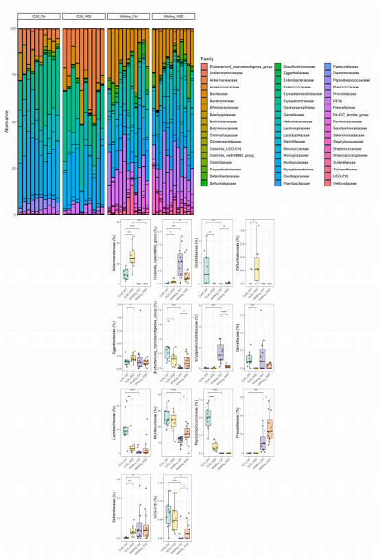
Figure 3. Impact of high-salt food consumption on bacterial families of CLM (n = 10/group) and wildling mice (n = 11 for wildling Ctrl and n = 12 for wildling HSD). Total composition at family level is represented by bar plot per each individual (on top) and boxplot for specific families (bottom); statistical comparisons were performed between groups by Wilcoxon-test. * p ≤ 0.05; ** p ≤ 0.01; *** p ≤ 0.001; **** p ≤ 0.0001.

Figure 4. Changes in bacterial genera in CLM (n = 10/group) and wildling mice (n = 11 for wildling Ctrl and n = 12 for wildling HSD). Overall relative abundance contribution at the genus level is plotted as a circular bar plot per each individual (on top) and a boxplot for specific genera (bottom); statistical comparisons were performed between groups by Wilcoxon-test. * p ≤ 0.05; ** p ≤ 0.01; *** p ≤ 0.001; **** p ≤ 0.0001.
3.3. HSD Affects Predictive Microbial Functions in CLM but Not in Wildling Mice
PICRUSt 2 output did not detect any significant difference between microbial community functions of wildling HSD vs. untreated wildling mice for both KEGG Orthology and MetaCyc pathway annotations, with the only exception of HSD-induced increased function on recG gene for an ATP-dependent helicase from the KEGG Orthology (Figure 5A). HSD impact on CLM was characterized by a significant decrease of predictive functions for KEGG Orthology, among them the gene spp (sucrose-6-phosphatase) and pfkA (phosphofructokinase 1), both involved in starch and sucrose metabolism, which is in line with previous findings [28] (Figure 5A). In addition, the gut microbiota of HSD-fed CLM was characterized by decreased predictive functions of genes involved in membrane transport (feoB for iron transport, AB 2P AB 2 permease protein, AB 2A AB 2 ATP binding protein), glutamine biosynthesis (glnA), LacI family transcriptional regulator (lacI, galR) and transketolase (tktA, tktB) (Figure 5A). For MetaCyc pathways, HSD significantly enriched the CLM gut microbiota of predictive functions associated with nitrate reduction (denitrification pathway), galactose degradation (D-galactarate degradation, super pathway of D-glucarate and D-galactarate degradation), phenyl-propanoate degradation, fatty acid salvage, succinate degradation to butanoic acid and amino acid degradation (aromatic amine degradation, L-leucine degradation) (Figure 5B). Furthermore, in line with previous findings [28], HSD gut microbiota in CLM lost predictive functions for ammino acid biosynthesis (super pathway of L-alanine biosynthesis, L-lysine biosynthesis), mixed acid fermentation, with additional novel signature lost like N-acetylglucosamine/N-acetyl-mannosamine/N-acetylneuraminate degradation and deoxyribonucleosides degradation (pyrimidine and purine degradation, inosine5phosphate biosynthesis III) (Figure 5B).

Figure 5. Cont.

Figure 5. Effect of HSD on gut predictive metagenomic functions in CLM (n = 10/group) and wildling (n = 11 for wildling Ctrl and n = 12 for wildling HSD) gut microbiota. PICRUSt2 output plotted as cuneiform plot for KEGG Orthology annotation (A) and MetaCyc pathways (B) expressed as log2 mean ratio of the predictive functions counts between HSD vs. Ctrl samples. All statistical comparisons were performed between Ctrl vs. HSD groups by Wilcoxon-test.
4. Discussion
Complex and diverse wildling gut microbiota is known to be more resilient to certain disease models [51] and dietary regimes, such as high-fat intake [54,55]. However, no previous study has evaluated the effects of high sodium intake on murine wild-derived gut microbiota. Here, we investigated for the first time how HSD affects wildling gut microbiota compared to CLM. Interestingly, our results demonstrated that compared to CLM, the wildling microbiome is more resistant to HSD disturbance at both compositional and predictive functional levels. It is well established that high salt intake could exacerbate the risk of various diseases, such as cardiovascular or autoimmune diseases, by altering gut microbiome composition and immune homeostasis [25,29,31,34,63–65]. In line with previous reports, HSD-induced shifts in gut microbiota in CLM were characterized by significant alterations in microbial diversity, composition, and predictive functions [28]. Health-promoting bacteria such as the Peptostreptococcaceae family and genera Lactobacillus, Roseburia, and Tuzzerella decreased in terms of relative abundance in CLM, while Akkermansia significantly increased in HSDfed groups. We also detected higher relative abundances of HSD in Defluvitaleaceae, Enterorhabdus, and Parasutterella. Interestingly, the genus Parasutterella is a core component of the gut microbiota of both CLM and humans, where it behaves as an asaccharolytic and producer of succinate [66]. Both Enterorhabdus from the Eggerthellaceae family and Parasutterella from the Sutterellaceae family are known to be enriched in patients with IBD [67,68], further indicating how HSD may affect disease development. However, interestingly, wildling mice did not show a similar entity of HSD-induced microbial shifts, such as CLM. Despite this, wildling diversity significantly increased on HSD for observed OTUs and Chao1 metrics, and only a few taxa were involved in the HSD disturbance of wildling gut microbiota, among them an increase of Anaerovorax, coupled with a decrease of Erysipelatoclostridium, Roseburia, and Lachnospiraceae UCG-004 genus. Roseburia was the only bacterial signature commonly shared between HSD groups compared with the corresponding controls, despite HSD-fed CLM still being characterized by a higher abundance of this bacteria compared to HSD-fed wildling mice. Of note, butyrate-producing bacteria such as Roseburia were shown to have lower relative abundance in patients with ulcerative colitis [69] and this reduction was also observed to be correlated with IBD genetic risk of human subjects [70]. This is in line with previous findings, where shifts in bacterial genera such as Roseburia or Lactobacillus were found to be associated with a risk of hypertension, possibly promoted by a Western diet [71]. The bacterial composition of the gut is also associated with gut motility and physiology [72].

cistanche benefits for men-strengthen immune system
The genus Anaerovorax has previously been observed in mice with abnormal gut physiology and reduced motility [73]; however, the enrichment of Anaerovorax in HSD for wildling mice may lead to a different role of this taxa in the context of gut homeostasis and proper function. In line with previous findings, we observed an increase in the genus Akkermansia in the HSD group of CLM [28], while the gut microbiota of wildling mice was depleted of this genus, which is also consistent with earlier studies on this model [51,53–55]. Although the genus Akkermansia is a potential probiotic due to its positive effect on improving host immunological and metabolic profiles (e.g., in obesity and type 2 diabetes) [42,74–77], the role of this genus is still unclear due to its negative correlation with clinical outcomes in colorectal cancer [78], Parkinson’s disease [79,80] and multiple sclerosis patients [81]. Consistent with our previous results obtained with MetaCyc pathways [28], CLM upon HSD showed decreased predictive microbial functions associated with starch and sucrose metabolism for KEGG orthology. However, the minor shifts in the gut bacterial composition of HSD-fed wildling mice failed to induce any significant variations in predictive bacterial functions, indicating that wildling-derived gut microbiota and metabolic/ecological networks are much more stable and might adapt much more easily to HSD-induced dietary variations compared to CLM gut ecosystems, which warrants further investigation. Worth mentioning also is the possible influence of the gut fungal community on the gut bacterial network upon differential dietary regimes. Earlier studies have already suggested how potential interactions between bacteria and fungi are implicated in host immune system homeostasis and disease development [82–85]. In this context, CLM is further limited by their lower bacterial complexity compared to wildling mice, which may hinder the establishment of a diverse gut mycobiota [54]. Future studies will be able to determine the contribution of gut fungal communities in settings of gut microbiota and host immunity by using the wildling model. In summary, our study provides data on how high sodium intake affects a natural, wild-derived gut microbial ecosystem in comparison to a domesticated gut bacterial community of CLM. Our study demonstrated that HSD does not affect bacterial taxa and gut microbiota in wildling mice in the same way as it does for a domesticated gut microbiota from CLM. This divergence, as previously stated for other dietary regimens or conditions such as high-fat diets [54,55], indicates that future research is needed in natural murine model systems to recapitulate and estimate the impact of dietary interventions on more complex gut ecosystems, as in humans.

cistanche tubulosa-improve immune system
References
1. Candela, M.; Biagi, E.; Turroni, S.; Maccaferri, S.; Figini, P.; Brigidi, P. Dynamic efficiency of the human intestinal microbiota. Crit. Rev. Microbiol. 2015, 41, 165–171. [CrossRef] [PubMed]
2. Candela, M.; Biagi, E.; Maccaferri, S.; Turroni, S.; Brigidi, P. Intestinal microbiota is a plastic factor responding to environmental changes. Trends Microbiol. 2012, 20, 385–391. [CrossRef] [PubMed]
3. Boets, E.; Gomand, S.V.; Deroover, L.; Preston, T.; Vermeulen, K.; De Preter, V.; Hamer, H.M.; Van den Mooter, G.; De Vuyst, L.; Courtin, C.M. Systemic availability and metabolism of colonic-derived short-chain fatty acids in healthy subjects: A stable isotope study. J. Physiol. 2017, 595, 541–555. [CrossRef] [PubMed] 4. Tan, J.; McKenzie, C.; Potamitis, M.; Thorburn, A.N.; Mackay, C.R.; Macia, L. The role of short-chain fatty acids in health and disease. Adv. Immunol. 2014, 121, 91–119. [PubMed]
5. Kumar, J.; Rani, K.; Datt, C. Molecular link between dietary fiber, gut microbiota, and health. Mol. Biol. Rep. 2020, 47, 6229–6237. [CrossRef] [PubMed]
6. Bilotta, A.J.; Cong, Y. Gut microbiota metabolite regulation of host defenses at mucosal surfaces: Implication in precision medicine. Precis. Clin. Med. 2019, 2, 110–119. [CrossRef]
7. Rooks, M.G.; Garrett, W.S. Gut microbiota, metabolites, and host immunity. Nat. Rev. Immunol. 2016, 16, 341–352. [CrossRef]
8. Rodríguez, J.M.; Murphy, K.; Stanton, C.; Ross, R.P.; Kober, O.I.; Juge, N.; Avershina, E.; Rudi, K.; Narbad, A.; Jenmalm, M.C. The composition of the gut microbiota throughout life, with an emphasis on early life. Microb. Ecol. Health Dis. 2015, 26, 26050. [CrossRef]
9. Arrieta, M.-C.; Stiemsma, L.T.; Amenyogbe, N.; Brown, E.M.; Finlay, B. The intestinal microbiome in early life: Health and disease. Front. Immunol. 2014, 5, 427. [CrossRef]
10. Chung, W.S.F.; Walker, A.W.; Louis, P.; Parkhill, J.; Vermeiren, J.; Bosscher, D.; Duncan, S.H.; Flint, H.J. Modulation of the human gut microbiota by dietary fibers occurs at the species level. BMC Biol. 2016, 14, 3. [CrossRef]
11. Danneskiold-Samsøe, N.B.; Barros, H.D.d.F.Q.; Santos, R.; Bicas, J.L.; Cazarin, C.B.B.; Madsen, L.; Kristiansen, K.; Pastore, G.M.; Brix, S.; Júnior, M.R.M. Interplay between food and gut microbiota in health and disease. Food Res. Int. 2019, 115, 23–31. [CrossRef] 12. Scott, K.P.; Duncan, S.H.; Flint, H.J. Dietary fiber and the gut microbiota. Nutr. Bull. 2008, 33, 201–211. [CrossRef]
13. Donohoe, D.R.; Garge, N.; Zhang, X.; Sun, W.; O’Connell, T.M.; Bunger, M.K.; Bultman, S.J. The microbiome and butyrate regulate energy metabolism and autophagy in the mammalian colon. Cell Metab. 2011, 13, 517–526. [CrossRef]
14. Gomaa, E.Z. Human gut microbiota/microbiome in health and diseases: A review. Antonie Van Leeuwenhoek 2020, 113, 2019–2040. [CrossRef]
15. Requena, T.; Martínez-Cuesta, M.C.; Peláez, C. Diet and microbiota linked in health and disease. Food Funct. 2018, 9, 688–704. [CrossRef] [PubMed]
16. Ericsson, A.C.; Franklin, C.L. The gut microbiome of laboratory mice: Considerations and best practices for translational research. Mamm. Genome 2021, 32, 239–250. [CrossRef] [PubMed]
17. Beresford-Jones, B.S.; Forster, S.C.; Stares, M.D.; Notley, G.; Viciani, E.; Browne, H.P.; Boehmler, D.J.; Soderholm, A.T.; Kumar, N.; Vervier, K. The Mouse Gastrointestinal Bacteria Catalogue enables translation between the mouse and human gut microbiotas via functional mapping. Cell Host Microbe 2022, 30, 124–138.e8. [CrossRef]
18. Fava, F.; Rizzetto, L.; Tuohy, K. Gut microbiota and health: Connecting actors across the metabolic system. Proc. Nutr. Soc. 2019, 78, 177–188. [CrossRef]
19. David, L.A.; Materna, A.C.; Friedman, J.; Campos-Baptista, M.I.; Blackburn, M.C.; Perrotta, A.; Erdman, S.E.; Alm, E.J. Host lifestyle affects human microbiota on daily timescales. Genome Biol. 2014, 15, R89. [CrossRef]
20. Tanaka, M.; Nakayama, J. Development of the gut microbiota in infancy and its impact on health in later life. Allergol. Int. 2017, 66, 515–522. [CrossRef]
21. David, L.A.; Maurice, C.F.; Carmody, R.N.; Gootenberg, D.B.; Button, J.E.; Wolfe, B.E.; Ling, A.V.; Devlin, A.S.; Varma, Y.; Fischbach, M.A. Diet rapidly and reproducibly alters the human gut microbiome. Nature 2014, 505, 559–563. [CrossRef] [PubMed]
22. García-Montero, C.; Fraile-Martínez, O.; Gómez-Lahoz, A.M.; Pekarek, L.; Castellanos, A.J.; Noguerales-Fraguas, F.; Coca, S.; Guijarro, L.G.; García-Honduvilla, N.; Asúnsolo, A. Nutritional components in Western diet versus Mediterranean diet at the gut microbiota–immune system interplay. Implications for health and disease. Nutrients 2021, 13, 699. [CrossRef] [PubMed]
23. Soverini, M.; Rampelli, S.; Turroni, S.; Schnorr, S.L.; Quercia, S.; Castagnetti, A.; Biagi, E.; Brigidi, P.; Candela, M. Variations in the post-weaning human gut metagenome profile as a result of Bifidobacterium acquisition in the western microbiome. Front. Microbiol. 2016, 7, 1058. [CrossRef] [PubMed]
24. Manzel, A.; Muller, D.N.; Hafler, D.A.; Erdman, S.E.; Linker, R.A.; Kleinewietfeld, M. Role of “Western diet” in inflammatory autoimmune diseases. Curr. Allergy Asthma Rep. 2014, 14, 404. [CrossRef] [PubMed]
25. Kleinewietfeld, M.; Manzel, A.; Titze, J.; Kvakan, H.; Yosef, N.; Linker, R.A.; Muller, D.N.; Hafler, D.A. Sodium chloride drives autoimmune disease by the induction of pathogenic TH17 cells. Nature 2013, 496, 518–522. [CrossRef] [PubMed]
26. Haase, S.; Wilck, N.; Kleinewietfeld, M.; Müller, D.N.; Linker, R.A. Sodium chloride triggers Th17 mediated autoimmunity. J. Neuroimmunol. 2019, 329, 9–13. [CrossRef] [PubMed]
27. Hernandez, A.L.; Kitz, A.; Wu, C.; Lowther, D.E.; Rodriguez, D.M.; Vudattu, N.; Deng, S.; Herold, K.C.; Kuchroo, V.K.; Kleinewietfeld, M. Sodium chloride inhibits the suppressive function of FOXP3+ regulatory T cells. J. Clin. Investig. 2015, 125, 4212–4222. [CrossRef]
28. Hamad, I.; Cardilli, A.; Corte-Real, B.F.; Dyczko, A.; Vangronsveld, J.; Kleinewietfeld, M. High-Salt Diet Induces Depletion of Lactic Acid-Producing Bacteria in Murine Gut. Nutrients 2022, 14, 1171. [CrossRef]
29. Wilck, N.; Matus, M.G.; Kearney, S.M.; Olesen, S.W.; Forslund, K.; Bartolomaeus, H.; Haase, S.; Mähler, A.; Balogh, A.; Markó, L. Salt-responsive gut commensal modulates TH 17 axis and disease. Nature 2017, 551, 585–589. [CrossRef]
30. Wei, Y.; Lu, C.; Chen, J.; Cui, G.; Wang, L.; Yu, T.; Yang, Y.; Wu, W.; Ding, Y.; Li, L. High salt diet stimulates gut Th17 response and exacerbates TNBS-induced colitis in mice. Oncotarget 2017, 8, 70. [CrossRef]
31. He, F.J.; Li, J.; MacGregor, G.A. Effect of longer-term modest salt reduction on blood pressure. Cochrane Database Syst. Rev. 2013, 346, f1325. [CrossRef] [PubMed]
32. Hu, L.; Zhu, S.; Peng, X.; Li, K.; Peng, W.; Zhong, Y.; Kang, C.; Cao, X.; Liu, Z.; Zhao, B. High salt elicits brain inflammation and cognitive dysfunction, accompanied by alternations in the gut microbiota and decreased SCFA production. J. Alzheimer’s Dis. 2020, 77, 629–640. [CrossRef]
33. Tubbs, A.L.; Liu, B.; Rogers, T.D.; Sartor, R.B.; Miao, E.A. Dietary salt exacerbates experimental colitis. J. Immunol. 2017, 199, 1051–1059. [CrossRef]
34. Muller, D.N.; Wilck, N.; Haase, S.; Kleinewietfeld, M.; Linker, R.A. Sodium in the microenvironment regulates immune responses and tissue homeostasis. Nat. Rev. Immunol. 2019, 19, 243–254. [CrossRef] [PubMed]
35. Burr, A.H.; Bhattacharjee, A.; Hand, T.W. Nutritional modulation of the microbiome and immune response. J. Immunol. 2020, 205, 1479–1487. [CrossRef] [PubMed]
36. Roca-Saavedra, P.; Mendez-Vilabrille, V.; Miranda, J.M.; Nebot, C.; Cardelle-Cobas, A.; Franco, C.M.; Cepeda, A. Food additives, contaminants, and other minor components: Effects on human gut microbiota—A review. J. Physiol. Biochem. 2018, 74, 69–83. [CrossRef]
37. Côrte-Real, B.F.; Hamad, I.; Hornero, R.A.; Geisberger, S.; Roels, J.; Van Zeebroeck, L.; Dyczko, A.; van Gisbergen, M.W.; Kurniawan, H.; Wagner, A. Sodium perturbs mitochondrial respiration and induces dysfunctional Tregs. Cell Metab. 2023, 35, 299–315.e298. [CrossRef] [PubMed]
38. Zagato, E.; Pozzi, C.; Bertocchi, A.; Schioppa, T.; Saccheri, F.; Guglietta, S.; Fosso, B.; Melocchi, L.; Nizzoli, G.; Troisi, J. Endogenous murine microbiota member Faecalibaculum rodentium and its human homolog protect from intestinal tumor growth. Nat. Microbiol. 2020, 5, 511–524. [CrossRef] [PubMed]
39. Mao, G.; Li, S.; Orfila, C.; Shen, X.; Zhou, S.; Linhardt, R.J.; Ye, X.; Chen, S. Depolymerized RG-I-enriched pectin from citrus segment membranes modulates gut microbiota, increases SCFA production, and promotes the growth of Bifidobacterium spp., Lactobacillus spp. and Faecalibaculum spp. Food Funct. 2019, 10, 7828–7843. [CrossRef]
40. Miranda, P.M.; De Palma, G.; Serkis, V.; Lu, J.; Louis-Auguste, M.P.; McCarville, J.L.; Verdu, E.F.; Collins, S.M.; Bercik, P. High salt diet exacerbates colitis in mice by decreasing Lactobacillus levels and butyrate production. Microbiome 2018, 6, 57. [CrossRef]
41. Chen, L.; He, F.J.; Dong, Y.; Huang, Y.; Wang, C.; Harshfield, G.A.; Zhu, H. Modest sodium reduction increases circulating short-chain fatty acids in untreated hypertensives: A randomized, double-blind, placebo-controlled trial. Hypertension 2020, 76, 73–79. [CrossRef] [PubMed]
42. Lukovac, S.; Belzer, C.; Pellis, L.; Keijser, B.J.; de Vos, W.M.; Montijn, R.C.; Roeselers, G. Differential modulation by Akkermansia muciniphila and Faecalibacterium prausnitzii of host peripheral lipid metabolism and histone acetylation in mouse gut organoids. MBio 2014, 5, e01438-14. [CrossRef] [PubMed]
43. Dao, M.C.; Everard, A.; Aron-Wisnewsky, J.; Sokolovska, N.; Prifti, E.; Verger, E.O.; Kayser, B.D.; Levenez, F.; Chilloux, J.; Hoyles, L. Akkermansia muciniphila and improved metabolic health during a dietary intervention in obesity: Relationship with gut microbiome richness and ecology. Gut 2016, 65, 426–436. [CrossRef] [PubMed]
44. Llewellyn, S.R.; Britton, G.J.; Contijoch, E.J.; Vennaro, O.H.; Mortha, A.; Colombel, J.-F.; Grinspan, A.; Clemente, J.C.; Merad, M.; Faith, J.J. Interactions between diet and the intestinal microbiota alter intestinal permeability and colitis severity in mice. Gastroenterology 2018, 154, 1037–1046.e1032. [CrossRef]
45. Berber, K.; Gerdes, L.A.; Cekanaviciute, E.; Jia, X.; Xiao, L.; Xia, Z.; Liu, C.; Klotz, L.; Stauffer, U.; Baranzini, S.E. Gut microbiota from multiple sclerosis patients enables spontaneous autoimmune encephalomyelitis in mice. Proc. Natl. Acad. Sci. USA 2017, 114, 10719–10724. [CrossRef]
46. Sanchez, J.M.S.; DePaula-Silva, A.B.; Libbey, J.E.; Fujinami, R.S. Role of diet in regulating the gut microbiota and multiple sclerosis. Clin. Immunol. 2022, 235, 108379. [CrossRef]
47. Mak, I.W.; Evaniew, N.; Ghert, M. Lost in translation: Animal models and clinical trials in cancer treatment. Am. J. Transl. Res. 2014, 6, 114.
48. Payne, K.J.; Crooks, G.M. Immune-cell lineage commitment: Translation from mice to humans. Immunity 2007, 26, 674–677. [CrossRef]
49. Seok, J.; Warren, H.S.; Cuenca, A.G.; Mindrinos, M.N.; Baker, H.V.; Xu, W.; Richards, D.R.; McDonald-Smith, G.P.; Gao, H.; Hennessy, L. Genomic responses in mouse models poorly mimic human inflammatory diseases. Proc. Natl. Acad. Sci. USA 2013, 110, 3507–3512. [CrossRef] [PubMed]
50. Nguyen, T.L.A.; Vieira-Silva, S.; Liston, A.; Raes, J. How informative is the mouse for human gut microbiota research? Dis. Model. Mech. 2015, 8, 1–16. [CrossRef]
51. Rosshart, S.P.; Vassallo, B.G.; Angeletti, D.; Hutchinson, D.S.; Morgan, A.P.; Takeda, K.; Hickman, H.D.; McCulloch, J.A.; Badger, J.H.; Ajami, N.J. Wild mouse gut microbiota promotes host fitness and improves disease resistance. Cell 2017, 171, 1015–1028.e1013. [CrossRef] [PubMed]
52. Suzuki, T.A.; Phifer-Rixey, M.; Mack, K.L.; Sheehan, M.J.; Lin, D.; Bi, K.; Nachman, M.W. Host genetic determinants of the gut microbiota of wild mice. Mol. Ecol. 2019, 28, 3197–3207. [CrossRef] [PubMed]
53. Maurice, C.F.; CL Knowles, S.; Ladau, J.; Pollard, K.S.; Fenton, A.; Pedersen, A.B.; Turnbaugh, P.J. Marked seasonal variation in the wild mouse gut microbiota. ISME J. 2015, 9, 2423–2434. [CrossRef] [PubMed]
54. Rosshart, S.P.; Herz, J.; Vassallo, B.G.; Hunter, A.; Wall, M.K.; Badger, J.H.; McCulloch, J.A.; Anastasakis, D.G.; Sarshad, A.A.; Leonardi, I. Laboratory mice born to wild mice have natural microbiota and model human immune responses. Science 2019, 365, eaaw4361. [CrossRef] [PubMed]
55. Hild, B.; Dreier, M.S.; Oh, J.H.; McCulloch, J.A.; Badger, J.H.; Guo, J.; Thefaine, C.E.; Umarova, R.; Hall, K.D.; Gavrilova, O. Neonatal exposure to a wild-derived microbiome protects mice against diet-induced obesity. Nat. Metab. 2021, 3, 1042–1057. [CrossRef] [PubMed]
56. Caporaso, J.G.; Lauber, C.L.; Walters, W.A.; Berg-Lyons, D.; Lozupone, C.A.; Turnbaugh, P.J.; Fierer, N.; Knight, R. Global patterns of 16S rRNA diversity at a depth of millions of sequences per sample. Proc. Natl. Acad. Sci. USA 2011, 108, 4516–4522. [CrossRef] [PubMed]
57. Bolyen, E.; Rideout, J.R.; Dillon, M.R.; Bokulich, N.A.; Abnet, C.C.; Al-Ghalith, G.A.; Alexander, H.; Alm, E.J.; Arumugam, M.; Asnicar, F. Reproducible, interactive, scalable and extensible microbiome data science using QIIME 2. Nat. Biotechnol. 2019, 37, 852–857. [CrossRef]
58. Callahan, B.J.; McMurdie, P.J.; Rosen, M.J.; Han, A.W.; Johnson, A.J.A.; Holmes, S.P. DADA2: High-resolution sample inference from Illumina amplicon data. Nat. Methods 2016, 13, 581–583. [CrossRef]
59. Lozupone, C.; Lladser, M.E.; Knights, D.; Stombaugh, J.; Knight, R. UniFrac: An effective distance metric for microbial community comparison. ISME J. 2011, 5, 169–172. [CrossRef]
60. Oksanen, J.; Simpson, G.; Blanchet, F.; Kindt, R.; Legendre, P.; Minchin, P.; O’Hara, R.; Solymos, P.; Stevens, M.; Szoecs, E.; et al. Vegan: Community Ecology Package. Version 2.6-4. 11 October 2022. Available online: https://CRAN.R-project.org/package= vegan (accessed on 26 November 2022).
61. Segata, N.; Izard, J.; Waldron, L.; Gevers, D.; Miropolsky, L.; Garrett, W.S.; Huttenhower, C. Metagenomic biomarker discovery and explanation. Genome Biol. 2011, 12, R60. [CrossRef]
62. Douglas, G.M.; Maffei, V.J.; Zaneveld, J.R.; Yurgel, S.N.; Brown, J.R.; Taylor, C.M.; Huttenhower, C.; Langille, M.G. PICRUSt2 for prediction of metagenome functions. Nat. Biotechnol. 2020, 38, 685–688. [CrossRef]
63. Neal, B.; Wu, Y.; Feng, X.; Zhang, R.; Zhang, Y.; Shi, J.; Zhang, J.; Tian, M.; Huang, L.; Li, Z. Effect of salt substitution on cardiovascular events and death. N. Engl. J. Med. 2021, 385, 1067–1077. [CrossRef]
64. Arroyo Hornero, R.; Hamad, I.; Côrte-Real, B.; Kleinewietfeld, M. The impact of dietary components on regulatory T cells and disease. Front. Immunol. 2020, 11, 253. [CrossRef] [PubMed]
65. Wu, G.D.; Chen, J.; Hoffmann, C.; Bittinger, K.; Chen, Y.Y.; Keilbaugh, S.A.; Bewtra, M.; Knights, D.; Walters, W.A.; Knight, R.; et al. Linking long-term dietary patterns with gut microbial enterotypes. Science 2011, 334, 105–108. [CrossRef] [PubMed]
66. Ju, T.; Kong, J.Y.; Stothard, P.; Willing, B.P. Defining the role of Parasutterella, a previously uncharacterized member of the core gut microbiota. ISME J. 2019, 13, 1520–1534. [CrossRef] [PubMed]





