Low Interferon-γ Levels in Cord And Peripheral Blood Of Pregnant Women Infected With SARS-CoV-2
Nov 23, 2023
Abstract:
COVID-19 is characterized by the immune system’s overreaction resulting in a ‘cytokine storm’, consisting of a massive release of cytokine into the bloodstream, leading to the local and systemic inflammatory response. This clinical picture is further complicated in case of infection of patients with a peculiar immunological status, such as pregnancy. In this paper, we focused on Interferon-γ, which plays a pivotal immunomodulatory role in normal pregnancy and fetal development, as well as in defense against pathogens. In this study, we compared the levels of Interferon-γ and the Interferon autoantibodies of the peripheral and cord blood of pregnant women with confirmed mild COVID-19 and healthy pregnant women. The Interferon-γ was significantly lower both in the peripheral and cord blood of SARS-CoV-2-positive mothers, suggesting that infection can affect the fetal microenvironment even without severe maternal symptoms. In conclusion, further studies are needed to clarify whether lower levels of Interferon-γ due to SARS-CoV-2 infection affect the development or infection susceptibility of infants born to SARS-CoV-2-infected mothers.

cistanche tubulosa-improve immune system
Keywords:
SARS-CoV-2; pregnancy; Interferon-γ; anti-Interferon antibody; immune system; cord blood
1. Introduction
Since its identification in China in late 2019, the SARS-CoV-2 epidemic has spread rapidly worldwide, affecting millions of people, pushing the World Health Organization (WHO) to declare the COVID-19 outbreak a global pandemic. The real extent of this health emergency is difficult to assess due to the swift increase in reported cases and the high number of undiagnosed asymptomatic cases (from 18% to 33%) [1,2], making it difficult to find an effective strategy to limit the SARS-CoV-2 spread [3–5]. The clinical spectrum of COVID-19 ranges from asymptomatic or paucisymptomatic forms to full-blown clinical presentations, characterized by acute respiratory failure requiring mechanical ventilation, septic shock, and multiple organ failure. When present, symptoms are caused by an immune system’s overactivation, resulting in a ‘cytokine storm’ characterized by the release of high levels of cytokines, especially IL-6 and TNF-α, into the circulation, leading to a local and systemic inflammatory response [6,7]. The onset in the majority of symptomatic patients is commonly characterized by fever, cough, and shortness of breath and less commonly by sore throat, anosmia, dysgeusia, anorexia, nausea, myalgia, and diarrhea.

cistanche tubulosa-improve immune system
Regarding the laboratory abnormalities, both leukopenia and leukocytosis, lymphopenia, thrombocytopenia, abnormal renal function parameters and elevated values of C-reactive protein levels (CRP), lactate dehydrogenase (LDH), D-dimer, erythrocyte sedimentation rate and procalcitonin have been reported [8–10]. This clinical picture becomes even more complicated in patients with a peculiar immunological status such as pregnancy, characterized by multiple developmental stages, each with unique immunological requirements, with the aim of protecting the fetus by preventing its identification as a non-self. Interferons (IFNs) play a pivotal immunomodulatory role in normal pregnancy and development, as well as in defense against pathogens [11,12], so it is possible to suppose that COVID-19 could affect the pregnancy course even in the case of asymptomatic infection. In addition, symptomatic pregnant mothers respond to infections differently, due to the combined effects of an altered general immune response and the peculiarity of a fetal–placement immune system, leading to an unpredictable disease [13,14]. Moreover, the respiratory changes that occur physiologically during pregnancy increase the susceptibility to severe COVID-19, per se [15]. To assess whether SARS-CoV-2 infection affects maternal and fetal IFN secretion, we compared IFNγ levels in maternal blood and in cord blood in a pregnant population affected by COVID-19.
2. Materials and Methods
2.1. Study Population
Women and their newborns admitted to University Hospital Federico II between February and June 2021 were included in this study. Cases were pregnant women with SARS-CoV-2 infection hospitalized in the COVID-19 Unit of the Mother and Child Department and controls were pregnant women with uneventful pregnancies admitted to the Mother and Child Department. This population was mostly asymptomatic. Exclusion criteria were maternal age < 18, unvaccinated patients, patients admitted in active labor, multiple gestation, and premature rupture of membranes. All patients were informed of the purpose and procedures of the study and signed a written consent form. Clinical and anamnestic data were collected at admission and reported on a dedicated dataset. The study was conducted according to the ethical guidelines of the Helsinki Declaration of the World Medical Association and was approved by the ethics committee (protocol 140/20/ES2COVID19) of the University of Naples Federico II.
2.2. Sample Collection
Maternal whole blood samples and maternal serum samples were collected at admission for each patient using BD vacutainer (Becton Dickinson, Oxfordshire, UK) blood collection tubes containing EDTA and BD vacutainer with no additives, respectively; all patients were asked to respect a 12 h fast before blood collection. The whole blood sample was immediately analyzed for white cell count, while the serum sample was separated from blood cells and immediately stored at −80 ◦C. Soon after the delivery and before the afterbirth, an arterial cord blood serum sample was collected and stored, as reported for maternal serum samples.

cistanche tubulosa-improve immune system
2.3. Biochemical Determination
White blood cell count was performed using the Siemens Advia 2120i hematology analyzer (Siemens Healthcare, Munich, Germany) according to the manufacturer’s recommendations. C-Reactive Protein was evaluated on Architect c16000 (C-Reactive Protein assay, Abbott Diagnostics, Chicago, IL, USA) according to the manufacturer’s recommendations. SARS-CoV-2 infection was assessed by molecular analysis (RT-PCR) of the nasopharyngeal swab [16]. In detail, swabs were evaluated by Alinity m SARS-CoV-2 assay (Abbott Molecular Inc., Des Plaines, IL, USA), a real-time reverse-transcriptase polymerase chain reaction (rRT-PCR) test performed on the fully automated Alinity m system. The assay detects SARS-CoV-2 RNA from a volume of 500 µL. The Alinity m system automatically performs sample preparation, RT-PCR assembly, amplification, detection, results calculation, and reporting. The Alinity m SARS-CoV-2 assay is a dual target assay (RNA-dependent RNA polymerase (RdRp) and nucleocapsid (N) genes) to detect the presence of the SARS CoV-2 RNA. A sequence unrelated to SARS-CoV-2—from the pumpkin plant Cucurbita pepo—serves as a process control.
2.4. Determination of Interferon-γ (IFN-γ) and Anti-IFN-γ Antibodies
Sera were analyzed for IFN-γ and anti-IFN-γ antibodies performing two different ELISA assays; Human IFN-γ Uncoated ELISA quantitative detection of human IFN-γ INVITROGEN (Thermo Fisher Scientific, Eugene, OR, USA) and Human Anti-Interferon antibody ELISA Kit; (MyBioSource San Diego, CA, USA) in accordance with the manufacturers’ instructions.
2.5. Statistical Analysis
Data were analyzed with the GraphPad Prism 5 software package. Data were tested for normality using the D’Agostino–Pearson normality test. For not normally distributed data we used nonparametric tests. Statistical analysis was performed by an unpaired two-tailed t-test or two-tailed Mann–Whitney test, as indicated in figure legends. Simple linear regression analysis of the data to assess the relationship between maternal and fetal IFN-γ and between maternal IFN-γ and lymphocytes were considered statistically significant when the p-value was either equal to or less than 0.05.
3. Results
3.1. Characteristics of the Study Population
This study involved 25 mothers with COVID-19 and 24 controls. The main clinical and demographic characteristics of the study population are described in Table 1. There was no significant difference in terms of maternal age, comorbidity, and parity.
Table 1. Characteristics of Study Population.

Table 1. Cont.

3.2. Characteristics of the Newborns
All the included newborns were appropriate for their gestational age and they did not develop neonatal complications. Furthermore, there was no significant difference in neonatal length, weight, and head circumference between the two groups (Table 2).
Table 2. Characteristics of the newborns. Anthropometric characteristics of newborns are expressed as mean (± SD).

3.3. Effect of SARS-CoV-2 Infection on Blood Cell Count and CRP in Pregnant Women
We analyzed white blood cell counts and we measured the levels of CRP. In our study population, we did not find any significant difference between white blood cell count, neutrophils, monocytes, and CRP (Table 3) in SARS-CoV-2-positive mothers compared to controls. Conversely, the lymphocyte count was significantly lower in pregnant women affected by COVID-19 (Figure 1) when compared to controls (p-value 0.0093).

Figure 1. Lymphocyte count in maternal blood. Lymphocytes were counted in EDTA blood using an ADVIA 2 analyzer. Lymphocyte concentration is expressed as 1000/µL. Box plots denote median and 25th to 75th percentiles (boxes) and min-to-max whiskers. The p-value was evaluated using an unpaired t-test.
Table 3. Laboratory parameters in maternal blood.

3.4. Measurement of IFN-γ and Anti IFN-γ Antibodies in Serum and Cord Blood of Mothers Affected by COVID-19
To determine if the decrease in lymphocytes is mirrored by lower levels of serum type II Interferon, we measured IFN-γ levels in the maternal and cord blood of infected mothers. As shown in Figure 2a, IFN-γ levels were significantly lower in both the maternal and cord blood of COVID19-positive mothers, suggesting that the maternal decrease in lymphocyte number affects maternal IFN-γ levels, which might lead to lower IFN-γ levels in cord blood.
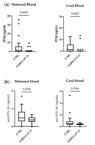
Figure 2. Serum IFN-γ in maternal blood and cord blood (a) and serum IFN antibodies (b). Box plots denote the median and 25th to 75th percentiles (boxes) and Tukey whiskers. The p-value was evaluated using an unpaired t-test and Mann–Whitney test.
In addition, to evaluate the potential involvement of an autoimmune mechanism explaining lower IFN-γ levels observed in SARS-CoV-2-positive mothers, we measured the anti-interferon autoantibody levels in the maternal and cord blood (Figure 2b). In both the maternal and cord blood, the anti-interferon antibodies were significantly lower in SARS-CoV-2-positive mothers compared to controls, indicating that autoantibodies are not involved in the IFN-γ decrease.
3.5. Correlation between IFN-γ Cord Blood and Maternal Blood
To assess whether the IFN-γ levels in cord blood reflect the maternal IFN-γ levels, we performed a simple linear regression, as shown in Figure 3a. The IFN-γ displayed a significantly positive correlation (p-value < 0.0001), suggesting that blood IFN-γ levels mirrored the maternal values.

Figure 3. Simple linear regression of (a) IFN-γ levels (pg/mL) in maternal blood and cord blood; (b) lymphocyte count (1000/µL) and IFN-γ levels (pg/mL) in maternal blood. Each subject is represented on a scatter plot.
3.6. Correlation between Maternal IFN-γ and Maternal Lymphocyte Count
To further confirm that the lower levels of IFN-γ are caused by the depletion of maternal lymphocytes, we carried out a simple linear regression between maternal IFN-γ and lymphocyte count. IFN-γ positively correlated with the number of lymphocytes, suggesting that the lymphocyte reduction following the COVID-19 infection could lead to
4. Discussion
In our cohort, study we showed that IFN-γ is, significantly, decreased in COVID-19 pregnant women compared to controls in both maternal and cord blood samples (Figure 4).

Figure 4. Schematic representation of lymphocyte and IFN-γ decrease in maternal and cord blood. Parts of the figure were drawn using pictures from Servier Medical Art by Servier, licensed under a Creative Commons Attribution 3.0 Unported License
COVID-19 was described as a trigger that leads to the fast activation of innate immune cells, with a profound cytokine response, resembling a hyper-inflammatory state. Tanacan et al., in fact, described pregnant women with COVID-19 as people with an alert immune system in which neutrophils, monocytes, and lymphocytes were increased, as well as CRP [17]. In addition, the progression of the severe form does not seem to be exclusively related to viral load, but it may include defective Interferon responses [18]. A dysregulated inflammatory response to SARS-CoV-2 represents the main cause of disease severity and death in COVID-19 patients [19]. It is characterized by acute lymphopenia, elevated levels of circulating cytokines, and substantial mononuclear cell infiltration in the lung, spleen, kidney, lymph nodes, and hearth [20]. In patients affected by SARS-CoV-2, the proinflammatory response and the cytokine storm play a pivotal role in clinically relevant consequences for the host. When the immune system is not as able to counteract the virus and to conclude the inflammatory response, the aberrant production of the cytokines leads to macrophage hyperactivity, leading to fever, anemia, and organ malfunction. At some point, the cytokine storm becomes unstoppable, leading to irreversible end-organ dysfunction and even death [21,22]. These studies examined patients with severe disease, whereas our results are more likely related to the large portion of asymptomatic cases or those with mild symptoms. Other studies conjecture, in fact, that the identification of specific cytokines as indicators of disease severity might improve clinical management of COVID-19 patients, having a great impact on diagnostic and therapeutic decision-making [23].

cistanche benefits for men-strengthen immune system
Click here to view Cistanche Enhance Immunity products
【Ask for more】 Email:cindy.xue@wecistanche.com / Whats App: 0086 18599088692 / Wechat: 18599088692
According to the literature, lymphocyte count decreased in our study population [24,25]. However, the mechanism leading to lymphopenia during COVID-19 still needs to be elucidated. It may be due to the direct contribution of the virus or redistribution of WBC via chemotaxis or apoptosis [26–28]. Nevertheless, the reduction of IFN-γ is likely due to lymphopenia both in maternal and, subsequently, cord blood. Physiologically, Interferon (IFN) signaling plays an important role in restricting congenital infections, as well as in the physiology of healthy pregnancies. IFNs are best known for their antiviral activities: in mouse models, in fact, IFN signaling has an antiviral effect at the maternal–fetal interface and transplacental transmission [29–31]. In addition, IFN-γ inhibits the secretion of granulocyte/macrophage colony-stimulating factor, which promotes trophoblast growth and differentiation during normal pregnancy [32]. IFN-γ also inhibits trophoblast cell outgrowth, and trophoblast cell invasion is accelerated in mice with a genetic deficiency in the IFN-γ or the IFN-γ receptor [33].
Our results showed that serum IFN-γ auto-Ab concentration (including both neutralizing and non-neutralizing antibodies) is decreased both in maternal serum and cord blood. Conversely, we expected an inverse proportionality between IFN-γ and IFN-γ auto-Ab concentration, as previously described [34,35]. This unexpected finding could be explained with two possible hypotheses or with their combination. The first hypothesis, the most likely, could be that IFN-γ auto-Ab was not produced due to the lack of the antigenic stimulus (low concentration of IFN-γ) [36]. The second one could be related to the immunological status of pregnant women [37,38]. Whatever the reason, the low concentration of both IFN-γ and IFN-γ auto-Ab could be relevant to assess the clinical outcome. It has already been described, in fact, that the association of the low concentration of IFN-γ, the presence of neutralizing IFN-γ auto-Ab, and the increased susceptibility to non-tuberculous mycobacteria disseminated infection in vulnerable hosts [39–41]. Thus, further studies are required to clarify whether children born to SARS-CoV-2-infected women will have worse outcomes than the general population.
Regarding neonatal outcomes, regarding neonatal length, birthweight, head circumference, and Apgar score, we observed no significant differences between cases and controls. These findings are consistent with the literature, particularly in reference to the alpha variant of SARS-CoV-2 [42–44]. There are differences in the response of the immune system (in the mother and fetus) and symptoms among SARS-CoV-2 variants [45,46]. Nowadays, the most prevalent strain is omicron, but in the first semester of 2021, the most widespread variant was the alpha one; the B.1.1.7 lineage [47–49]. Thus, in our study population, the most represented variant was the alpha one. Further studies are needed to explore neonatal outcomes for other SARS-CoV-2 variants. Our study has some limitations. Firstly, we were not able to investigate the outcomes of children born from women infected with SARS-CoV-2. Secondly, further studies are needed to clarify the molecular mechanism involved in the decrease in IFN-γ and IFN-γ auto-Ab.

cistanche tubulosa-improve immune system
5. Conclusions
In conclusion, our findings showed that Interferon-γ was significantly lower both in the peripheral and cord blood of SARS-CoV-2-positive mothers. Thus, infection can affect the fetal microenvironment even without severe maternal symptoms, suggesting the need for long-term follow-up of newborns born from SARS-CoV-2-infected pregnant mothers.
References
1. Mizumoto, K.; Kagaya, K.; Zarebski, A.; Chowell, G. Estimating the asymptomatic proportion of coronavirus disease 2019 (COVID-19) cases on board the Diamond Princess cruise ship, Yokohama, Japan, 2020. Eurosurveillance 2020, 25, 2000180. [CrossRef] [PubMed]
2. Nishiura, H.; Kobayashi, T.; Miyama, T.; Suzuki, A.; Jung, S.-M.; Hayashi, K.; Kinoshita, R.; Yang, Y.; Yuan, B.; Akhmetzhanov, A.R.; et al. Estimation of the asymptomatic ratio of novel coronavirus infections (COVID-19). Int. J. Infect. Dis. 2020, 94, 154–155. [CrossRef] [PubMed]
3. Forouzesh, M.; Rahimi, A.; Valizadeh, R.; Dadashzadeh, N.; Mirzazadeh, A. Clinical display, diagnostics and genetic implication of novel Coronavirus (COVID-19) epidemic. Eur. Rev. Med. Pharmacol. Sci. 2020, 24, 4607–4615. [PubMed]
4. Chakraborty, I.; Maity, P. COVID-19 outbreak: Migration, effects on society, global environment and prevention. Sci. Total Environ. 2020, 728, 138882. [CrossRef] [PubMed]
5. Gorbalenya, A.E.; Baker, S.C.; Baric, R.S.; de Groot, R.J.; Drosten, C.; Gulyaeva, A.A.; Haagmans, B.L.; Lauber, C.; Leontovich, A.M.; Neuman, B.W.; et al. Severe acute respiratory syndrome-related coronavirus: The species and its viruses—A statement of the Coronavirus Study Group. BioRxiv 2020. [CrossRef]
6. Wang, J.; Jiang, M.; Chen, X.; Montaner, L.J. Cytokine storm and leukocyte changes in mild versus severe SARS-CoV-2 infection: Review of 3939 COVID-19 patients in China and emerging pathogenesis and therapy concepts. J. Leukoc. Biol. 2020, 108, 17–41. [CrossRef]
7. Azkur, A.K.; Akdis, M.; Azkur, D.; Sokolowska, M.; van de Veen, W.; Bruggen, M.-C.; O’Mahony, L.; Gao, Y.; Nadeau, K.; Akdis, C.A. Immune response to SARS-CoV-2 and mechanisms of immunopathological changes in COVID-19. Allergy 2020, 75, 1564–1581. [CrossRef]
8. Zhu, J.; Zhong, Z.; Ji, P.; Li, H.; Li, B.; Pang, J.; Zhang, J.; Zhao, C. Clinicopathological characteristics of 8697 patients with COVID-19 in China: A meta-analysis. Fam. Med. Community Health 2020, 8, e000406. [CrossRef]
9. Coopersmith, C.M.; Antonelli, M.; Bauer, S.R.; Deutschman, C.S.; Evans, L.E.; Ferrer, R.; Hellman, J.; Jog, S.; Kesecioglu, J.; Kissoon, N.; et al. The Surviving Sepsis Campaign: Research Priorities for Coronavirus Disease 2019 in Critical Illness. Crit. Care Med. 2021, 49, 598–622. [CrossRef]
10. Gupta, A.; Madhavan, M.V.; Sehgal, K.; Nair, N.; Mahajan, S.; Sehrawat, T.S.; Bikdeli, B.; Ahluwalia, N.; Ausiello, J.C.; Wan, E.Y.; et al. Extrapulmonary manifestations of COVID-19. Nat. Med. 2020, 26, 1017–1032. [CrossRef]
11. Mor, G.; Aldo, P.; Alvero, A.B. The unique immunological and microbial aspects of pregnancy. Nat. Rev. Immunol. 2017, 17, 469–482. [CrossRef]
12. Murphy, S.P.; Tayade, C.; Ashkar, A.A.; Hatta, K.; Zhang, J.; Croy, B.A. Interferon Gamma in Successful Pregnancies. Biol. Reprod. 2009, 80, 848–859. [CrossRef] [PubMed]
13. Rasmussen, S.A.; Smulian, J.C.; Lednicky, J.A.; Wen, T.S.; Jamieson, D.J. Coronavirus Disease 2019 (COVID-19) and pregnancy: What obstetricians need to know. Am. J. Obstet. Gynecol. 2020, 222, 415–426. [CrossRef] [PubMed]
14. Mor, G.; Cardenas, I. The Immune System in Pregnancy: A Unique Complexity. Am. J. Reprod. Immunol. 2010, 63, 425–433. [CrossRef] [PubMed]
15. LoMauro, A.; Aliverti, A. Respiratory physiology in pregnancy and assessment of pulmonary function. Best Pract. Res. Clin. Obstet. Gynaecol. 2022, 85, 3–16. [CrossRef]
16. De Luca, C.; Gragnano, G.; Conticell, F.; Cennamo, M.; Pisapia, P.; Terraccino, D.; Malapelle, U.; Montella, E.; Triassi, M.; Troncone, G.; et al. Evaluation of a fully closed real-time PCR platform for the detection of SARS-CoV-2 in nasopharyngeal swabs: A pilot study. J. Clin. Pathol. 2022, 75, 551–554. [CrossRef]
17. Tanacan, A.; Yazihan, N.; Erol, S.A.; Anuk, A.T.; Yetiskin, F.D.Y.; Biriken, D.; Ozgu-Erdinc, A.; Keskin, H.L.; Tekin, O.M.; Sahin, D. The impact of COVID-19 infection on the cytokine profile of pregnant women: A prospective case-control study. Cytokine 2021, 140, 155431. [CrossRef] [PubMed]
18. Hadjadj, J.; Yatim, N.; Barnabei, L.; Corneau, A.; Boussier, J.; Smith, N.; Péré, H.; Charbit, B.; Bondet, V.; Chenevier-Gobeaux, C.; et al. Impaired type I interferon activity and inflammatory responses in severe COVID-19 patients. Science 2020, 369, 718–724. [CrossRef]
19. Mehta, P.; McAuley, D.F.; Brown, M.; Sanchez, E.; Tattersall, R.S.; Manson, J.J.; HLH Across Speciality Collaboration, UK. COVID-19: Consider cytokine storm syndromes and immunosuppression. Lancet 2020, 395, 1033–1034. [CrossRef]
20. Xu, Z.; Shi, L.; Wang, Y.; Zhang, J.; Huang, L.; Zhang, C.; Liu, S.; Zhao, P.; Liu, H.; Zhu, L.; et al. Pathological findings of COVID-19 associated with acute respiratory distress syndrome. Lancet Respir. Med. 2020, 8, 420–422. [CrossRef]
21. Chakraborty, R.K.; Burns, B. Systemic Inflammatory Response Syndrome, in StatPearls; StatPearls Publishing LLC.: Treasure Island, FL, USA, 2022.
22. Yazdanpanah, F.; Hamblin, M.R.; Rezaei, N. The immune system and COVID-19: Friend or foe? Life Sci. 2020, 256, 117900. [CrossRef] [PubMed]
23. Cabaro, S.; D’Esposito, V.; Di Matola, T.; Sale, S.; Cennamo, M.; Terracciano, D.; Parisi, V.; Oriente, F.; Portella, G.; Beguinot, F.; et al. Cytokine signature and COVID-19 prediction models in the two waves of pandemics. Sci. Rep. 2021, 11, 1–11. [CrossRef] [PubMed]
24. Chen, N.; Zhou, M.; Dong, X.; Qu, J.; Gong, F.; Han, Y.; Qiu, Y.; Wang, J.; Liu, Y.; Wei, Y.; et al. Epidemiological and clinical characteristics of 99 cases of 2019 novel coronavirus pneumonia in Wuhan, China: A descriptive study. Lancet 2020, 395, 507–513. [CrossRef] [PubMed]
25. Li, C.K.-F.; Wu, H.; Yan, H.; Ma, S.; Wang, L.; Zhang, M.; Tang, X.; Temperton, N.; Weiss, R.A.; Brenchley, J.M.; et al. T Cell Responses to Whole SARS Coronavirus in Humans. J. Immunol. 2008, 181, 5490–5500. [CrossRef]
26. Huang, C.; Wang, Y.; Li, X.; Ren, L.; Zhao, J.; Hu, Y.; Zhang, L.; Fan, G.; Xu, J.; Gu, Z.; et al. Clinical features of patients infected with 2019 novel coronavirus in Wuhan, China. Lancet 2020, 395, 497–506. [CrossRef]
27. Wang, D.; Hu, B.; Hu, C.; Zhu, F.; Liu, X.; Zhang, J.; Wang, B.; Xiang, H.; Cheng, Z.; Xiong, Y.; et al. Clinical Characteristics of 138 Hospitalized Patients With 2019 Novel Coronavirus—Infected Pneumonia in Wuhan, China. JAMA 2020, 323, 1061–1069. [CrossRef]
28. Curbelo, J.; Bueno, S.L.; Galván-Román, J.M.; Ortega-Gómez, M.; Rajas, O.; Fernández-Jiménez, G.; Vega-Piris, L.; Rodríguez Salvanes, F.; Arnalich, B.; Díaz, A.; et al. Correction: Inflammation biomarkers in blood as mortality predictors in community acquired pneumonia admitted patients: Importance of comparison with neutrophil count percentage or neutrophil-lymphocyte ratio. PLoS ONE 2019, 14, e0212915. [CrossRef]
29. Lazear, H.M.; Schoggins, J.W.; Diamond, M.S. Shared and Distinct Functions of Type I and Type III Interferons. Immunity 2019, 50, 907–923. [CrossRef]
30. Casazza, R.L.; Lazear, H.M.; Miner, J.J. Protective and Pathogenic Effects of Interferon Signaling During Pregnancy. Viral Immunol. 2020, 33, 3–11. [CrossRef] [PubMed]
31. Fasouliotis, S.J.; Spandorfer, S.D.; Witkin, S.S.; Schattman, G.; Liu, H.C.; Roberts, J.E.; Rosenwaks, Z. Maternal serum levels of interferon-gamma and interleukin-2 soluble receptor-alpha predict the outcome of early IVF pregnancies. Hum. Reprod. 2004, 19, 1357–1363. [CrossRef]
32. Ain, R.; Canham, L.N.; Soares, M.J. Gestation stage-dependent intrauterine trophoblast cell invasion in the rat and mouse: Novel endocrine phenotype and regulation. Dev. Biol. 2003, 260, 176–190. [CrossRef]
33. Ashkar, A.A.; Di Santo, J.P.; Croy, B.A. Interferon-gamma contributes to the initiation of uterine vascular modification, decidual integrity, and uterine natural killer cell maturation during normal murine pregnancy. J. Exp. Med. 2000, 192, 259–270. [CrossRef] [PubMed]
34. Browne, S.K.; Zaman, R.; Sampaio, E.P.; Jutivorakool, K.; Rosen, L.B.; Ding, L.; Pancholi, M.J.; Yang, L.M.; Priel, D.L.; Uzel, G.; et al. Anti-CD20 (rituximab) therapy for anti-IFN-gamma autoantibody-associated nontuberculous mycobacterial infection. Blood 2012, 119, 3933–3939. [CrossRef] [PubMed]
35. Nagamura, N.; Imada, T. Anti-interferon-gamma autoantibody related disseminated nontuberculous mycobacteriosis with pathological features of immunoglobulin G4-related disease. Immunol. Med. 2022, 45, 48–53. [CrossRef] [PubMed]
36. Hsieh, H.-S.; Gong, Y.-N.; Chi, C.-Y.; Hsieh, S.-Y.; Chen, W.-T.; Ku, C.-L.; Cheng, H.-T.; Lin, L.; Sung, C.M. Gut microbiome profiles and associated metabolic pathways in patients of adult-onset immunodeficiency with anti-interferon-gamma autoantibodies. Sci. Rep. 2022, 12, 1–11. [CrossRef]
37. Ivashkiv, L.B. IFNγ: Signalling, epigenetics, and roles in immunity, metabolism, disease and cancer immunotherapy. Nat. Rev. Immunol. 2018, 18, 545–558. [CrossRef]
38. La Rocca, C.; Carbone, F.; Longobardi, S.; Matarese, G. The immunology of pregnancy: Regulatory T cells control maternal immune tolerance toward the fetus. Immunol. Lett. 2014, 162, 41–48. [CrossRef]
39. Aoki, A.; Sakagami, T.; Yoshizawa, K.; Shima, K.; Toyama, M.; Tanabe, Y.; Moro, H.; Aoki, N.; Watanabe, S.; Koya, T.; et al. Clinical Significance of Interferon-gamma Neutralizing Autoantibodies Against Disseminated Nontuberculous Mycobacterial Disease. Clin. Infect. Dis. 2018, 66, 1239–1245. [CrossRef]
40. Ishii, T.; Tamura, A.; Matsui, H.; Nagai, H.; Akagawa, S.; Hebisawa, A.; Ohta, K. Disseminated Mycobacterium avium complex infection in a patient carrying autoantibody to interferon-gamma. J. Infect. Chemother. 2013, 19, 1152–1157. [CrossRef]
41. Koya, T.; Tsubata, C.; Kagamu, H.; Koyama, K.-I.; Hayashi, M.; Kuwabara, K.; Itoh, T.; Tanabe, Y.; Takada, T.; Gejyo, F. Anti interferon-gamma autoantibody in a patient with disseminated Mycobacterium avium complex. J. Infect. Chemother. 2009, 15, 118–122. [CrossRef]
42. Deng, J.; Ma, Y.; Liu, Q.; Du, M.; Liu, M.; Liu, J. Association of Infection with Different SARS-CoV-2 Variants during Pregnancy with Maternal and Perinatal Outcomes: A Systematic Review and Meta-Analysis. Int. J. Environ. Res. Public Health 2022, 19, 15932. [CrossRef] [PubMed]
43. Minisha, F.; Farrell, T.; Abuyaqoub, S.; Rahim, A.A.; Ahmed, H.; Omer, M.; Abraham, M.; Teunissen, F.; Gassim, M. Q-PRECIOUS group Maternal risk factors of COVID-19-affected pregnancies: A comparative analysis of symptomatic and asymptomatic COVID-19 from the Q-PRECIOUS registry. Qatar Med. J. 2022, 2022, 52. [CrossRef] [PubMed]
44. Asalkar, M.R.; Thakkarwad, S.M.; Bacchewad, R.P.; Sharma, N.H. Perinatal Outcome in Maternal COVID-19 Infection at a Tertiary Care Institute- A cross Sectional Study. J. Obstet. Gynecol. India 2022, 1–9. [CrossRef]
45. Liu, H.; Shi, J.; Fok, K.L.; Chen, H. The N501Y Mutation of SARS-CoV-2 Spike Protein Impairs Spindle Assembly in Mouse Oocytes. Reprod. Sci. 2022, 29, 2842–2846. [CrossRef] [PubMed]
46. Yamasoba, D.; Kimura, I.; Nasser, H.; Morioka, Y.; Nao, N.; Ito, J.; Uriu, K.; Tsuda, M.; Zahradnik, J.; Shirakawa, K.; et al. Virological characteristics of the SARS-CoV-2 Omicron BA.2 spike. Cell 2022, 185, 2103–2115.e19. [CrossRef] [PubMed]
47. Washington, N.L.; Gangavarapu, K.; Zeller, M.; Bolze, A.; Cirulli, E.T.; Schiabor Barrett, K.M.; Larsen, B.B.; Anderson, C.; White, S.; Cassens, T.; et al. Emergence and rapid transmission of SARS-CoV-2 B.1.1.7 in the United States. Cell 2021, 184, 2587–2594.e2587. [CrossRef]
48. Marjanovic, S.; Romanelli, R.J.; Ali, G.-C.; Leach, B.; Bonsu, M.; Rodriguez-Rincon, D.; Ling, T. COVID-19 Genomics UK (COG-UK) Consortium: Final Report. Rand. Health Q. 2022, 9, 24. [CrossRef]
49. Volz, E.; Mishra, S.; Chand, M.; Berrett, J.C.; Johnson, R.; Geidelberg, L.; Hinsley, W.R.; Laydon, D.J.; Dabrera, G.; O’Toole, A.; et al. Assessing transmissibility of SARS-CoV-2 lineage B.1.1.7 in England. Nature 2021, 593, 266–269. [CrossRef]