

Part 1:Why Cytokinin Plant Hormones Have Neuroprotective Activity in In Vitro Models Of Parkinson’s Disease?
Mar 22, 2022
Contact: joanna.jia@wecistanche.com / WhatsApp: 008618081934791
Pls click here to Part 2

Cistanche has a very good neuroprotective effect
Gabriel Gonzalez 1,2 , Jiˇrí Grz 1, Cosimo Walter D’Acunto 1, Petr Ka ˇnovsk2 and Miroslav Strnad1,2,*
1. Laboratory of Growth Regulators, Institute of Experimental Botany of the Czech Academy of Sciences,
and Faculty of Science, Palacký University, Šlechtitel ˚u 27, CZ-78371 Olomouc, Czech Republic;
Gonzalez.gabriel@seznam.cz (G.G.); jiri.gruz@upol.cz (J.G.); waldacun@gmail.com (C.W.D.)
2. Department of Neurology, University Hospital Olomouc and Faculty of Medicine and Dentistry,
Palacký University Olomouc, CZ-775 20 Olomouc, Czech Republic; Petr.Kanovsky@fnol.cz
* Correspondence: miroslav.strnad@upol.cz; Tel.: +420-585-634-850
Abstract: Cytokinins are adenine-based phytohormones that regulate key processes in plants, such as cell division and differentiation, root and shoot growth, apical dominance, branching, and seed germination. In preliminary studies, they have also shown protective activities against human neurodegenerative diseases. To extend knowledge of the protection (protective activity) they offer, we investigated activities of natural cytokinins against salsolinol (SAL)-induced toxicity (a Parkinson’s disease model) and glutamate (Glu)-induced death of neuron-like dopaminergic SH-SY5Y cells. We found that kinetin-3-glucoside, cis-zeatin riboside, and N6-isopentenyl adenosine were active in the SAL-induced PD model. In addition, trans-, cis-zeatin, and kinetin along with the iron chelator deferoxamine (DFO) and the necroptosis inhibitor necrostatin 1 (NEC-1) significantly reduced cell death rates in the Glu-induced model. Lactate dehydrogenase assays revealed that the cytokinins provided lower neuroprotective activity than DFO and NEC-1. Moreover, they reduced apoptotic caspase-3/7 activities less strongly than DFO. However, the cytokinins had very similar effects to DFO and NEC-1 on superoxide radical production. Overall, they showed protective activity in the SAL-induced model of parkinsonian neuronal cell death and the Glu-induced model of oxidative damage mainly by reduction of oxidative stress.
Keywords: cytokinin; phytohormone; neuroprotection; neuron-like SH-SY5Y cells; cytotoxicity; salsolinol; glutamate; oxidative stress; Parkinson’s disease
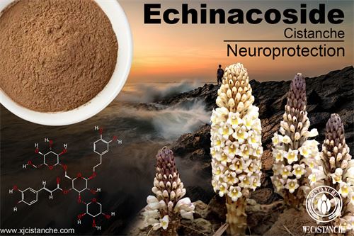
cistanche plant on the effects on Neuroprotection
1. Introduction
Parkinson’s disease (PD) is the second most common motor-related neurodegenerative disease, and numbers of globally diagnosed cases are expected to rise from 6 million in 2015 to more than 12 million by 2040 [1]. It is characterized by motor symptoms linked with specific degeneration and loss of approximately 30–70% of dopaminergic (DA) neurons in the substantia nigra pars compacta and their projections to the striatum [2,3]. Some, of many, known molecular hallmarks of PD include enhanced oxidative and nitrosative stress, mitochondrial dysfunction [4–7], excitotoxicity [8], ubiquitin/proteasomal system dysfunction [9], and neuroinflammation [10]. Current treatments have various adverse side-effects and only offer symptomatic relief [11], so there are intense efforts to develop drugs with efficient curative effects on degenerating DA neurons. Resources that may aid such efforts include natural compounds that tend to have fewer side effects. Inter alia, substances from Ginkgo biloba (ginkgetin, ginkgolide, bilobalide), ginseng (ginsenosides), and flavonoids (baicalein, kaempferol, rutin, and luteolin) have demonstrated broad protective activity in several in vitro models (including the human neuroblastoma cell line SH-SY5Y) and in vivo models of PD induced by 1,1’-dimethyl-4,4’-bipyridinium dichloride (paraquat), 1-methyl- 4-phenyl-1,2,3,6-tetrahydropyridine (MPTP), 1-methyl-4-phenylpyridinium (MPP+), and 6-hydroxydopamine (6-OHDA) [12].
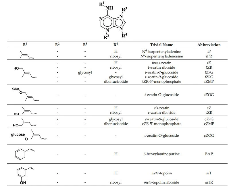
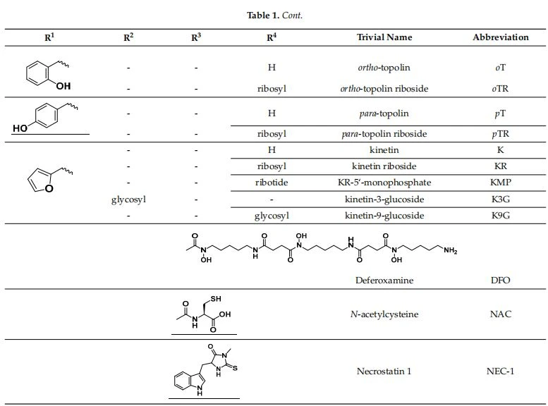
The study presented here focused on the effects of a class of natural phytohormones called cytokinins (CKs), and their metabolites, which are well-known regulators of cell division, growth, differentiation, and leaf senescence in plants [13]. Structurally, CKs are adenine derivatives substituted at the N6-position with either a prenyl (isopentenyl) or aromatic sidechain. Natural forms include 6-(E)-4-hydroxy-3-methyl but-2-enylaminopurine (trans- zeatin, tZ), its 6-(Z)-isomer (cis-zeatin, cZ), N6-isopentenyl adenine (iP), 6-benzyl amino purine (BAP), 6-furfurylaminopurine (kinetin, K), and ortho-, meta-, and para-hydroxylated or methoxylated derivatives of BAP, called topolins (oT, mT, pT, MeoT, MemT, MepT). Various 9-ribosides, 9-nucleotides, as well as 7-, 9, and O-glucosides of these forms also commonly occur, as shown in Table 1. In addition to their native roles in plants, CKs have shown potent ant-oxidant activity towards reactive oxygen species (ROS) that provides protection in several in vitro stress models of aging-associated disorders [14].
Table 1. Structures of cytokinins and the positive control agents.
In particular, CKs reportedly have cytoprotective activity in models such as H2O2- induced cell death of human fibroblasts [15] and D-galactose-induced glycoxidative stress in rat astrocytes [16]. More importantly, in this context, they have shown neuroprotective effects in models related to neurodegenerative diseases such as familial PD, proteasome inhibitor MG 132-induced or H2O2-induced toxicity in SH-SY5Y cells [17], Glu-induced oxidative damage of HT22 mouse hippocampal neuronal cells [18], and the PC12 cell model of Huntington’s disease [19]. Other studies indicate that CKs’ protective activities involve both direct [20,21] and indirect [15,16,22] modulation of cellular redox systems. In addition to CKs’ characteristic antioxidant activities, they reportedly have regulatory effects in mitochondria that enhance neuronal viability [17]. Moreover, K can stabilize mitochondrial membrane potential and increase ATP production, thereby mitigating Glu-induced death of HT22 cells [18]. However, despite findings regarding their effects in several models,there is limited knowledge of CKs’ protective activity in the most common (sporadic) form of PD.
To address the knowledge gap described above, we systematically evaluated the effects of natural CKs and their metabolites in two in vitro models: A salsolinol (SAL)-induced model of PD and glutamate (Glu)-induced model of oxidative damage in neuron-like SH-SY5Y cells. This line was used because of its dopaminergic phenotype, sensitivity to dopaminergic toxins such as SAL, and convenient formation of relatively stable populations of differentiated neuronal cells with reduced proliferation rates following 48 h exposure to 10 uM all-trans retinoic acid (ATRA) [23–25].
Neuron-like cells were exposed to the endo/exotoxin SAL to mimic PD pathology via dysfunction of cellular redox system: Depletion of the glutathione (GSH), and inhibition of both anti-oxidant enzyme (Cu/Zn superoxide dismutase and catalase) activities and mitochondrial complexes (I and II), leading to apoptosis and necrosis [26]. In the other model, Glu induces potentially lethal oxidative damage by disruption of the redox sys- tem. Both models in the SH-SY5Y cell line have been previously used in neuroprotection studies [26,27].
Cytoprotective and/or antioxidant activities related to degenerative disorders of K, iP, BAP, iPR, tZR, and their free bases have been tested, and (as outlined above) some CKs have been found to have protective activities in neuronal cells. However, no previously published studies have examined the structure-neuroprotective activity relationship (SAR) of natural CKs (Table 1). Therefore, this study was undertaken to examine neuroprotective (anti-parkinsonian) activities of almost all known naturally occurring CKs in the selected SAL- and Glu-induced models of neurodegeneration. First, we evaluated each of the CKs’ oxygen radical absorbance capacity (ORAC) and (in safety tests) cytotoxicity towards neuron-like SH-SY5Y cells. Then, we evaluated the compounds’ neuroprotective effects and influence on oxidative stress levels by measuring superoxide (O2 .) production (dihy- droethidium, DHE assay) and apoptotic caspase-3,7 activities. The results provide the first reported systematic indications of the relationships between natural CKs’ structures and neuroprotective activities.

Cistanche herba has a very good neuroprotective effect
2. Results and Discussion
2.1. Cytokinins’ Oxygen Radical Absorbance Capacity (ORAC)
As neurodegenerative diseases are associated with elevated oxidative stress, antioxidant activity plays a key role in the defenses of neuronal cells. To assess CKs’ biological potential in this respect, antioxidant capacity was determined by ORAC, which is commonly used to determine substances’ antioxidant capacity [28]. Antioxidant capacity was expressed as Trolox equivalents (TE), which determines the effectiveness (lower to higher) of compounds than Trolox on an equimolar basis. The results, presented in Table 1, show that topolins (oT, mT, and pT) and their ribosides (oTR, mTR, pTR) have high antioxidant activities, which are probably closely related to the electron-rich system of their C6-hydroxy benzyl amino substituent. Despite their high ORAC values, the topolins did not have high neuroprotective activity. However, several heteroaromatic CKs including K (N6-furfurylaminopurine) and non-aromatic cis-zeatin-O-glucoside (cZOG), which has a 4-hydroxy-3-methylbut-2-en-1-yl)amino substituent, also showed high antioxidant capacity (Table 2). Other CK metabolites—including kinetin-3-glucoside (K3G), kinetin riboside 5<-monophosphate (KMP), kinetin-9-glucoside (K9G), and trans-zeatin riboside-5<- monophosphate (tZMP)—had moderate antioxidant activity. All the others had detectable capacity except BAP. These results confirm previous findings that IP, pT, K can act as direct radical scavengers, but conflict with the previously reported activity of BAP in the ORAC test [20,21]. To conclude, these compounds have potential in the treatment of neurodegenerative diseases associated with increased oxidative stress [29].
cistanche stem benefits on anti-Alzheimer's disease
2.2. Differentiation of SH-SY5Y Cells
To study CKs’ neuroprotective effects, SH-SY5Y neuroblastoma cells (chosen for reasons already described [23]) were differentiated by exposure to 10 uM ATRA for 48 h as previously described [23,24]. They were then stained using a membrane staining kit to examine morphological differences between undifferentiated and differentiated cells. As shown in Figure 1A, the neuron-like differentiated cells grew less densely, were more prolonged, and produced more neurites (indicated by yellow arrows in the figure) than the undifferentiated cells. These morphological changes associated with differentiation have been previously observed, even after shorter exposure (24 h) to ATRA [24,30]. More importantly, the number of neurites rises dramatically to a level when they can create a neurite network. For this reason, cell viability was measured to compare the rate of proliferation of undifferentiated and differentiated SH-SY5Y cells. The viability of undifferentiated SH-SY5Y was taken as the maximum rate of proliferation. The results present in
Figure 1B show that the proliferation rate (assessed by Calcein AM viability assay) of SH-SY5Y was reduced by 23% after 48 h ATRA treatment.

Figure 1. (A) Fluorescent micrographs of SH-SY5Y cells with membranes stained using a Neurite outgrowth kit (Invitro- gen™): Control, undifferentiated cells (exposed to mock treatment solution: <0.1% DMSO); Cells differentiated by exposure to 10 uM all-trans retinoic acid (ATRA) for 48 h. Bars = 50 um. (B) Proliferation rates of undifferentiated and differentiated SH-SY5Y cells: numbers of viable cells after 48 h exposure to <0.1% DMSO and 10 uM ATRA, respectively. Data were obtained from five independent experiments with triplicate cultures: asterisks show the significance of differences in numbers of viable cells (as percentages of numbers of undifferentiated cells) between the cultures: * p < 0.05.
Table 2. Oxygen radical absorbance capacity (ORAC) of the tested cytokinins (CKs) expressed as Trolox equivalents (TE) on an equimolar basis. Names, abbreviations, and structures of the CKs are presented in Figure 1.

2.3. Cytotoxicity of Cytokinins towards Neuron-like SH-SY5Y Cells
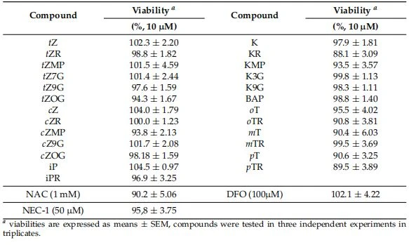
In tests of the CKs’ potential cytotoxicity with the Calcein AM viability assay [31] most showed low toxicity towards the neuron-like SH-SY5Y cells. The decrease of viability below 90% was considered as a threshold for neurotoxic effect. The only two exceptions were KR (11.9%) and pTR (10.5%), in accordance with previous findings that some cytokinin metabolites, particularly ribosides, may have cytotoxic effects [32]. Other ribosides, such as cZR, iPR, oTR, mTR, caused no apparent reduction in the neuron-like SH-SY5Y cells’ viability (Table 3). DFO [33,34] and NEC-1 [35,36] used as positive controls in our in vitro model were also proved by other studies on SH-SY5Y cells to be non-toxic. In conclusion, mainly derivatives KR and pTR showed lower viability than 90% and were therefore considered less interesting for further evaluation in both in vitro models of neurodegeneration.
Table 3. Cell viability of neuron-like SH-SY5Y cells after exposure to cytokinins for 24 h. Viability is expressed as a percentage of DMSO control.
2.4. Identification of Neuroprotective Cytokinins in the SAL-induced Model of PD
For these tests, neuronal SH-SY5Y cells were differentiated for 48 h then co-treated with 500 uM SAL and each CK at three concentrations (0.1, 1, 10 uM). As shown by the dotted line in Figure 2A, application of the neurotoxin SAL at 500 uM reduced the viability of differentiated SH-SY5Y cells, according to the Calcein AM assay, by 30%. N-acetylcysteine (NAC) was used as a positive control in these tests due to its previously reported neuroprotective effect in the same SH-SY5Y cell-based in vitro model [37]. Concentrations of 10, 100, and 1000 uM NAC were used to induce partial or almost complete recovery in the SAL model. NAC was able to increase cell viability at 100 uM and 1 mM concentration, corresponding to 83.39 ± 1.74% and 89.21 ± 2.89%, respectively. NAC’s protective activity at 100 uM (indicated by the dashed line in Figure 2A) was used as a potency threshold for selecting CKs for further tests. According to this setup, the biologically significant neuro-protective activities have been observed with K3G at 10 uM (81.84 ± 2.36%), cZR at 0.1 uM (81.14 ± 2.30%) and 1 uM (81.53 ± 2.24%) and iPR at 1 uM (82.43 ± 2.51%). Thus, iPR and cZR were effective neuroprotectants at lower micro or sub-micromolar concentrations than NAC. The cytokinin screening also revealed that many other metabolites can moderately increase the viability of differentiated SH-SY5Y cells exposed to SAL. However, some tested CKs (including tZR, tZMP, mT, mTR, pT, and pTR) had a very little protective effect.
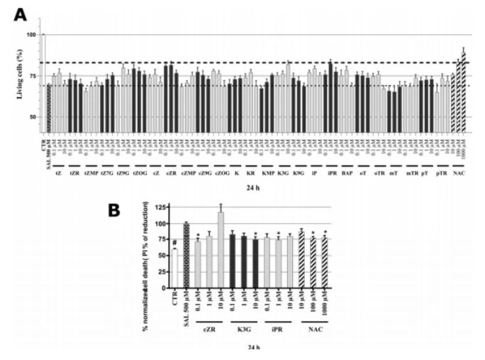
Figure 2. (A) Neuroprotective activity of cytokinins and N-acetylcysteine (NAC) in SAL-induced model of PD on neuron-like SH-SY5Y cells. The dashed line shows the NAC effect threshold at which cytokinins were selected for further testing; the dotted line then counts the number of living cells in the Calcein AM assay after treating the cells with 500 uM SAL; healthy control cells (CTR, DMSO < 0.1%). Triplicates in at least three separated days. (B) Normalized SH-SY5Y cell death after propidium iodide staining. Triplicates in at least five independent days. * P compared with the vehicle with 500 uM SAL, # P compared with the vehicle without 500 uM SAL.
To confirm, the most active natural CKs’ anti-PD activities, overall cell death rates were quantified by propidium iodide (PI) staining, which (in contrast to cell metabolism-based viability tests) only labels cells with impaired membrane integrity, dying cells, and already dead cells [38]. Results were normalized with respect to the cell death rate following treatment with SAL alone (set as 100%). As shown in Figure 2B, the NAC positive control substance significantly reduced cell death rates at both 100 and 1000 uM (to 77.3 ± 2.21% and 77.5 ± 4.44%, respectively). Overall, NAC proved to be a neuroprotective agent with comparable activities to those recorded in other studies in a dose-dependent manner (in the 50–500 uM range) for SH-SY5Y cells [37]. The PI assay also showed that the CKs cZR, K3G, and iPR have protective activities, especially cZR, which reduced the cell death rate to 71.6 ± 5.08% at 0.1 uM. In contrast to cZR, K3G had reversed dose-dependent effects, with maximum activity at 10 uM (reducing the cell death rate to 75.0 ± 3.69%) and NPR's activity peaked at 1 uM (reducing the rate to 73.9 ± 4.99%). Taken together, as shown in Figure 2, CKs provided a comparable neuroprotective activity to 100 uM NAC according to both the viability and cytotoxicity assays. Moreover, effective concentrations of CKs such as cZR and iPR were much lower than those of NAC, in the sub-micromolar and micromolar ranges. Previous observations obtained following double staining with PI and annexin V/PI indicate that K may reduce apoptosis [39], thus we also investigated the effects of CKs and NAC on oxidative stress and caspase-3,7 activation (a well-known apoptosis marker).
2.5. Cytokinins Decrease SAL-induced Superoxide Radical Formation
Oxidative stress (OS) is a key pathological contributor to several neurodegenerative diseases, and both SAL (at > 100 uM) and tetrahydroisoquinolines are potent OS inducers [26,40]. Thus, we also measured the formation of superoxide (a ROS and important OS marker) in the presence of SAL with and without selected CKs or NAC. To ensure that SAL caused sufficient OS damage in SH-SY5Y cells to detect responses, cells were exposed to 500 uM SAL for 24 h, as in previous work [37] and in accordance with findings presented above. The cells were then stained by dihydroethidium (DHE) to detect superoxide radical formation [41,42]. As can be seen in Figure 3A, cells were visually observed after labeling with DHE (which provides red fluorescence signals following reaction with superoxide). SAL induced a clear rise in DHE fluorescence, relative to levels in control and NAC-treated cells. Moreover, three CKs (cZR, K3G, and iPR) had similar visual effects to NAC (100 uM) on DHE fluorescence. Furthermore, the spectrophotometric quantification with respect to levels detected in cells treated by SAL alone (set as 100%), was in line with microscopy observation. As shown in Figure 3B, the normalized superoxide level in healthy control cells (CTR) was less than 39%, and the positive control substance NAC provided moderate- to-complete reduction of SAL-induced ROS production at 100 and 1000 uM (to 76.3 ± 4.33 and 44.3 ± 5.12%), suggesting that glutathione (GSH) depletion plays a key role in the model [26]. Interestingly, SAL induced dramatic reductions in GSH contents of SH-SY5Y cells accompanied by elevation of OS, to levels similar to those previously observed in a study that also recorded NAC-mediated effects on cell viability, cell death, and glutathione contents [43]. Results presented here show that NAC also reduced superoxide radical formation to basal levels (i.e., levels in DMSO-treated controls). CK ribosides were tested at active concentrations (0.1–1 uM) along with K3G, and significantly reduced the cells’ superoxide radical contents to the following levels (relative to those of cells treated with SAL alone): cZR 80.34 ± 5.99% at 0.1 uM; K3G 77.1 ± 4.89% at 10 uM; iPR 79.2 ± 5.91% at 1 uM, comparable to the effects of 100 uM NAC. Collectively, the orthogonal demonstrations strongly indicate that potent anti-OS activity plays a key role in the protective effects of NAC and CKs in the SAL-induced PD model. A correlation between OS amelioration and neuroprotection has also been noted by other authors [29], and several studies have found that K and BAP can directly ameliorate OS activities [44] through the formation of complexes with Cu2+ ions, resulting in superoxide dismutase-like activity [45,46]. However, CKs have also been described as indirect antioxidants with effects mediated by induction of the nuclear factor erythroid 2-related factor 2 (NRF2) antioxidant response pathway (iPR) [22] or partial restoration of glutathione peroxidase and SOD activities (K) [16]. In addition, K reportedly has neuroprotective activities against OS injury induced by H2O2 in SH- SY5Y cells [17]. Both types of reported anti-ROS activity of CKs could potentially explain to effects of cZR, K3G, and iPR in the reduction of superoxide radicals in the SAL-induced SH-SY5Y cell PD model [47–50].

Figure 3. (A) Microphotographs showing SAL-induced oxidative stress and oxidative stress-reducing activities of cytokinins in human differentiated neuron-like SH-SY5Y cells visualized by fluorescence microscopy following dihydroethidium (DHE) labeling. Bars = 50 um. The images show cells treated with DMSO solution (controls), 500 uM salsolinol (SAL) alone, and combinations of 500 uM SAL and 1000 uM NAC (+NAC), 0.1 uM cZR (+cZR); 10 uM K3G (+K3G), 1uM iPR (+iPR) for 24 h before staining with DHE. (B) SAL-induced superoxide radical formation and cytokinin or N-acetylcysteine (NAC) protective activity. The graph shows the quantification of DHE stained cells using the Infinite M200 Pro microplate reader (Tecan, Austria). Triplicates in at least five independent days. * P compared with the vehicle with 500 uM SAL, # P compared with the vehicle without 500 uM SAL.





