

Part Ⅱ Improvement in The Blood Urea Nitrogen And Serum Creatinine Using New Cultivation Of Cordyceps Militaris
May 09, 2023
Results
The control and nephrectomy mice were treated with and without C. militaris mycelia, once a day by oral gavage for 30 days. .e sample number in each treatment was 4–6 mice per group. .e percent increase in body weight and the values of blood urea nitrogen (BUN), serum creatinine, total protein, and uric acid at day 30 are summarized in Figure 1. At the end of the experiments, the percent increase in body weight was lower in uremic mice who underwent partial nephrectomy (Nx, 0.66 ± 2.58%) than in the control mice (Sham, 6.41 ± 2.92%), revealing that uremic mice experienced growth retardation during the experimental period. Body weights from the uremic mice with and without C. militaries treatments are shown in Figures 1(a) and 2. .e results indicated that the body weight in HKS, HKS + A, or CM + A group was decreased compared to that of the Nx group at day 30 (Figure 1(a)). However, none of the C. militaris-treated groups exhibited obvious differences in body weight compared with the uremic control mice during the experimental period. .ese data in Figure 2 suggest that the treatment of C. militaris mycelia did not significantly affect the weight of the uremic mice.


Plasma biochemical values are presented in Figures 1(b), 1(c), and 3. As expected, the BUN and serum creatinine were significantly higher (2.83-fold and 1.63-fold, respectively) in the uremic mice than in the Nx control mice, demonstrating that this uremic mouse model precisely reflects progressive CKD (Figures 1(b) and 1(c)). .e levels of BUN had a pattern of decreasing on day 10 in uremic mice fed with C. militaris mycelia cultured in HKS (41%), HKS + A (41%), and CM + A (34%) media compared with the uremic control mice (Nx) (Figure 3(a)). .e level of serum creatinine was reduced on day 10 in the HKS + A (35%) group and on day 30 in the HKS (14%) and CM + A (13%) groups (Figure 3(b)). .e level of serum creatinine increased in the HKS (93%) and CM (41–53%) groups on day 10 (Figure 3(b)). Differences in body weight, total protein, and uric acid were not detected between the sham and Nx mice (data not shown). In addition, the total protein levels were similar in different groups indicating that C. militaris treatment from different cultivations could prevent the progression of CKD (Figure 1(d)). .e uric acid also had similar levels, except for the CM group, which had higher uric acid levels (Figure 1(e)).


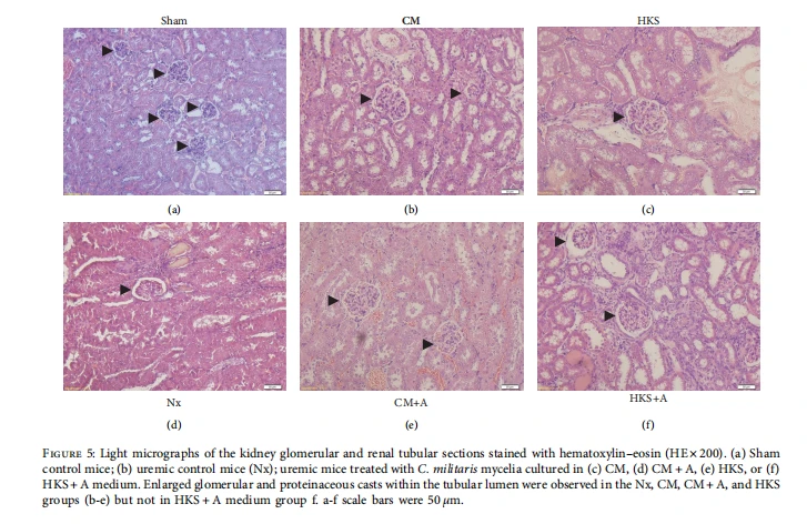
To compare the effects of different C. militaries mycelia treated groups on the kidney, and histological data from the healthy and uremic mice were examined. Figure 4 shows the renal glomerular cross-sectional areas in the sham and uremic mice with or without C. militaris mycelia treatments. .e mean the glomerular cross-sectional area was obtained by calculating the mean area of approximately 8 to 15 individual glomeruli using the program Image J. .ere are no statistical significances between the uremic groups. In the sham control group, renal tissue sections exhibited normal morphology (Figure 5(a)), whereas the renal tissue sections from the uremic mice exhibited enlarged glomeruli and dilated renal tubules surrounded by flat epithelial cells (Figures 5(b)–5(f )). .e glomerular cross-sectional area was significantly larger in the uremic mice (1.74-fold) than in the healthy (sham) mice. An irregular arrangement of tubules and colloid casts in the lumen of the tubules was also present in the injured kidney. .e enlargement of the remnant glomeruli demonstrates that the remnant kidney in this uremic mice model may be the result of long-lasting high hemodynamic pressure and the overloading of metabolic waste.
Discussion
Recently, artificial cultures of C. militaris mycelia are a popular research topic [22–27]. For example, researchers have investigated different media to artificially culture C. militaris mycelia, such as silkworm pupa, solid rice medium, germinated soybean medium, and soybean broth [34–39]. Many studies have investigated different concentrations of carbon and nitrogen in the media to explore the best conditions for the growth of C. militaris mycelia [40–45]. Previous studies have shown that C. militaris mycelia contain specific pharmacologically active components, such as cordycepin, polysaccharides, ergosterol, and mannitol, and that they can be used effectively for various medicinal purposes or as functional foods [46–48]. Differences in the structural characterizations, immunomodulation of polysaccharides, and the antioxidant activity of C. militaris mycelia grown on different media have also been explored [49]. .e current study focused on the renoprotection properties of C. militaris mycelia in vivo and compared the effects of C. militaris mycelia incubated in new media. .e results exhibited that mice with kidney injury treated with C. militaris mycelia cultivated on HKS or CM + A medium had signs of improvement in their kidney function. .e HKS medium contains yeast extract and two-fold more peptone than the CM medium; moreover, it has obvious glucose scarcity. .e HKS medium is a high-nitrogen medium, whereas the CM medium is a high-carbon medium. We are the pioneer investigators in to use of HKS and CM media for the artificial culturing of C. militaris mycelia to understand the differential effects that a high-nitrogen medium (HKS) and a high-carbon medium (CM) have on the biosynthetic changes of C. militaris mycelia.
Vitamin A is a group of fat-soluble organic substances that include retinol, retinal, retinoic acid, several pro-vitamin A carotenoids, and beta-carotene [50]. Carotenoids, which are vitamin A precursors, are isoprenoid molecules synthesized de novo by photosynthetic plants, fungi, and algae and are responsible for the orange, yellow, and (some of the) red colors of various fruits and vegetables. Vitamin A and its related derivatives have been shown to inhibit several biological functions, including tumor growth, angiogenesis, metastasis, and cell proliferation, as well as induce cell apoptosis and differentiation [51, 52]. .e presence of vitamin A in the medium has stimulated the biosynthesis of β-carotene in Phycomyces blakesleeanus [53], suggesting that vitamin A is a strong activator of carotenogenesis in Phycomyces. As shown in Figure 3(b), on day 30 in the CM + A group, C. militaris mycelia started to exert an inhibitory effect on serum creatinine. By contrast, treatment with C. militaris mycelia in the CM group (without vitamin A) had no inhibitory effect on serum creatinine levels. In addition, the inhibitory effect was not observed in C. militaris mycelia when vitamin A was added to the KHS medium. .erefore, the function of vitamin A in fungi is unclear. We suspect that vitamin A may provide the material for C. militaris mycelia to synthesize secondary metabolites and thereby affect cell apoptosis. .e role of vitamins in media should be further studied to support the industrial production of C. militaris mycelia [54].

Click here to buy Cistanche products
Dong et al. exhibited the effect of C. militaris mycelia on BUN and serum creatinine levels in streptozotocin-induced diabetic rats [55]. .e inhibition effects on BUN, creatinine, uric acid, and protein revealed the protection of C. militaris extracts against diabetic nephropathy. .ey presumed that the C. militaris extract has great potential for diabetes treatment. Yu et al. investigated the renal injury-reducing effect of Cordyceps militaris treatment in type 2 diabetic nephropathy mice [56]. .ey reported that blood glucose, renal dysfunction markers (e.g., serum creatinine and kidney-to-body weight ratio), and pathological alterations in renal tissues were significantly mitigated and ameliorated after treatment. Herein, we demonstrated that the levels of BUN in uremic mice were inhibited by C. militaris mycelia cultured in the HKS, HKS + A, or CM + A medium after 10 days, and in the HKS or CM + A medium after 30 days (Figure 3(a)). In addition, we demonstrated that the levels of serum creatinine were inhibited by C. militaris mycelia cultured in the HKS + A medium after 10 days, and in the HKS or CM + A medium after 30 days (Figure 3(b)). We found that C. militaris mycelia powders harvested from the HKS and CM + A media have a stronger effect on decreasing the BUN and serum creatinine levels to remedy kidney injury in CKD mice. C. militaris mycelia harvested from different media may exert different compounds that have various protective effects on CKD. Taken together, treatment with C. militaris mycelia may improve the biochemistry index of CKD, but it could not repair the damages in this uremic mice model.
Khan et al. exploited emulgel with aspirin for topical application [57]; this concept could be further applied in our study. We propose that C. militaris mycelia can be loaded in micro/nanobeads for controlled drug release applications. In addition, Fattepur et al. and Zharif et al. use the ethanolic extract of plants to study the toxicological, pharmacological activity, or synergistic effect of antibiotics [58, 59]. .e bioactive compounds’ extraction was a good idea for our further study. .erefore, we propose that the ethanolic extraction of C. militaris mycelia would be used and studied in the future.
We propose that future studies would be focused on the detailed molecular mechanism of immunomodulation activity, the histopathological changes in kidney tissues, and the new formulation of C. militaris mycelia.

Cistanche's effects
Conclusion
We have successfully observed the renoprotective effects of C. militaris mycelia in new media on mice with CKD. Kidney surgery was performed on young mice (approximately 30 g) to induce uremia. .is is the first report using four new media (HKS, HKS + A, CM, and CM + A medium) for C. militaris mycelia. Each medium of mycelial C. militaries on CKD exhibits a specific effect on the BUN, serum creatinine, body weight, total protein, and uric acid. .e results revealed that the HKS medium of mycelial C. militaris has the strongest effect on decreasing the BUN and serum creatinine levels to remedy kidney injury in CKD mice. .e HKS + A medium of mycelial C. militaris was effective on the 10th day, but not suitable for long-term use (e.g., 30 days). .e CM medium of mycelial C. militaris was not an expert in inhibiting the effects on BUN or serum creatinine. .e CM + A medium of mycelial C. militaris has an acceptable effect on uremic mice. We presume that C. militaris mycelia cultivated in the HKS or CM + A medium have the potential to be a new functional food for CKD.
The Effectiveness of Cistanche on Chronic Kidney Disease
Chronic Kidney Disease (CKD) is a major public health problem worldwide, affecting millions of people globally. CKD is characterized by a gradual decline in kidney function, often leading to end-stage renal disease (ESRD). Despite advances in conventional medicine, no effective treatment for preventing or delaying the progression of ESRD currently exists. As such, interest has been placed on alternative therapies like traditional Chinese medicine (TCM).
Cistanche is a medicinal plant with a long history of use in TCM. It has been used for centuries to tonify the kidneys, improve male sexual function, and enhance longevity. Recent research indicates that Cistanche extract may hold promise as an alternative therapy for CKD.
Research suggests that Cistanche extract has potent antioxidant and anti-inflammatory properties, which can help protect against oxidative stress and inflammation in the kidneys. Studies have shown that Cistanche may improve the glomerular filtration rate (GFR), reduce proteinuria and hematuria, and prevent kidney fibrosis. These effects are thought to be due to various bioactive compounds present in Cistanche, including phenylethanoid glycosides, iridoids, lignans, and polysaccharides.

Standardized Cistanche
In animal studies, Cistanche extract has been shown to delay the progression of CKD and improve renal function. For instance, one study found that treatment with Cistanche extract significantly reduced urinary protein levels and attenuated structural damage to the kidneys in animals with induced CKD. Another animal study found that Cistanche extract improved GFR, decreased proteinuria and renal tubular epithelial cell apoptosis, and reduced kidney fibrosis in rats with CKD.
Clinical trials also provide evidence supporting the potential efficacy of Cistanche in improving kidney function in patients with CKD. In a clinical trial conducted on 62 participants with stage 3 CKD, those given Cistanche extracts capsules exhibited significant improvement in kidney function compared to those given a placebo. The administration of Cistanche extract increases the glomerular filtration rate, decreases proteinuria, and improved renal function.
However, some questions remain regarding the ideal dosing regimen for Cistanche extract and its long-term safety profile. Further research is necessary to determine optimal dosages and the exact mechanisms of action involved in the therapeutic effects of Cistanche on CKD.
Despite the promising results, there are still limitations that must be taken into account when considering Cistanche extract as an alternative therapy for CKD. Firstly, most studies conducted so far have been preclinical or clinical pilot studies with small sample sizes and variable study designs. Additionally, many of these studies have been primarily conducted on animal models rather than human trials. More extensive, high-quality research is required to establish the safety and efficacy of Cistanche as a treatment for CKD. Moreover, there is also a need for standardized preparation of Cistanche to ensure uniform quality and efficacy.
In conclusion, Cistanche extract has been shown to have beneficial effects on kidney function and may offer a promising alternative therapy for patients suffering from CKD. While further large-scale clinical trials are needed before definitive recommendations can be made about Cistanche extract's therapeutic potential, early data are promising and suggest that this traditional Chinese medicine could be used as part of a holistic approach to treating CKD. As always, patients need to consult with their healthcare provider regarding any alternative therapies they are considering taking to avoid adverse effects and ensure safety.

Cistanche extract
References
[36] Y. Ji, A. Su, G. Ma et al., “Comparison of bioactive constituents and effects on gut microbiota by in vitro fermentation between Ophicordyceps sinensis and Cordyceps militaris,” Journal of Functional Foods, vol. 68, Article ID 103901, 2020.
[37] P. Aramwit, N. Bang, J. Ratanavaraporn, T. Nakpheng, and T. Srichana, “An anti-cancer cordycepin produced by Cordyceps militaris growing on the dead larva of Bomby more silkworm,” Journal of Agricultural Science, vol. 6, pp. 41–53, 2014.
[38] X. Li, Q. Liu, W. Li et al., “A breakthrough in the artificial cultivation of Chinese cordyceps on a large-scale and its impact on science, the economy, and industry,” Critical Reviews in Biotechnology, vol. 39, no. 2, pp. 181–191, 2019.
[39] F. Wu, H. Yan, X. Ma et al., “Comparison of the structural characterization and biological activity of acidic polysaccharides from Cordyceps militaris cultured with different media,” World Journal of Microbiology and Biotechnology, vol. 28, no. 5, pp. 2029–2038, 2012.
[40] Y. Ohta, J.-B. Lee, K. Hayashi, A. Fujita, D. K. Park, and T. Hayashi, “In vivo anti-influenza virus activity of an immunomodulatory acidic polysaccharide isolated from Cordyceps militaries grown on germinated soybeans,” Journal of Agricultural and Food Chemistry, vol. 55, no. 25, pp. 10194– 10199, 2007.
[41] S. K. Das, M. Masuda, M. Hatashita, A. Sakurai, and M. Sakakibara, “Optimization of culture medium for cordycepin production using Cordyceps militaris mutant obtained by ion beam irradiation,” Process Biochemistry, vol. 45, no. 1, pp. 129–132, 2010.
[42] Y.-X. Gu, Z.-S. Wang, S.-X. Li, and Q.-S. Yuan, “Effect of multiple factors on accumulation of nucleosides and bases in Cordyceps militaris,” Food Chemistry, vol. 102, no. 4, pp. 1304–1309, 2007.
[43] X.-B. Mao, T. Eksriwong, S. Chauvatcharin, and J.-J. Zhong, “Optimization of carbon source and carbon/nitrogen ratio for cordycepin production by submerged cultivation of medicinal mushroom Cordyceps militaris,” Process Biochemistry, vol. 40, no. 5, pp. 1667–1672, 2005.
[44] B. Chen, Y. Sun, F. Luo, and C. Wang, “Bioactive metabolites and potential mycotoxins produced by cordyceps fungi: a review of safety,” Toxins, vol. 12, no. 6, p. 410, 2020.
[45] V. Kaushik, A. Singh, A. Arya, S. C. Sindhu, A. Sindhu, and A. Singh, “Enhanced production of cordycepin in Ophiocordyceps sinensis using growth supplements under submerged conditions,” Biotechnology Reports, vol. 28, Article ID e00557, 2020.
[46] A. Suparmin, T. Kato, H. Takemoto, and E. Y. Park, “Metabolic comparison of aerial and submerged mycelia formed in the liquid surface culture of Cordyceps militaris,” Microbiology open, vol. 8, no. 9, Article ID e00836, 2019.
[47] H.-W. Lou, Y. Zhao, H.-B. Tang, et al., “Transcriptome analysis of Cordyceps militaris reveals genes associated with carotenoid synthesis and identification of the function of the Cmtns gene,” Frontiers in Microbiology, vol. 10, p. 2105, 2019.
[48] L. Wu, H. Sun, Y. Hao et al., “Chemical structure and inhibition on α-glucosidase of the polysaccharides from Cordyceps militaris with different developmental stages,” International Journal of Biological Macromolecules, vol. 148, pp. 722–736, 2020.
[49] P. H. Leung, S. Zhao, K. P. Ho, and J. Y. Wu, “Chemical properties and antioxidant activity of exopolysaccharides from the mycelial culture of Cordyceps sinensis fungus Cs-HK1,” Food Chemistry, vol. 114, no. 4, pp. 1251–1256, 2009.
[50] A. S. Hombali, J. A. Solon, B. T. Venkatesh, N. S. Nair, and J. P. Peña-Rosas, “Fortification of staple foods with vitamin A for vitamin A deficiency,” Cochrane Database of Systematic Reviews, vol. 5, Article ID CD010068, 2019.
[51] F. Alizadeh, A. Bolhassani, A. Khavari, S. Z. Bathaie, T. Naji, and S. A. Bidgoli, “Retinoids and their biological effects against cancer,” International Immunopharmacology, vol. 18, no. 1, pp. 43–49, 2014.
[52] N. Bushue and Y.-J. Y. Wan, “Retinoid pathway and cancer therapeutics,” Advanced Drug Delivery Reviews, vol. 62, no. 13, pp. 1285–1298, 2010.
[53] M. J. Ruiz-Hidalgo, E. P. Benito, G. Sandmann, and A. P. Eslava, “.e phytoene dehydrogenase gene of Phycomyces: regulation of its expression by blue light and vitamin A,” Molecular and General Genetics MGG, vol. 253, no. 6, pp. 734–744, 1997.
[54] N. Chitsaz, L. Dehghani, M. Rezvani, V. Shaygannejad, F. Khorvash, and N. Esmalian-Afyouni, “.e comparative assessment of Vitamin D supplement on glycemic indices in autoimmune diseases,” Biointerface Research in Applied Chemistry, vol. 6, pp. 1627–1630, 2016.
[55] Y. Dong, T. Jing, Q. Meng, et al., “Studies on the antidiabetic activities of Cordyceps militaris extract in diet-streptozotocin-induced diabetic Sprague-Dawley rats,” BioMed Research International, vol. 2014, Article ID 160980, 11 pages, 2014.
[56] S.-H. Yu, N. K. Dubey, W.-S. Li et al., “Cordyceps militaris treatment preserves renal function in type 2 diabetic nephropathy mice,” PLoS One, vol. 11, no. 11, Article ID e0166342, 2016.
[57] J. Khan, S. Norfarhani, R. K. Sahu, et al., “Development and evaluation of topical emulgel of Aspirin using different polymeric bases,” Research Journal of Pharmacy and Technology, vol. 13, no. 12, pp. 6300–6304, 2020.
[58] S. Fattepur, K. C. Nilugal, T. T. Darshan, et al., “Toxicological and pharmacological activity of ethanolic extracts of Catharanthus roseus in experimental animals,” International Journal of Medical Toxicology & Legal Medicine, vol. 21, no. 3- 4, pp. 141–144, 2018.
[59] N. Sharif, F. Santosh, C. N. Kiran, A. Fadli, A. Ibrahim, and G. Nizam, “Synergistic effect of ethanolic extract of melastoma malabataricum leaves and antibiotics,” International Journal of Medical Toxicology & Legal Medicine, vol. 21, no. 3-4, pp. 167–170, 2018.
Chih-Hui Yang , 1,2,3Wen-Shuo Kuo,4 Jun-ShengWang,2Yi-Ping Hsiang,3,5Yu-Mei Lin,1,6 Yi-Ting Wang , 1,6 Fan-Hsuan Tsai,6 Chun-Ting Lee,6,7 Jiun-Hua Chou,1 Huei-Ya Chang,1 Lung-Shuo Wang , 6,8 Shu-Chi Wang , 6,9 and Keng-Shiang Huang 6
1 Department of Biological Science and Technology, I-Shou University, Kaohsiung, Taiwan
2 Taiwan Instrument Research Institute, National Applied Research Laboratories, Hsinchu, Taiwan
3 Pharmacy Department of E-Da Hospital, Kaohsiung, Taiwan
4 School of Chemistry and Materials Science, Nanjing University of Information Science and Technology, Nanjing, China
5 Department of Biotechnology and Chemical Engineering, I-Shou University, Kaohsiung, Taiwan
6 School of Chinese Medicine for Post-Baccalaureate, I-Shou University, Kaohsiung, Taiwan
7 Amulette Chinese Medicine Clinic, Taipei, Taiwan
8 Department of Chinese Medicine, Sin-Lau Hospital, Tainan, Taiwan
9 School of Medicine for International Students, I-Shou University, Kaohsiung, Taiwan





