Employment Of Phenolic Compounds From Olive VegetationWater in Broiler Chickens: Effects On Gut Microbiota And On The Shelf Life Of Breast Fillets Part 3
Mar 12, 2022
Please contact oscar.xiao@wecistanche.com for more information
2.7.Lipid Oxidation
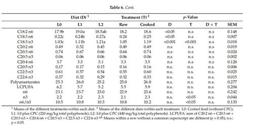
Lipid oxidation was studied through the measure of the secondary(TBARs)and primary(D232 and D270) oxidation compounds(Table 7). No significant differences were noted among the three experimental groups, although it is worth noting that there was a slight increase in the TBARs values for chickens in the L2 group with respect to those in the L1 and L0 groups, while D232 showed the opposite trend. These two analytical indicators were significantly correlated (TBARs vs. D232 r=-0.5,p<0.001 and TBARs vs. D270r=-0.37,p<0.05). The cooking treatment caused decreases in the primary oxidation products (p<0.01), which were followed by increases in the secondary ones (p <0.001). No interactions between diet and cooking were observed. On the contrary, [31]and [45] observed a significant reduction in TBARs in the breasts from chickens whose diets were supplemented with either Olive cake or OMWW. Similarly, [46] observed a reduction in TBARs in the quadriceps femoris of chickens whose diets were enriched with OMWW permeate and the retentate.

Please click here to know more
Oke et al. [16] added different volumes of olive leaf extract(5, 10, and 15 mL/liter) to the drinking water supplied to Abor acre strain broilers, observing a significant effect on the reduction of blood levels of malondialdehyde(MDA). Ibrahim et al. [42], observed a significant reduction in the MDA concentration in chicken breast obtained by adding the diet with increasing levels of fermented or enzymatically fermented dried olive pomace. Unlike other studies, a protective effect against muscular fat oxidation in muscle tissue was not observed in the present one. There are many factors that generate variability and differences between studies. Apart from the animal breed and the length of the fattening period, other factors such as the nutrient composition of the diet, the feed intake, the environmental temperature together with other stressful conditions can lead to very different results that often are difficult to compare. Not last the type of matrix with which the diet is enriched with phenols(olive-cake, olive pomace, olive leaf extract, vegetative water extract, just to name a few) due to the concentration of the active compounds and the presence of additional substances with a synergistic or protective action present in the same matrix, can significantly influence the antioxidant action both in vivo and in meat tissue after slaughter [47]. The potential health benefits from integrating antioxidant additives in fresh meat and meat products are not always proven [48]. On the contrary, many primary and secondary lipid and protein oxidation compounds, such as hydroperoxides, epoxides, 4-hydroxynonenal, malonaldehyde are recognized as potential carcinogens or can affect cellular signal transduction as is the case of carbonyl compounds [49]. In the case of fresh meat, the possibility of increasing the antioxidant potential through the rationing of animals is a very suggestive prospect, especially in the use of natural rather than synthetic substances. In fact, unlike meat-based products where technology offers different possibilities of intervention to increase the antioxidant potential, in fresh meat the only alternative to intervention through the diet of animals is that of a treatment aimed at the surface of the product that however must be consistent with the legislation in force. The residual quantity of phenols measured in the breast meat of the present study is unlikely to cause direct health benefits but indirectly can do. The cooking of meat, for instance, causes a net loss of the tissue antioxidant defenses, therefore the meat tissue enrichment by compounds that are still active even after heat treatment, as is the case of phenols, is of great application interest in view of an increase in the shelf life and safety of the product itself. The reason why neither in video nor ex video effects were observed as a consequence of the feeding trial with CPC, deserves further experimental investigations, especially regarding the interaction between dietary polyphenols and the intestinal microbiota of the chicken on which it leverages a significant part of the potential biochemical effects of phenols [9,10].

3. Materials and Methods 3.1. Experimental Facilities
The trial was undertaken in the poultry house of the Experimental Farm of the University of Padova (Legnaro, Padova, Italy)after 6 months of downtime. The poultry house was equipped with a cooling system, forced ventilation, radiant heating, and controlled light systems. A total of 12wire-net pens (2.5×2.4 m;6 m2)were used, each equipped with five nipple drinkers(distance: 20cm) and a circular feeder (diameter: 30 cm) for manual distribution of feed. Each pen had a concrete floor covered with wood shaving litter(depth 5 cm,2.5 kg/m).
The light was provided 24 h/day for the first 2 days after the chickens had arrived at the poultry house. Then, the number of hours of light was progressively reduced until an 18L:6D photoperiod was achieved, which was then maintained from 12 days of age onwards.
Cistanche can improve immunity
3.2. Animals, Experimental Groups, and In Vivo Recordings
The study was approved by the Ethical Committee for Animal Experimentation (Organismo per la Protezione del Benessere Animale; OPBA)of the University of Padova (project 17/2016;No 154392 of the 10 May 2016).All animals were handled according to the principles of Directive 2010/63/EU[50] regarding the protection of animals used for experimental and other scientific purposes. Researchers involved in animal handling were either animal specialists(PhD or Master's degree in Animal Science) and/or veterinary practitioners.
A total of 144 1-day-old chicks(Ross 308)were delivered by a commercial truck, in compliance with Council Regulation (EC) no.1/2005 [51], to the experimental facilities of the University. All chicks had been vaccinated against Marek's disease, infectious bronchitis, and Newcastle disease at the hatchery. Chicks were individually weighed on the day of their arrival, identified by a leg mark, randomly allocated among the 12 pens (12 birds per pen and 10 birds/m²), and then weighed once per week to measure their live weight until commercial slaughtering. Pen feed consumption was measured daily during the trial. Three commercial diets were administered in crumble form during the trial (Martini SpA Longiano, Forli-Cesena, Italy): diet P1 from 0to 23 d, diet P2 from 24 to 37 d, and diet P3 from 38 d until slaughtering at 48 days (see Supplementary materials for analytical data, Table S1). The crude phenolic concentrate (CPC) was obtained as described by [12], briefly, OVWs were treated using enzymes with pectinase and hemicellulosic activities and then subjected to microfiltration, ultrafiltration, and reverse osmosis.CPC was added to the diets as follows:(i)L0 Control diet (P3), (ii)L1 Control diet plus CPC(220 mg/kg feed as theoretical total phenols), (i) L2 Control diet plus CPC(440 mg/kg feed as theoretical total phenols). The actual content of individual phenolic compounds of CPC and of the diets was reported in Table S2. Each diet was replicated in four pens (homogeneous for initial live weight and variability). The CPC was supplied from 24 days (d) of age until commercial slaughter at 48 d (see Supplementary Material for the phenolic composition of CPC). All animals were fed ad libitum throughout the trial, and the supplemented diets were prepared weekly.
To determine the composition of the gut microbial community, cloacal swabs were collected (4 animals/pen)at 23,34, and 44 d of age for a total of 12 animals (4 on each diet)who were tested three times (36 swab samples collected in total).
3.3. Commercial Slaughtering, and Carcass and Meat Quality Recording
At 48 days of age, after feed and water withdrawal (for 7 and 2 h, respectively), all chickens were slaughtered in a commercial slaughterhouse. Chickens were individually weighed before crating. All chickens from a pen were loaded into a transport cage (height, 62.5cm ×160 cm ×25.0 cm; floor area, 1 m²). Loading and transport from the experimental facilities to the commercial slaughterhouse and lairage before slaughter took approximately 3 h. Chickens were slaughtered according to the standard practices of the commercial slaughterhouse. Carcasses were recovered after 2 h of refrigeration at 2°C and individually weighed to measure the slaughter dressing percentage [52].
Twenty-four hours after slaughter, carcasses were dissected to obtain the pectoralis major muscles separated from the breasts [53].
During the trial, there was a recorded loss of 17.4%(25 chickens) due to mortality. Of the 119 carcasses,72 were assigned to microbiological analyses during the shelf-life period (Section 2.4 explains the establishment of the breast microbiota composition by culture-dependent and-independent methods), while the remaining 47 were frozen (-40°C) and subsequently used for the evaluation of the proximate compositions and lipid oxidation rates of raw and cooked meat (paragraph 3.5).
3.4.Shelf-life Evaluation
3.4.1.Preparation of Packaged Chicken Breast
P major muscles were individually packaged in low-density polyethylene trays wrapped in a 12 μm-thick PVC film (Weigel, KOEX412, Gruppo Fabbri, Vignola, Modena,Italy)and stored at4±1°C.in a refrigerated cabinet (Majolo" Plus 100 Seasoning Con-troller, Majolo, Cadoneghe, Padova, Italy). Exposure to light was fixed from 8:00 to 20:00 using a 36 W fluorescent lamp [54]. The storage settings were designed with the intent to mimic the refrigeration conditions during the sale. The packages were randomly sampled at 24,72,120, 180, 216, and 264 h from the slaughter date
3.4.2. Sensory Analysis
A sensory evaluation of fresh breasts was performed according to [37]using a demerit 3-point scoring system (1=not acceptable,2=acceptable, 3=good quality). The synthetic sensory index(SI) was calculated as [(2×odour score+2× color score+1× texture score)/5], with a score of 1.8 acting as the threshold to define spoiled samples. The sensory analysis was performed by eight trained panelists.
3.4.3.Microbiological Analysis of Breast Meat during the Shelf-Life Period
Microbiological analyses were performed according to the procedures described by [38]. Pectoralis major muscles (about 2 cm long ×1 cm deep × 2 cm wide) were aseptically excised along each breast to obtain a representative sample of 25 per breast. Samples were mixed in a stomacher bag with 225 mL of buffered peptone water and analyzed after the appropriate dilutions. For DNA extraction, 1 mL of each homogenate was collected in 2 mL Eppendorf tubes and centrifugated at 13,500×g for 1 min (Eppendorf centrifuge 5425, Hamburg, Germany). Then, the supernatant was discarded and the pellet was frozen at -80 °C. Several microbial targets were investigated to describe the dynamics of the microbial population during the shelf-life period as follows:The total viable count (TVC) and total psychrotrophic count (TPC) were evaluated on Plate Count Agar(Biokar Diagnostics, Beauvais, France), and plates were incubated at 30 °C for 72 h or at 4°C for 10 days. Enterobacteriaceae were enumerated with Violet Red Bile Glucose Agar (Biokar Diagnostics, Beauvais, France) at 37 °C for 24 h. Lactic acid bacteria (LAB) were analyzed on De Man, Rogosa, and Sharpe Agar(Biokar Diagnostics, Beauvais, France)under anaerobic conditions at 30℃ for 48 h. The Pseudomonas spp. the count was evaluated on Pseudomonas Agar Base supplemented with cetrimide, fucidine, and cephaloridine at 25 °C for 48 h(Oxoid Ltd., Basingstoke, Hampshire, UK). The H2S-producing bacteria (putative Shewanella spp.) count was carried out on iron agar(Lyngby, Laboratorios Conda, Torrejon de Ardoz, Spain) at 25°C for 48 h. Results are reported as log10 CFU/g meat.
3.4.4.pH and Drip Loss
A pH meter (Portamess 910, Knick, Berlin, Germany), equipped with a specific electrode (Mettler Toledo, Milano, Italy), was used to measure pH values by insertion in the pectoralis major muscles in triplicate on their ventral side. Drip loss was evaluated in accordance with the method presented by [55] using the bag method. Approximately 2.5 cm slices cut from the surface of dorsal breasts were inserted into polyethylene bags and suspended overnight at 2±1℃C. The drip loss(%)was calculated by the following equation: [(initial weight - final weight)/initial weight].
3.4.5. Phenol Concentration in the Diet and in the Breast Muscle
The extraction of phenols was conducted on 5g of minced diet sample mixed with 50 mL of methanol/water (80/20(o/o)solution containing 20mg/L of butylated hydroxy-toluene (BHT), the mixture was homogenized using a rod disperser (IKA, T50 Ultra-Turrax, Werke, Staufen, Germany)for 1 min at 7000 rpm, centrifuged at 9327×g for 10 min and the supernatant recovered. The procedure was repeated twice, and the collected extract was then concentrated by a rotary evaporator(Buchi Rotavapor, R-210, Switzerland)until reaching a final volume of 20 mL.An SPE Bond Elut Jr-C18,1 g cartridges(Agilent Technologies, Santa Clara, CA, USA), previously activated with 10 mL of methanol and 10 mL of water, was loaded with 1 mL of aqueous extract the elution was performed with 50 mL of methanol. After solvent removal under vacuum, the phenolic extract was solubilized in 1 mL of a solution composed of methanol/water(50:50 v/v) and filtered through a polyvinylidene fluoride (PVDF)syringe filter(0.2 mm). The qualitative and quantitative analysis of the phenolic compounds of the extract was conducted according to [31] by
HPLC (Mod. 1100 Agilent Technologies, Santa Clara, CA, USA), equipped with a C18 column (Spherisorb ODS-1(250 mm × 4.6 mm)5 um particle size, supplied by Waters S.p.A.(Milan, Italy). The phenolic compounds were detected by using the DAD set at 280 nm. The quantification of polyphenols was determined by using single calibration curves for each compound and the results are expressed as mg kg-1. The hydroxytyrosol (3,4-DHPEA) was obtained from Cabru S.p.A.(Milan, Italy), tyrosol p-HPEA was purchased from Merck Life Science S.r.l. (Milan, Italy), and verbascoside was taken from Extrasynthese(Genay, France). The dialdehyde form of the de-carboxymethyl oleuropein aglycone (3,4-DHPEA-EDA) was extracted from virgin olive oil according to the procedure reported from [56].
A total of 10grams of meat were mixed with50 mL of methanol and water(80/20,o/ø)containing formic acid 0.2% and 20 mg/L of BHT. The solution was homogenized using a rod disperser (IKA, T50 Ultra-Turrax, Werke, Staufen, Germany)for 1 min at 7000 rpm, centrifuged at 4146×g for 10 min and the supernatant recovered. The operation was repeated twice, and the two aliquots of the extract were collected and the methanol was removed by rotary evaporator(Buchi Rotavapor, R-210, Switzerland). Twenty mL of the aqueous extract was loaded to SPE Bond Elut HF Mega BE-C18,5g cartridges(Agilent Technologies, Santa Clara, CA, USA), previously activated with 20 mL of methanol and 20 mL of water; the elution was performed with 50 mL of methanol. After solvent removal under vacuum, the phenolic extract was recovered with 0.5 ml of a solution composed of methanol/water (50∶50 o/ø) and filtered through a polyvinylidene fluoride (PVDF)syringe filter (0.2 mm). The HPLC analyses of the phenolic extracts were conducted according to [56] with the same equipment as reported above. The hydroxytyrosol was detected by using the fluorescence detector (FLD) operated at an excitation wavelength of 280 nm and an emission of 313 nm. The quantification of hydroxytyrosol was made using the calibration curve and the results are expressed as μg/kg.
3.5. Proximate Composition, Cooking Loss, and Fatty Acid Analysis
The proximate composition, fatty acid profile, and oxidative stability were evaluated on both raw (right side P. major)and cooked (left side P. maijor)samples. After thawing, the cooking loss was evaluated according to[53] with the following modifications: The left half of each breast was weighed, vacuum packed, and cooked in a water bath at 80 °C for 45 min. After that, the breasts were blast chilled up to 4°C and kept in the refrigerator (4°C) for 72 h before being unpacked and weighed again to calculate the cooking loss as [(raw meat weight-cooked meat weight)/raw meat weight]×100). The muscle, both raw and cooked, was finely ground (twice 2500rpm×10 s, Retsch, Dusseldorf, Germany) and submitted to moisture, crude protein, and ash [57] analysis, while the crude fat content was determined using the method presented by [58]. Briefly, 200 mL of dichloromethane/methanol 1:1 mixture was added to 5g of the sample, homogenized at 11,000/min for 1 min (Ultraturrax T25 Basic, Ika Werke. Staufen, Germany), and incubated at 60 C in an oven(PID system, M120-VF, MPN instruments, Bernaggio, MB, Italy) for 20 min. Subsequently, another 100 mL of dichloromethane was added, and after homogenization(11,000/min for 1 min)the sample was filtered (rapid filter paper) and 100 mL of KCl 1 M were added to the permeate. After centrifugation(AvantiJ-E, Beckman Coulter, Brea, CA, USA) at 1000×g for 30 m at 4 °C, the supernatant was discharged and the organic extract was filtered through anhydrous sodium sulphate. An aliquot (around 10 g) was used for gravimetric determination of the fat percentage, the remainder was distilled (Rotavapor"R-210, Büchi, Essen, Germany), and the anhydrous fat was used for further analysis. Fatty acid methyl esters (FAMEs)were prepared by weighing 0.015 g of anhydrous fat into a glass tube with a screw cap [39], which was solubilized with 1 mL of 3 M methanolic HCl(Sigma-Aldrich, St. Louis, MO, USA) and incubated for 2 h at 60°C. Then,1 mL of deionized water was added and, after mixing, the FAMEs were recovered in 1 mL of n-hexane. FAMEs (1 μL)were analyzed by a gas chromatograph (Shimadzu Italia Srl, model 2014, Milano, Italy)equipped with a flame ionization detector (set at 250°C) and a split-splitless injector(set at 280°C). The thermal chamber, which held the capillary column(Supelco SP-2560;100 m ×0.25 mm ×0.2 um), was kept at 60°C for 8 min. The temperature was then increased to 120°Cfor 1 min at a rate of 10°C/min, and then from 120 to 240°C at a rate of 2.5℃C/min. The final isotherm was kept at 240°C for 20 min. Fatty acids were identified using an external standard mixture (37 Component FAME Mix, Supelco, Germany). Analyses were done in duplicate.
3.6.Lipid Oxidation
Primary oxidation compounds were determined as conjugated dienes by spectrophotometric analysis in the ultraviolet field using a modified version of the method indicated in the annex of the Council Reg.EU/2015/1833 [59]. Briefly,0.01±0.001g of anhydrous fatwas weighed into a 10 mL graduated flask and dissolved by spectrophotometric grade cyclohexane up to the mark. A UV/Vis spectrophotometer (Mod. 7800 Jasco UV/VIS, Oklahoma City, OK, USA)was used to determine the specific extinction values at 232 (con-jugated dienes) and 270 nm (conjugated trienes)using the same solvent as the reference. Secondary products of lipid oxidation were determined according to the method presented by [60] with modifications, as follows: in a centrifuge tube, 8 mL of an aqueous solution of 5%(m/v) trichloroacetic acid and 5 mL of n-hexane were added to approximately 2g of the homogenized sample. After homogenization with Ultraturrax for the 30s at high speed. the sample was centrifuged for 3 min at 3000×g at 4C(Eppendorf 5810R; Eppendorf, Hamburg, Germany), and the supernatant was removed. The sample was filtered through a rapid paper filter, and 2.5 ml of the filtrate was added to 2.5 mL of an aqueous solution of 0.02 M thiobarbituric acid and incubated for 35 min at 95℃C in a thermostated bath (labor ED-13; Seelbach, Baden-Württemberg, Germany). After cooling, the absorbance was taken using a spectrophotometer set at a wavelength of 532 nm. Data are expressed as milligrams of malondialdehyde per kilogram of meat. The measurements were carried out in duplicate on both cooked and raw chicken breasts.
3.7.DNA Extraction and NGS Library Preparation
DNA was extracted using a DNeasy PowerSoil DNA Isolation Kit (Qiagen, Hilden, Germany). The homogenate pellets of breast meat were defrosted with 600 uL of Lysis buffer provided by the kit. Cloacal swabs were cut with sterile scissors, put into a microtube containing 600uL of Lysis buffer and 6 uL of 2-mercaptoethanol (Sigma-Aldrich, St. Louis, MO, USA). Tubes were mixed well by using vortex for 5 min and swabs were removed. For both cloacal samples and breast meat pellet a total of 100 mg of sterile 0.1 mm beads (BioSpec, Bartlesville, OK, USA) was added, and a bead mill (TissueLyser, Qiagen, Hilden, Germany)was used to carry out disruption in high-speed shaking steps(2×30 s at 30 Hz). A total of 40 uL of proteinase K(Qiagen, Hilden, Germany) was added and incubated at 56°C for 90min. After this step, the DNeasy PowerSoil9DNA Isolation Kit manufacturer's instructions were followed. The concentration and purity of DNA were analyzed using a spectrophotometer(NanoDrop ND-1000, Thermo Scientific, Waltham, MA, USA).
A two step-amplification approach was used to construct the 16S DNA library. The samples were amplified in 20-μL reactions, each composed of 5 μL of diluted DNA,0.4 uM of each primer(Table1), 0.25 mM deoxynucleotide(dNTP),1× Phusion HF buffer, and 1 U Phusion High-Fidelity DNA polymerase (New England BioLabs, Inc., Ipswich, MA, USA). PCR was conducted in a 2720 Thermal Cycler(Applied Biosystems, Foster City, CA, USA)with 25 cycles of 95°C for 30 s,60°Cor 49℃C for 30 s,72℃C for 45s.and a final extension of 7 min at 72°C. Three PCR replicates were performed per sample.
Products were purified using the SPRIselect Purification Kit(Beckman Coulter Life Sciences, Indianapolis, IN, USA) and, after bead purification, the target band was confirmed using 1.8% agarose gel. Barcodes were introduced by a second PCR analysis with platform-specific barcode-bearing primers. Each 50-uL PCR reaction contained the PCR product, 0.2 μMof each primer(Table 1),0.3 mMdNTP,1×Phusion HFbuffer, and1UPhusion high-fidelity DNA polymerase. Ten cycles of the PCR profile described above were performed.
PCR products were purified using the SPRIselect purification kit (Beckman Coulter Life Sciences, Indianapolis, IN, USA). After the final purification, each sample was quantified using a Qubit 2.0 Fluorometer (Invitrogen, Life Technologies, Monza, Italy). The amplicon library quality was tested using an Agilent 2100 Bioanalyzer(Agilent Technologies, Palo Alto, CA, USA). Libraries were sequenced using the Illumina MiSeq platform with a paired-end 300-cycle run (Macrogen Inc., Seoul, Korea).
4. Conclusions
The enrichment of the diets of chickens with phenols obtained by the filtration and concentration of oil mill vegetation water for 24 days was not associated with differences in animal growth, the feed conversion rate, or the carcass yield. The study of the intestinal microbiota showed a significant effect of feeding time but not of diet per se on the alternation of microbial groups. On the other hand, the microbiota composition of the meat during the shelf-life period showed greater growth of Pseudomonas in the samples of chickens that consumed feed enriched with phenols. Finally, no differences were observed among diets with respect to the analytical markers (TBARs and conjugate dienes)of the lipid oxidation process. Instead, hydroxytyrosol was identified in the muscle tissue of the samples from chickens that consumed feed with added phenols, showing dose-dependent intestinal absorption. Since the oxidative process mainly occurs in the membrane structure of the muscle cells, further studies are necessary to better understand the distribution of HT in the cellular and extracellular compartments.
This article is extracted from Molecules 2021, 26, 4307. https://doi.org/10.3390/molecules26144307 https://www.mdpi.com/journal/molecules