Synergistic Anti‑Skin Hyperpigmentation Effect Of Cistanche‑Derived Phenylethanoid Glycosides Combined With Glabridin
Sep 23, 2026
Abstract
Skin hyperpigmentation disorders, such as melasma, freckles and post‑inflammatory hyperpigmentation (PIH), are frequently triggered by ultraviolet‑B (UVB) radiation, cutaneous injury and local skin inflammation. Excess melanin deposition impairs skin appearance and negatively influences patient psychological wellbeing. Conventional depigmenting agents such as hydroquinone primarily target tyrosinase with single‑mode action and carry well‑documented safety risks. Multi‑target natural botanical blends have emerged as promising alternatives for modern functional cosmetic formulation.
Phenylethanoid glycosides (PhGs) are signature bioactive constituents isolated from Cistanche deserticola, a well‑known medicinal herb from arid desert regions. Glabridin (Gla), a prenylated isoflavan extracted from Glycyrrhiza glabra, is widely recognized as a high‑potency skin‑brightening natural compound. This research systematically investigated the synergistic anti‑hyperpigmentation performance of PhGs combined with glabridin. In‑vitro biochemical assays including tyrosinase inhibition, DPPH and ABTS⁺ free‑radical‑scavenging tests were applied for preliminary mass‑ratio screening. Three cellular models were further constructed: UVB‑irradiated B16F10 mouse melanoma cell pigmentation model; lipopolysaccharide (LPS)‑stimulated HaCaT keratinocyte inflammatory model; AAPH‑induced HaCaT oxidative‑stress model. Indicators covering intracellular tyrosinase activity, melanin content, pro‑inflammatory cytokine release (IL‑6, TNF‑α), superoxide dismutase (SOD) and catalase (CAT) enzyme activity were quantified. Combination‑index (CI) calculation was implemented to quantitatively identify synergistic interaction between the two botanical extracts.
Among tested proportions, mass ratios m(PhGs):m(Gla)=1:1, 5:1 and 10:1 exhibited prominent tyrosinase‑suppressing and antioxidant synergy. At total concentration of 0.4 g/L in pH 6.8 phosphate‑buffered saline (PBS), tyrosinase inhibition rates reached 94.37 % (1:1), 92.93 % (5:1), and 88.06 % (10:1). Corresponding DPPH radical‑scavenging percentages were 89.44 %, 88.72 %, 88.10 %; ABTS⁺ cation radical‑scavenging capacities achieved 100.13 %, 100.01 %, 99.87 %. At working concentration 25 μg/mL, all three compound formulations displayed no significant cytotoxicity toward B16F10 and HaCaT cell lines. The 1:1 blended group generated superior comprehensive efficacy: residual intracellular tyrosinase activity dropped to 23.80 %, relative melanin content decreased to 30.90 %. Moreover, PhGs/Gla (1:1) exerted strongest suppression on IL‑6 and TNF‑α secretion and maximally restored SOD and CAT antioxidant enzyme activity. Experimental outcomes confirmed that PhGs paired with glabridin achieve synergistic anti‑hyperpigmentation outcomes through triple pathways: inhibiting melanin biosynthesis, mitigating skin inflammation and counteracting oxidative stress. The 1:1 mass ratio delivers optimal integrated biological performance. This study provides experimental evidence supporting the application of Cistanche phenylethanoid glycosides‑glabridin composite materials in clean‑beauty, plant‑derived whitening skincare raw‑material development.
Keywords: Cistanche deserticola phenylethanoid glycosides; glabridin; synergistic effect; anti‑hyperpigmentation; antioxidant; anti‑inflammation; cosmetic botanical raw materials
1. Introduction
Excessive melanin accumulation in epidermal layers results in heterogeneous skin pigmentation phenotypes, including melasma, ephelides and post‑inflammatory hyperpigmentation (PIH)Google for.... Multiple extrinsic and intrinsic driving factors contribute to pathological melanin overproduction. UVB ultraviolet irradiation represents the primary environmental trigger; skin trauma and chronic local inflammatory responses further amplify pigmentary disorders. It is well‑established that cross‑talk between epidermal keratinocytes and melanocytes dominates pigment progression. Upon external stimulation, activated keratinocytes liberate reactive oxygen species (ROS) and pro‑inflammatory mediators. These signaling molecules diffuse and act on adjacent melanocytes, up‑regulating tyrosinase catalytic activity, accelerating melanin synthesis and promoting melanosome transportation toward keratinocytes, finally producing visible pigmented lesions on skin surface. Accordingly, effective intervention for hyperpigmentation cannot merely focus on tyrosinase inhibition. Multi‑modal therapeutic strategies should simultaneously cover melanogenesis inhibition, anti‑inflammatory intervention and oxidative‑stress neutralization.
Traditional synthetic skin‑lightening actives such as hydroquinone deliver depigmenting efficacy mainly by direct tyrosinase suppression. Nevertheless, single‑target mechanisms limit practical performance, and long‑term topical hydroquinone application may trigger skin irritation, contact dermatitis and potential safety hazards, constraining cosmetic‑grade usage scope. Natural plant‑derived bio‑ingredients attract growing interest from cosmetic formulators and consumers for their mild profiles and multi‑path pharmacological actions. Compound blends of different botanical extracts are capable of producing synergistic biological effects, enhancing overall efficacy while lowering effective dosage requirements, which opens new technical directions for anti‑pigmentation cosmetic‑raw‑material researchGoogle
Cistanche deserticola, commonly named "desert ginseng", is a classic tonic herb in traditional Chinese medicine. Modern phytochemical investigations reveal that phenylethanoid glycosides (PhGs) constitute its principal pharmacologically‑active fraction, featuring echinacoside and acteoside as representative compounds. Published pharmacological research demonstrates that PhGs possess robust antioxidant capacity, anti‑inflammatory bioactivity and moderate tyrosinase‑inhibitory potential, implying prospective application prospects in dermatological skincare materials. Glabridin is a characteristic isoflavonoid compound isolated from Glycyrrhiza glabra root, well‑known within cosmetic industry as "whitening gold". It potently suppresses melanogenesis and concurrently modulates cutaneous inflammatory cascades. From traditional‑medicine theoretical perspective, Cistanche nourishes kidney essence and tonifies yang, while licorice roots tonify spleen and clear heat‑toxin. Combined utilization conforms to compound compatibility principles of herbal dermatological therapies.
Mechanistic hypothesis holds that PhGs‑glabridin combination may regulate skin pigmentation via multi‑target and multi‑path modes: jointly inhibiting tyrosinase catalytic function to cut off melanin generation origin; quenching reactive‑oxygen‑species buildup; restraining pro‑inflammatory‑factor over‑release, so as to interrupt ROS‑ and cytokine‑mediated signal amplification for melanocyte activation. To date, few research papers have systematically reported synergistic anti‑hyperpigmentation evaluation for this binary mixture.
This paper sets out to explore combinatory biological effects between Cistanche phenylethanoid glycosides and glabridin. Biochemical in‑vitro enzyme‑inhibition and radical‑scavenging screening experiments were implemented first. Three representative cell models mimicking pigment deposition, skin inflammation and oxidative damage were constructed for multi‑dimensional efficacy assessment. Combination‑index methodology was applied to quantify synergy or antagonism interactions. We aimed to determine the optimal compound mass ratio, clarify triple‑mode functional mechanisms, and furnish experimental data support for developing natural synergistic skin‑brightening cosmetic raw‑materials.

2. Materials and Methods
2.1 Reagents, Cell Strains and Laboratory Instruments
B16F10 mouse melanoma cell line (STCC20013G‑1) was purchased from Saierbio Co., Ltd. Immortalized human epidermal keratinocyte HaCaT (iCell‑h066) was obtained from iCell Bioscience Inc. Cistanche phenylethanoid glycosides (PhGs) and glabridin (Gla) were commercially supplied by Shaanxi Tianxingjian Biochemical Technology Co., Ltd. Mushroom tyrosinase, arbutin, L‑tyrosine, L‑DOPA, DPPH, ABTS diammonium salt, L‑ascorbic acid (Vitamin C), AAPH, LPS, Triton X‑100, DMSO and other analytical‑grade chemical reagents were sourced from mainstream biochemical reagent vendors. Cell‑culture consumables included high‑glucose DMEM medium, fetal bovine serum, penicillin‑streptomycin mixed solution. ELISA assay kits targeting human IL‑6, TNF‑α, CAT and total‑SOD detection kits were procured from corresponding biotech manufacturers.
Key laboratory equipment contained Multiskan FC full‑wavelength microplate reader, Heracell 371 CO₂ incubator (Thermo Fisher); KQ‑200VDB ultrasonic cleaner; constant‑temperature water bath; DHG‑9246A blast‑drying oven; KN4006B UVB ultraviolet irradiation instrument with output irradiance of 8 mW/cm².
2.2 Sample Solution Preparation
Stock solutions of PhGs and glabridin were prepared in pH 6.8 PBS buffer or 50 % (v/v) ethanol‑water solvent, serially diluted to working concentrations: 0.001 g/L, 0.005 g/L, 0.025 g/L, 0.1 g/L, 0.4 g/L, 0.8 g/L, 1.2 g/L, 1.6 g/L, 2.0 g/L. Nine composite groups were configured at fixed total mass concentration of 0.4 g/L with varied mass proportions m(PhGs):m(Gla) = 40:1, 20:1, 10:1, 5:1, 1:1, 1:5, 1:10, 1:20, 1:40, designated as PhGs/Gla (40:1) to PhGs/Gla (1:40).
For cellular‑experiment assays, powder samples were dissolved in serum‑free DMEM medium to prepare series of working‑concentration solutions.

2.3 Biochemical In‑vitro Assays
2.3.1 Tyrosinase Inhibition Assay
50 μL sample solution, 50 μL pH 6.8 PBS and 50 μL 1 g/L L‑tyrosine substrate solution were sequentially added into each well on 96‑well microplate. Pre‑incubation proceeded under 37 °C water bath for 10 min. Subsequently, 50 μL 0.4 g/L mushroom tyrosinase solution (200 U/mL) was supplemented, and incubation continued at 37 °C for 10 min. Arbutin functioned as positive‑control substance, blank control adopted pH 6.8 PBS buffer. Absorbance value was measured under 490 nm wavelength on microplate reader. Tyrosinase‑inhibition‑rate calculation formula:
Tyrosinase inhibition rate (%) = {1 − [(A_reaction − A_reaction‑control) / (A_blank − A_blank‑control)]} × 100 %
Where: A_reaction: absorbance containing sample, substrate, PBS and tyrosinase; A_reaction‑control: absorbance of sample, substrate and PBS without enzyme; A_blank: absorbance of substrate, PBS and tyrosinase; A_blank‑control: absorbance only for substrate and PBS buffer.
2.3.2 DPPH Free‑Radical‑Scavenging Determination
DPPH powder was dissolved in anhydrous ethanol to prepare 0.2 mmol/L DPPH working liquid. PhGs, glabridin and Vitamin C reference substance were dissolved in 50 % ethanol‑water solvent to form concentration‑gradient stock solutions. Binary‑mixture samples with nine mass ratios were mixed evenly. Each well received 100 μL sample liquid plus 100 μL DPPH working solution. After thorough mixing, microplate was kept away from light at ambient temperature for 30 min. Absorbance reading was recorded at λ = 517 nm. Vitamin C acted as positive control, every experimental group repeated for 3 biological replicates. DPPH‑scavenging‑rate formula:
DPPH scavenging rate (%) = {1 − [(A₁ − A₃) / A₂]} × 100 %
A₁: absorbance of sample + DPPH working fluid; A₂: absorbance of 50 % ethanol + DPPH working fluid; A₃: absorbance of sample solution mixed with 50 % ethanol solvent.
2.3.3 ABTS⁺ Cation Radical‑Scavenging Assay
7.4 mmol/L ABTS stock solution and 2.6 mmol/L potassium persulfate solution were mixed equally, incubated in dark environment for 12 h to produce stable ABTS⁺ working reagent. Sample solutions were configured following protocols described in section 2.3.2. 40 μL sample solution and 160 μL ABTS⁺ working liquid were pipetted to microplate wells. After light‑shielded incubation for 6 min at room temperature, absorbance was measured at 734 nm. ABTS⁺ radical‑scavenging‑rate formula:
ABTS⁺ scavenging rate (%) = {1 − [(A₁ − A₃) / A₂]} × 100 %
A₁: absorbance of sample solution plus ABTS⁺ working solution; A₂: absorbance of deionized water mixed with ABTS⁺ working reagent; A₃: absorbance value for sample solution blended with pure deionized‑water solvent.

2.4 Cell‑Culture Procedures
B16F10 melanoma cells and HaCaT keratinocytes were cultured in high‑glucose DMEM medium supplemented with 10 % fetal‑bovine serum and 1 % penicillin‑streptomycin. Incubator parameters: 37 °C, 5 % volume fraction CO₂, relative humidity 70 %. Cell passaging and medium refreshment were performed according to cellular‑growth density observed under inverted optical microscope. Cells in logarithmic‑growth phase were collected for subsequent seeding onto 96‑well culture plates.
2.5 Establishment of Three Cellular Functional Models
2.5.1 UVB‑Induced B16F10 Pigmentation Model
After 1 h pre‑treatment with corresponding sample medium, cell‑layer surface was covered with PBS buffer. UVB irradiation intensity was set as 8 mW/cm², irradiation distance 3 cm, irradiation duration 110 s. Normal group received identical medium without UVB radiation intervention. Model control group obtained PBS pretreatment followed by equal‑dose UVB exposure without sample supplementation.
2.5.2 LPS‑Mediated HaCaT Inflammatory‑Activation Model
HaCaT cells were pre‑incubated with testing‑sample DMEM medium for 24 h. Afterwards, lipopolysaccharide LPS solution was supplemented to reach final concentration of 20 μg/mL, and co‑culture lasted another 24 h. Model‑group cells were challenged by LPS without sample pre‑treatment. Normal group maintained ordinary complete‑medium cultivation.
2.5.3 AAPH‑Triggered HaCaT Oxidative‑Stress Model
Cells received 24‑h sample pre‑incubation. AAPH reagent was added to culture medium, final concentration 25 mmol/L, continuing 24‑h incubation. Model‑control group was stimulated by AAPH without sample intervention. Normal‑control group grew under regular medium condition.
All cellular‑model groups contained three technical replicate wells.

2.6 Cytotoxicity Test (CCK‑8 Method)
Cell suspension was adjusted to density 1 × 10⁴ cells/mL and inoculated to 96‑well microplates. After cell adherence, gradient‑concentration sample medium was administrated, and incubation continued for 24 h, 48 h, 72 h. CCK‑8 detecting reagent mixed with fresh DMEM medium (volume fraction 10 %) replaced old medium, incubation proceeded for 60 min. Microplate reader measured absorbance at 450 nm wavelength. Calculation formula for relative cell‑viability percentage:
Cell viability (%) = [(A_sample − A_blank) / (A_0 − A_blank)] × 100 %
A_sample: absorbance of well containing cells, CCK‑8 reagent and sample medium; A_blank: absorbance for culture medium plus CCK‑8 without seeded cells; A_0: absorbance of cells and CCK‑8 reagent cultured with blank medium (no test samples).
2.7 Intracellular Tyrosinase‑Activity Quantification
After 48 h model‑establishment processing, cell supernatant was discarded. Cell monolayer was washed twice with pH 7.4 PBS. 50 μL 1 % Triton X‑100 lysis solution was added per well. Microplate was frozen at −80 °C refrigerator for 30 min, then thawed under room‑temperature condition for full cell‑membrane rupture. Pre‑warming treatment at 37 °C lasted 5 min. L‑DOPA substrate (10 mmol/L, 10 μL) was supplemented. Microplate was incubated for 1 h inside CO₂ incubator. Absorbance was detected at 475 nm for calculating relative intracellular tyrosinase‑activity.
2.8 Intracellular Melanin‑Content Measurement
NaOH cleavage method was adopted for melanin‑content detection. Post‑72‑h model‑intervention, culture supernatant was removed, cells rinsed twice using PBS buffer. 100 μL 1 mol/L NaOH aqueous solution containing 10 % DMSO was added. Microplate was incubated inside 90 °C oven for 60 min for complete melanin dissolution. Absorbance was read at 490 nm to compute relative melanin‑content percentage.

2.9 Detection of Inflammatory‑Factor and Antioxidant‑Enzyme Indicators
After 48 h model‑processing, HaCaT cells were scraped and collected, centrifuged to harvest cell supernatant. Commercial ELISA kits were utilized for quantitative determination of IL‑6 and TNF‑α pro‑inflammatory‑cytokine concentrations. For antioxidant‑enzyme testing, cell pellets were subjected to repeated freeze‑thaw cycles to achieve cell fragmentation. Centrifugation condition: 12 000 r/min, 4 °C, 10 min. Supernatant fraction was collected to assay SOD vitality and CAT enzyme content according to kit operational specifications.
2.10 Combination‑Index (CI) Analysis
Chou‑Talalay combination‑index (CI) algorithm was implemented to judge synergistic/additive/antagonistic interaction between PhGs and glabridin within tyrosinase‑inhibition and antioxidant assays:
CI = D₁/DX₁ + D₂/DX₂
D₁ and D₂: individual mass‑concentration of PhGs and glabridin inside compound solution, when reaching fixed biological‑effect level X. DX₁ and DX₂: required mass‑concentration for single PhGs or glabridin to produce identical X‑level biological effect. CompuSyn software completed CI‑value computation. Evaluation criteria: CI > 1 represents antagonistic interaction; CI ≈ 1 means additive effect; CI < 1 indicates significant synergistic‑enhancement effect.
2.11 Statistical Analysis
Experimental measurement outcomes were expressed as mean value ± standard deviation (Mean ± SD). GraphPad Prism 9.5.0 software performed statistical computation. One‑way analysis of variance (ANOVA) was used for multi‑group comparison. P‑value < 0.05 was regarded as statistically significant difference; P < 0.01 marked significant difference; P < 0.001 stood for extremely significant statistical difference.

3. Results and Discussion
3.1 In‑vitro Tyrosinase‑Inhibitory Performance of PhGs, Glabridin and Binary‑Mixture Samples
Half‑inhibitory concentration IC₅₀ values for single‑component PhGs PBS solution, glabridin PBS solution and positive‑control arbutin were (2.192 ± 0.038) g/L, (1.376 ± 0.025) g/L and (0.101 ± 0.003) μg/mL respectively. In‑vitro tyrosinase‑suppressing potency sequence: arbutin > glabridin > PhGs.
At total concentration of 0.4 g/L for compound PBS solution, PhGs/Gla (10:1), PhGs/Gla (5:1), PhGs/Gla (1:1) delivered tyrosinase‑inhibition rate of 88.06 %, 92.93 %, 94.37 %. Both 5:1 and 1:1 ratios surpassed inhibition efficiency of single‑component PhGs (36.92 %) and single‑component glabridin (92.71 %) at matching total mass‑concentration. The 1:1 group achieved inhibition magnitude comparable with positive‑control arbutin. According to CI‑value calculation results: m(PhGs):m(Gla) ranging from 40:1 to 1:1 generated CI < 1, confirming obvious synergistic enhancement on tyrosinase‑suppression. Conversely, proportion series from 1:5 up to 1:40 gave CI > 1, manifesting antagonistic interaction.
Phenolic hydroxyl groups constitute the core functional moiety for tyrosinase‑inhibitory capacity. PhGs contain ortho‑dihydroxyphenyl structural fragments capable of chelating copper ions within tyrosinase catalytic‑active pocket, competitively hindering substrate‑enzyme binding. Glabridin isoflavan skeleton carries 2‑OH and 4‑OH groups on its B‑ring, structurally analogous to L‑tyrosine / L‑DOPA aromatic hydroxyl‑substituted ring, enabling competitive occupation of enzyme‑active‑site domain. After compound blending, cumulative phenolic‑hydroxyl quantity rises; meanwhile, two molecules may attach to distinct binding sites of tyrosinase protein, jointly amplifying inhibitory efficiency.
3.2 Radical‑Scavenging Antioxidant Activity of Test Samples
DPPH‑scavenging IC₅₀ data: PhGs (21.135 ± 1.164 μg/mL), glabridin (55.537 ± 5.230 μg/mL), Vitamin C positive reference (2.852 ± 0.252 μg/mL). DPPH radical‑quenching‑ability ranking: Vitamin C > PhGs > glabridin. PhGs are abundant in caffeoyl‑group and 3,4‑dihydroxyphenylethanol moieties, with conjugated double‑bond systems strengthening free‑radical‑neutralizing performance. Glabridin flavonoid framework also supplies hydroxyl‑groups and extended conjugated‑ring architecture for stabilizing unpaired electrons from reactive‑oxygen‑species.
Three promising compound groups PhGs/Gla (1:1), (5:1), (10:1) presented excellent DPPH‑ and ABTS⁺‑scavenging performance. ABTS⁺‑scavenging rate nearly reached 100 %, indicating near‑complete elimination of cation free‑radicals. Different mass ratios produce varied molecular‑interaction states between two molecules, leading to disparities in hydroxyl‑group exposure status and resulting in divergence of antioxidant synergistic magnitude.
3.3 Cytotoxicity Evaluation of PhGs, Glabridin and Blended Preparations
CCK‑8 cytotoxicity testing demonstrated that PhGs within concentration scope 62.5 ~ 500 μg/mL and glabridin 6.25 ~ 25 μg/mL did not trigger obvious viability‑reduction for B16F10 and HaCaT cell lines. Under 25 μg/mL total mass‑concentration condition, three compound‑ratio groups PhGs/Gla (1:1, 5:1, 10:1) maintained high cell viability, showing no detectable cytotoxic effect against both cell strains. Consequently, 25 μg/mL was selected as safe working concentration for subsequent cellular‑function evaluation. For single‑agent groups, PhGs low‑/medium‑/high‑dosage was defined as 125 μg/mL, 250 μg/mL, 500 μg/mL; glabridin low‑/medium‑/high‑dosage: 6.25 μg/mL, 12.5 μg/mL, 25 μg/mL.
3.4 Influence on Intracellular Tyrosinase‑Activity and Melanin‑Content in UVB‑Stimulated B16F10 Cells
After UVB irradiation treatment, B16F10 model‑group exhibited statistically significant elevation in intracellular tyrosinase‑activity and melanin accumulation (P < 0.001), verifying successful construction of cellular pigmentation‑model. Both single‑agent PhGs and glabridin reduced tyrosinase‑activity and melanin level following concentration‑dependent tendency.
At unified total concentration of 25 μg/mL, compound groups displayed superior biological outcomes relative to single‑component counterparts. Among three ratios, PhGs/Gla (1:1) delivered optimal effect: residual intracellular tyrosinase‑activity only 23.80 %, relative melanin‑content dropped to 30.90 %. Statistical comparison revealed significant difference compared with 5:1 and 10:1 compound groups. The 1:1 proportion manifested prominent synergistic‑amplification for restraining melanin biosynthesis inside melanocyte cells.
UVB‑irradiation provokes melanocyte‑intrinsic melanogenic signaling cascade, up‑regulating MITF transcription factor expression and further inducing tyrosinase protein over‑expression. Our cellular‑experiment outcomes validated that PhGs combined with glabridin could effectively reverse UVB‑induced tyrosinase activation and cut down melanin generation.
3.5 Anti‑Inflammatory Effect toward LPS‑Challenged HaCaT Keratinocytes
LPS stimulation strongly elevated IL‑6 and TNF‑α pro‑inflammatory‑cytokine secretion in HaCaT model‑group (P < 0.001), proving inflammatory‑model replication success. Individual PhGs and glabridin suppressed cytokine release in concentration‑dependent manner.
When comparing three compound ratios at 25 μg/mL, anti‑inflammatory potency sequence was PhGs/Gla (1:1) > PhGs/Gla (5:1) > PhGs/Gla (10:1). The 1:1 formulation generated maximum reduction for IL‑6 and TNF‑α release, exceeding the inhibitory effect obtained by separate application of PhGs or glabridin.
Molecular‑mechanism explanation: PhGs phenolic‑hydroxyl moiety scavenges inflammatory‑triggering free‑radicals; moreover PhGs can interfere NF‑κB and MAPK intracellular signaling cascades to restrain pro‑inflammatory‑gene transcription. Glabridin inhibits phosphorylation of stress‑activated kinase JNK and p38, blocking downstream inflammatory‑signal transduction. Single substance can merely partially suppress inflammatory‑signal‑pathway activation. Binary combination simultaneously intervenes multiple nodes within inflammatory‑signaling‑network, achieving synergistic anti‑inflammatory‑performance.
Cutaneous inflammation serves as critical upstream driver for PIH occurrence. Inflammatory cytokines secreted by keratinocytes act as paracrine signaling messengers, stimulating adjacent melanocytes to accelerate melanin production and melanosome transfer. Effective mitigation of epidermal inflammation is essential for preventing and improving hyperpigmentation lesions. Therefore, the anti‑inflammatory advantage of PhGs‑glabridin compound confers additional value for anti‑pigmentation cosmetic‑material application.
3.6 Antioxidant‑Enzyme‑Regulating Capacity under AAPH‑Induced Oxidative‑Stress Condition
AAPH oxidative‑stress model‑group showed obvious decline of intracellular SOD and CAT antioxidant‑enzyme vitality (P < 0.001). Treatment with PhGs or glabridin alone could partially restore SOD and CAT activity. Among compound‑ratio groups, PhGs/Gla (1:1) displayed most powerful capability to elevate SOD and CAT enzyme level inside HaCaT cells.
Skin exposed to ultraviolet radiation generates large‑quantity reactive‑oxygen‑species. Excess ROS not only directly oxidizes melanin‑synthesis‑related biomolecules, but also activates multiple melanogenic signaling pathways. Endogenous antioxidant‑enzyme system (SOD, CAT) constitutes major defense barrier to eliminate intracellular ROS. When oxidative‑stress over‑whelms intrinsic antioxidant capacity, ROS‑accumulation accelerates pigmentation progression. PhGs‑glabridin compound not only directly scavenges free‑radicals via phenolic‑hydroxyl chemical reaction, but also repairs cellular antioxidant‑enzyme‑system function, relieving oxidative‑stress damage from two dimensions. Different mass ratios change molecular‑interaction pattern between two natural molecules, resulting in discrepancy in final antioxidant‑regulating efficacy.
3.7 Comprehensive Mechanism Discussion for Synergistic Anti‑Hyperpigmentation
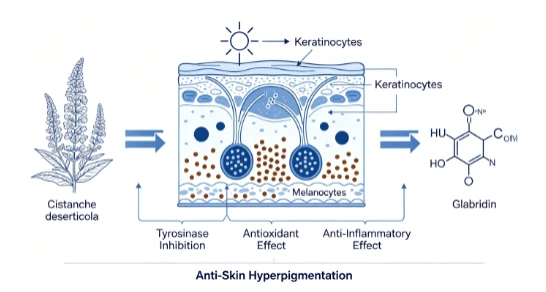
Skin hyperpigmentation pathological‑process forms a complex signaling network: UVB irradiation acts on both melanocytes and surrounding keratinocytes. Keratinocytes release ROS and pro‑inflammatory‑cytokines. These bio‑signals further activate melanocytes, up‑regulate tyrosinase activity, boost melanin biosynthesis, and facilitate melanosome transportation toward epidermal keratinocytes, finally leading to visible pigment deposition on skin surface. Accordingly, single‑target tyrosinase‑inhibition cannot fully reverse pathological pigmentation. Multi‑path intervention strategy should integrate melanogenesis‑inhibition, anti‑inflammation and antioxidant protection.
Our experimental results demonstrated that PhGs combined with glabridin realize multi‑modal intervention against hyperpigmentation:
Directly suppress tyrosinase catalytic‑activity inside melanocytes to inhibit melanin biosynthesis source;
Inhibit pro‑inflammatory‑cytokine over‑secretion from keratinocytes, cutting off paracrine inflammatory‑stimulation signal toward melanocytes;
Eliminate excess reactive‑oxygen‑species and recover endogenous SOD/CAT antioxidant‑enzyme activity, mitigating oxidative‑stress‑mediated pigment‑amplification.
The m(PhGs):m(Gla) = 1:1 ratio exhibited optimal comprehensive synergistic‑performance across biochemical enzyme‑inhibition, radical‑scavenging, melanocyte pigment‑model, keratinocyte inflammation‑model and oxidative‑stress‑model. This ratio out‑performed 5:1 and 10:1 groups in almost all testing indicators. Synergy may originate from complementary molecular‑structural characteristics: PhGs and glabridin possess distinct phenolic‑skeleton frameworks, interacting with different target proteins (tyrosinase, inflammatory‑signaling kinases, antioxidant‑enzyme‑related pathways). After compound blending, phenolic‑hydroxyl exposure status is optimized through intermolecular interaction, enhancing overall biological‑activity.
For cosmetic‑industry practical perspective, synergistic botanical‑compound raw‑materials bring two major benefits. First, synergistic‑effect enables reduction of required dosage for each single active‑ingredient, lowering formulation cost; second, multi‑path functional profile matches complex pathological‑mechanism of clinical‑observed skin hyperpigmentation such as PIH and melasma, offering theoretical‑basis for developing high‑efficacy natural skin‑brightening cosmetic formulas.
4. Conclusion
This research systematically evaluated synergistic anti‑hyperpigmentation bioactivity of Cistanche deserticola‑derived phenylethanoid glycosides (PhGs) combined with glabridin (Gla). Biochemical in‑vitro screening and three representative cellular‑models were utilized for multi‑dimensional efficacy characterization. Main experimental‑conclusions are summarized as follows:
Within in‑vitro biochemical assays for tyrosinase‑inhibition and free‑radical‑scavenging, compound formulations m(PhGs):m(Gla) = 1:1, 5:1, 10:1 presented favorable inhibitory‑performance and synergistic‑interaction. Under total concentration 0.4 g/L in pH 6.8 PBS, tyrosinase‑inhibition‑rates reached 94.37 % (1:1), 92.93 % (5:1), 88.06 % (10:1). DPPH‑radical‑scavenging‑rates: 89.44 %, 88.72 %, 88.10 %. ABTS⁺‑radical‑scavenging‑rates: 100.13 %, 100.01 %, 99.87 %.
At safe working‑concentration 25 μg/mL, PhGs/Gla (1:1), (5:1), (10:1) DMEM solutions displayed no cytotoxicity against B16F10 melanoma cells and HaCaT human keratinocytes. Among these groups, PhGs/Gla (1:1) achieved best integrated‑efficacy: intracellular tyrosinase‑activity decreased to 23.80 %, melanin‑content reduced to 30.90 %. It generated strongest inhibition on IL‑6, TNF‑α inflammatory‑cytokine release and maximal restoration for SOD and CAT antioxidant‑enzyme vitality. The 1:1 mass‑ratio group exhibited obvious synergistic‑enhancement effect superior to single‑agent groups and other compound‑ratio groups.
PhGs and glabridin binary mixture exerts anti‑skin‑hyperpigmentation effect through triple‑pathways: suppressing melanin biosynthesis, alleviating epidermal inflammation and counteracting oxidative‑stress damage. The optimal synergistic‑mass‑ratio m(PhGs):m(Gla) = 1:1. This study supplies experimental‑evidence and technical‑support for developing natural plant‑derived multi‑functional cosmetic raw‑materials for skin‑brightening and anti‑pigmentation applications.
Acknowledgements
This work was financially supported by National Natural Science Foundation of China (Grant No. 82160772). We thank Xinjiang Key Laboratory of Clinical Drug Research for experimental‑platform support.
References
[1] Nautiyal A, Wairkar S. Management of hyperpigmentation: Current treatments and emerging therapies[J]. Pigment Cell & Melanoma Research, 2021, 34(6):1000‑1014.
[2] Yardman‑Frank J M, Fisher D E. Skin pigmentation and its control: From ultraviolet radiation to stem cells[J]. Experimental Dermatology, 2021, 30(4):560‑571.
[3] Neagu N, Conforti C, Agozzino M, et al. Melasma treatment: A systematic review[J]. Journal of Dermatological Treatment, 2022, 33(4):1816‑1837.
[4] Hossain M R, Ansary T M, Komine M, et al. Diversified stimuli‑induced inflammatory pathways cause skin pigmentation[J]. International Journal of Molecular Sciences, 2021, 22(8):3970.
[5] Song X, Ni M T, Zhang Y, et al. Comparing the inhibitory abilities of epigallocatechin‑3‑gallate and gallocatechin gallate against tyrosinase and their combined effects with kojic acid[J]. Food Chemistry, 2021, 349(1):129172.
[6] Zolghadri S, Beygi M, Mohammad T F, et al. Targeting tyrosinase in hyperpigmentation: Current status, limitations and future promises[J]. Biochemical Pharmacology, 2023, 212:115574.
[7] Cicero A L, Delevoe C, Gilles‑Marsens F, et al. Exosomes released by keratinocytes modulate melanocyte pigmentation[J]. Nature Communications, 2015, 6:7506.
[8] Serre C, Busuttil V, Botto J M. Intrinsic and extrinsic regulation of human skin melanogenesis and pigmentation[J]. International Journal of Cosmetic Science, 2018, 40(4):328‑347.
[9] Irfan M, Shafeeq A, Siddiqui U, et al. A mechanistic approach for toxicity and risk assessment of heavy metals, hydroquinone and microorganisms in cosmetic creams[J]. Journal of Hazardous Materials, 2022, 433:128806.
[10] Zheng Y, Du X S, Zhang L, et al. Hydroquinone‑induced skin irritant reaction could be achieved by activating mast cells via mas‑related G protein‑coupled receptor X2[J]. Experimental Dermatology, 2023, 32(4):436‑446.
[11] Meng H, Liu X K, Li J R, et al. Bibliometric analysis of the effects of ginseng on skin[J]. Journal of Cosmetic Dermatology, 2022, 21(1):99‑107.
[12] Liu F, Qu L K, Li H, et al. Advances in biomedical functions of natural whitening substances in the treatment of skin pigmentation diseases[J]. Pharmaceutics, 2022, 14(11):2308.
[13] Wang T T, Zhang D, Wang Y J, et al. Synergistic skin care of Lonicera japonica polyphenols and ethanol extract of Vaccinium vitis‑idaea L.[J]. Fine Chemicals, 2023, 40(3):627‑637.
[14] Wu Y, Liu Q, Tang W, et al. Application of complexes of Salvia miltiorrhiza volatile oil and dandelion extract in cosmetics[J]. Fine Chemicals, 2022, 39(3):562‑568.
[15] Chen J R, Zhu X Y, Cui H Y, et al. Variations of active ingredients and antioxidant activities analysis of Cistanche deserticola from different breeds and parts in Xinjiang province[J]. Food Industry Science and Technology, 2024, 45(21):227‑233.
[16] Xiao L Y, Yao J, Miao Y Y, et al. Tubuloside B, isolated from Cistanche tubulosa, a promising agent against M1 macrophage activation via synergistically targeting Mob1 and ERK1/2[J]. Biomedicine & Pharmacotherapy, 2022, 153:113414.
[17] Xu H H. Study on the phenylethanoid glycosides of Herba cistanches transdermal delivery and inhibitory effects on melanin synthesis in B‑induced pigmentation in brown Guinea pig[D]. Urumqi: Xinjiang Medical University, 2020.
[18] Huang J, Xie C, Pan Z. Synergistic whitening, antioxidant and anti‑inflammatory activities of glabridin, resveratrol and poldatin complex[J]. Flavour Fragrance Cosmetics, 2024(3):99‑107,233.
[19] Wang S Q, Zhu Y Z, Zheng X B. Study and application of Chou‑Talalay method for anti‑tumor drug combination[J]. Chinese Journal of Modern Applied Pharmacy, 2013, 30(4):449‑453.
[20] Pillaiyar T, Manickam M, Namasivayam V. Skin whitening agents: Medicinal chemistry perspective of tyrosinase inhibitors[J]. Journal of Enzyme Inhibition & Medicinal Chemistry, 2017, 32(1):403‑425